. 2014 Sep 16;111(37):E3900-9.
doi: 10.1073/pnas.1323705111. Epub 2014 Sep 2.

One severe acute respiratory syndrome coronavirus protein complex integrates processive RNA polymerase and exonuclease activities

Affiliations

One severe acute respiratory syndrome coronavirus protein complex integrates processive RNA polymerase and exonuclease activities

Lorenzo Subissi et al. Proc Natl Acad Sci U S A. .

Abstract

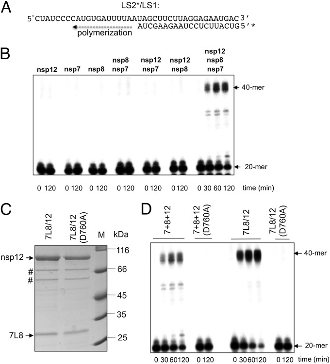
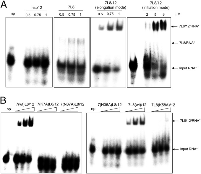
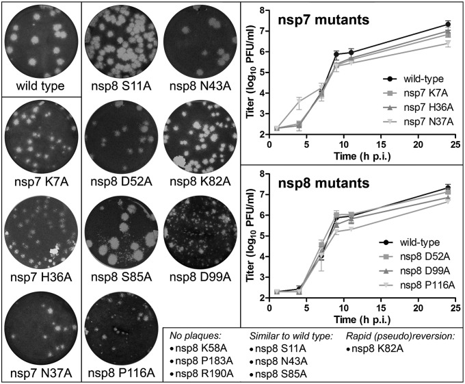
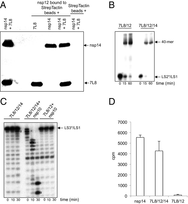
In addition to members causing milder human infections, the Coronaviridae family includes potentially lethal zoonotic agents causing severe acute respiratory syndrome (SARS) and the recently emerged Middle East respiratory syndrome. The ∼30-kb positive-stranded RNA genome of coronaviruses encodes a replication/transcription machinery that is unusually complex and composed of 16 nonstructural proteins (nsps). SARS-CoV nsp12, the canonical RNA-dependent RNA polymerase (RdRp), exhibits poorly processive RNA synthesis in vitro, at odds with the efficient replication of a very large RNA genome in vivo. Here, we report that SARS-CoV nsp7 and nsp8 activate and confer processivity to the RNA-synthesizing activity of nsp12. Using biochemical assays and reverse genetics, the importance of conserved nsp7 and nsp8 residues was probed. Whereas several nsp7 mutations affected virus replication to a limited extent, the replacement of two nsp8 residues (P183 and R190) essential for interaction with nsp12 and a third (K58) critical for the interaction of the polymerase complex with RNA were all lethal to the virus. Without a loss of processivity, the nsp7/nsp8/nsp12 complex can associate with nsp14, a bifunctional enzyme bearing 3'-5' exoribonuclease and RNA cap N7-guanine methyltransferase activities involved in replication fidelity and 5'-RNA capping, respectively. The identification of this tripartite polymerase complex that in turn associates with the nsp14 proofreading enzyme sheds light on how coronaviruses assemble an RNA-synthesizing machinery to replicate the largest known RNA genomes. This protein complex is a fascinating example of the functional integration of RNA polymerase, capping, and proofreading activities.

Keywords: processivity factor; replicative complex reconstitution.


Conflict of interest statement

The authors declare no conflict of interest.

Figures

Fig. 1.
Fig. 1.
SARS-CoV nsp12 polymerase activity is activated by nsp7 and nsp8. (A) Sequence of the RNA primer/template used in this study; the 20-nt primer LS2 was 5′-radiolabeled (marked by *) and annealed to the 40-nt template LS1. (B) Primer extension polymerase assays were performed using LS2*/LS1 as substrate and different combinations of separately purified nsp12, nsp8, and nsp7. RNA products were separated by denaturing gel electrophoresis (20% polyacrylamide/7 M urea) and analyzed by autoradiography. The positions of the primer (20-mer) and the full-length extension product (40-mer) are indicated. (C) WT or mutant (D760A) nsp12 were coexpressed in E. coli with the nsp7-L-nsp8 fusion protein (7L8). After purification of the 7L8/nsp12 complex on a Strep-Tactin column, analysis by 12% SDS/PAGE and Coomassie blue staining of the proteins constituting the complex (nsp12 WT or D760A mutant) was done. # indicates the position of E. coli protein contaminants (defined by MALDI-TOF analysis). (D) Comparison of primer extension polymerase activities of WT and mutant (D760A) nsp12 in the presence of nsp7 and nsp8. Nsp7, nsp8, and nsp12 were either purified and added separately (lanes labeled 7+8+12) or copurified from E. coli as described above (lanes labeled 7L8/12). The reactions were performed on RNA template LS2*/LS1 (see B). Primer conversion rates (at 60 min): 40% for 7+8+12; 67% for 7L8/12; and 0% for 7+8+12(D760A) and 7L8/12(D760A).
Fig. 2.
Fig. 2.
The 7L8/12 polymerase complex catalyzes de novo RNA synthesis. De novo RNA polymerase assays were performed using 500 nM 7L8/12 with 500 nM RNA template (3R), representing the 3′-terminal 339 nt of the SARS-CoV genome. Reaction products, collected at the indicated time points, were analyzed by denaturing agarose gel electrophoresis and autoradiography. Reaction products are identified on the Right of the gel, and radiolabeled RNA size marker (M) is shown to the Left of the panel.
Fig. 3.
Fig. 3.
The 7L8/12 polymerase complex processively replicates the 3R RNA template. (A) Schematic representation of the three experimental setups used, all including 500 nM of 7L8/12 polymerase complex and 100 nM of the 339-nt template 3R; the LS2/LS1 template (Fig. 1A) was used as a trap at 20 µM. NTPs* represents a mix of the four NTPs (500 µM GTP, UTP, CTP, and 50 μM ATP) and 0.17 µM [α-32P]ATP (0.5 µCi/µL). Nucleotide incorporation was followed in time as indicated in the three flow charts. (B) Reactions were started by the addition of NTPs*. RNA products were separated by denaturing acrylamide/urea gels and visualized by autoradiography. RNA products and residual radiolabeled ATP* are indicated on the Right of the autoradiograph.
Fig. 4.
Fig. 4.
Nsp7 and nsp8 confer RNA-binding capacity to the nsp12 RdRp. EMSAs were performed with 100 nM radiolabeled RNA template (LS2*/LS1) and increasing concentrations of the indicated proteins. With the exception of 7L8/12 in initiation mode, all of the other reactions were done with NTPs (500 μM CTP, UTP, GTP and 50 μM 3′-dATP) to facilitate formation of a stalled RNA/enzyme complex, and with 3′-dATP preventing synthesis of full-length RNA product. Reactions were incubated for 60 min at 30 °C and products were analyzed by native 6% PAGE. (A) No protein (np) control and increasing concentrations of nsp12, 7L8, and 7L8/12, as indicated above each panels. (B) No protein (np) control and increasing concentrations (0.5, 0.75, and 1 µM) of 7L8/12 polymerase complexes containing different nsp7 and nsp8 mutants, as indicated above each panel. 7L8/12 in elongation mode gave 42.5% ± 7.5% of shifted probe; 7L8, 9.0% ± 3.5%; 7L8(K58A)/12, 10.0% ± 6.5%, whereas nsp12 alone and the other mutants presented here did not show any ability to bind the probe (0%). These values are averages from quantification of three independent experiments.
Fig. 5.
Fig. 5.
Reverse-genetics analysis of the impact of selected SARS-CoV nsp7 and nsp8 mutations. Plaque phenotypes of viable but crippled mutants with engineered nsp7 and nsp8 mutations are illustrated on the Left. Plaque assays were performed with early progeny virus, harvested from cells at 18 h post full-length RNA transfection (Materials and Methods). On the Right, growth curves are shown for crippled nsp7 and nsp8 mutants and the WT parental virus. For these experiments, mutant virus was harvested from transfected cells at 42 h p.t. After titration of these virus stocks, fresh Vero-E6 cells were infected (M.O.I., 5), and virus production at the given time points was measured by plaque assay. Graphs display mean titers and SDs derived from three independent experiments. Nonviable mutants and mutants with a phenotype similar to WT virus are listed at the Bottom.
Fig. 6.
Fig. 6.
The SARS-CoV 7L8/12/14 complex possesses RdRp, ExoN, and N7-MTase activities. (A) Strep-tagged SARS-CoV nsp12 was bound to Strep-Tactin beads and incubated with 7L8, nsp14, or both simultaneously. After SDS/PAGE and Western blotting, his-tagged proteins (7L8 and nsp14) were revealed using an anti-His5-HRP antibody. (B) Time course primer extension polymerase assays were performed using either the 7L8/12 (500 nM) or the 7L8/12/14 (500 nM) complexes with LS2*/LS1 as primer*/template where LS2 was 5′-radiolabeled (marked by *). RNA products were separated in a denaturing polyacrylamide/urea gel and visualized by autoradiography. (C) Time course exoribonuclease assays were performed using the 7L8/12/14 (500 nM) complex in the absence or presence of 100 nM nsp10, and as control with 7L8/12 (500 nM) plus nsp10 (100 nM). The RNA substrate was a 40-nt RNA (LS1) annealed with 5′-radiolabeled LS3 primer carrying one noncomplementary base at its 3′ end (LS3*) and named LS3*/LS1. Digestion products were separated by denaturing polyacrylamide/urea gel electrophoresis and visualized by autoradiography (Fuji). The “α” symbol indicates RNA cleavage products. (D) AdoMet-dependent N7-MTase activity of the 7L8/12/14 complex. The different purified proteins or protein complexes (nsp14, 300 nM; 7L8/12/14, 300 nM; and 7L8/12, 300 nM) were incubated with substrate GpppAC4 RNA oligonucleotide in the presence of [3H]AdoMet. The methyl transfer to the capped RNA substrate was determined by using a filter-binding assay (as described in ref. 73). All experiments were done in triplicate (SDs are presented).

References

    1. Cameron CE, Götte M. In: Viral Genome Replication. Raney K, editor. New York: Springer; 2009.
    1. Kuhn RJ. Fields’ Virology. 5th Ed. Philadelphia: Lippincott Williams & Wilkins; 2007. pp. 1001–1022.
    1. Lai MMC, Perlman S, Anderson LJ. Fields’ Virology. 5th Ed. Philadelphia: Lippincott Williams & Wilkins; 2007. pp. 1305–1335.
    1. Lazarowitz SG. Fields’ Virology. 5th Ed. Philadelphia: Lippincott Williams & Wilkins; 2007. pp. 641–705.
    1. Domingo E, et al. Basic concepts in RNA virus evolution. FASEB J. 1996;10(8):859–864. - PubMed

Publication types

MeSH terms