. 2014 Sep 16;111(37):13433-8.
doi: 10.1073/pnas.1415357111. Epub 2014 Sep 2.

Activation of iNKT cells by a distinct constituent of the endogenous glucosylceramide fraction

Affiliations

Activation of iNKT cells by a distinct constituent of the endogenous glucosylceramide fraction

Patrick J Brennan et al. Proc Natl Acad Sci U S A. .

Abstract

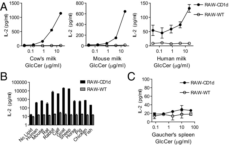
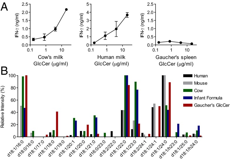
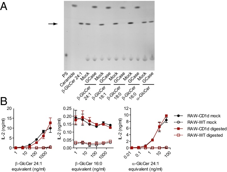
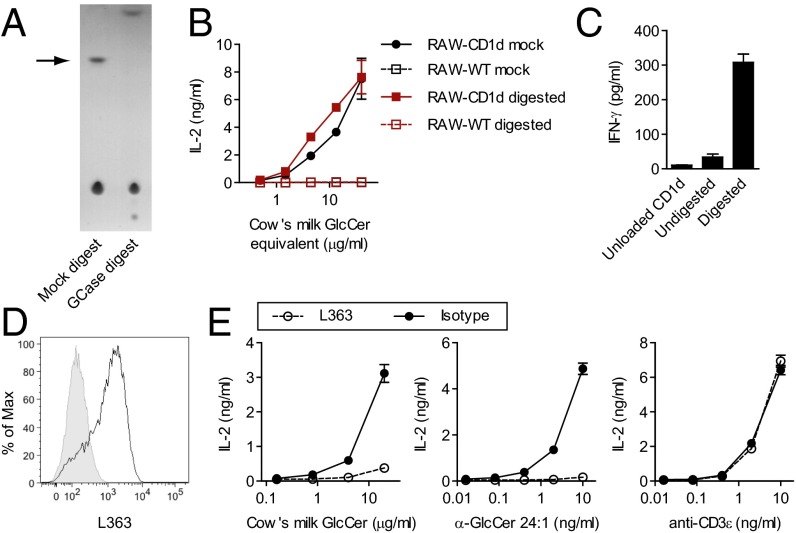
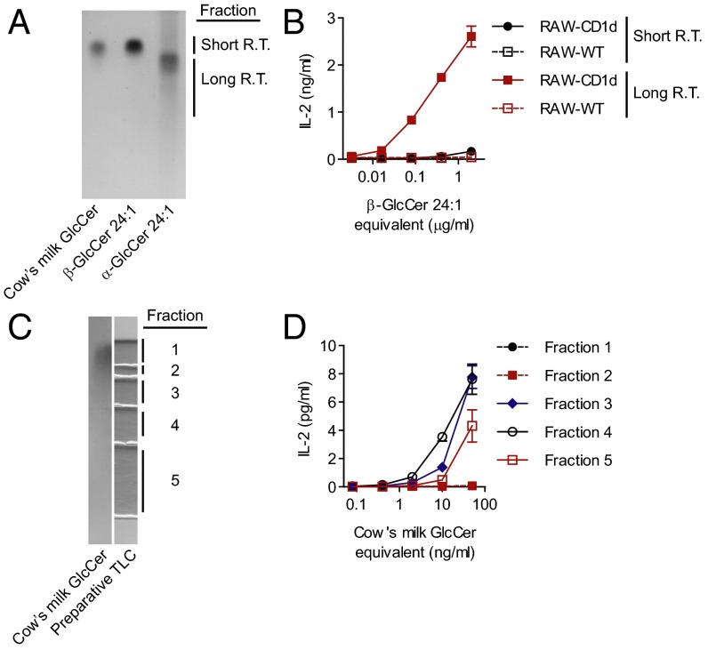
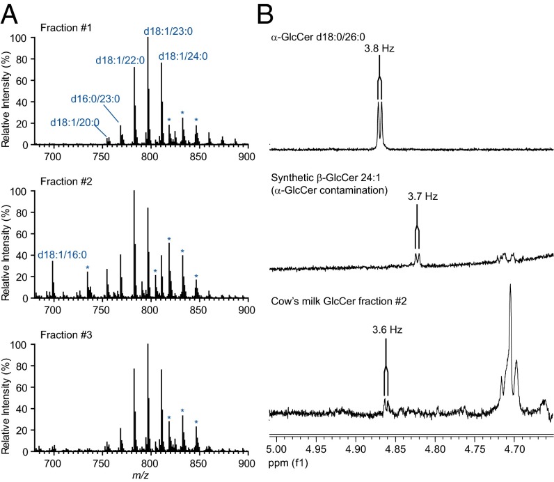
Invariant natural killer T (iNKT) cells are a specialized T-cell subset that recognizes lipids as antigens, contributing to immune responses in diverse disease processes. Experimental data suggests that iNKT cells can recognize both microbial and endogenous lipid antigens. Several candidate endogenous lipid antigens have been proposed, although the contextual role of specific antigens during immune responses remains largely unknown. We have previously reported that mammalian glucosylceramides (GlcCers) activate iNKT cells. GlcCers are found in most mammalian tissues, and exist in variable molecular forms that differ mainly in N-acyl fatty acid chain use. In this report, we purified, characterized, and tested the GlcCer fractions from multiple animal species. Although activity was broadly identified in these GlcCer fractions from mammalian sources, we also found activity properties that could not be reconciled by differences in fatty acid chain use. Enzymatic digestion of β-GlcCer and a chromatographic separation method demonstrated that the activity in the GlcCer fraction was limited to a rare component of this fraction, and was not contained within the bulk of β-GlcCer molecular species. Our data suggest that a minor lipid species that copurifies with β-GlcCer in mammals functions as a lipid self antigen for iNKT cells.

Keywords: CD1d; anomer; antigen presentation; dietary antigen; self-reactive.


Conflict of interest statement

The authors declare no conflict of interest.

Figures

Fig. 1.
Fig. 1.
Activity of glucosylceramide (GlcCer) fractions on mouse iNKT cells. (A) Purified GlcCer from cow’s milk, mouse milk, or human milk was cocultured with the DN32 iNKT cell hybridoma and RAW-CD1d or RAW-WT cells. Response to α-GlcCer d18:1/24:1 is shown for comparison in Fig. S1C. Please note the different scales of the axes between panels, reflecting different response magnitudes. (B) The GlcCer retention time fractions were purified from 500 μg of the indicated serum polar lipids and cocultured with DN32 and RAW-CD1d or RAW-WT cells. (C) GlcCer from the spleen of a human with Gaucher’s disease was cocultured with DN32 and RAW-CD1d or RAW-WT cells. Data presented are release of IL-2 by DN32 as measured by ELISA. Cow’s milk GlcCer was from three independent sources. Human milk GlcCer was tested from two individual donors and 10 pooled donors. Experiments were repeated at least twice, and a representative experiment is shown.
Fig. 2.
Fig. 2.
Activity of glucosylceramides (GlcCers) on human iNKT cells. (A) Purified GlcCer from cow’s milk, human milk, and Gaucher’s spleen were cocultured with a primary human iNKT cell line and human GM-CSF/IL-4-induced antigen-presenting cells. The activity of α-GlcCer d18:1/24:1 is shown for comparison in Fig. S1D. Data are ELISA for IFN-γ release. Three independent experiments were performed and a representative experiment is shown. (B) GlcCers from various mammalian milk sources (human, mouse, cow, infant formula) and Gaucher’s spleen were analyzed by collision-induced dissociation mass spectrometry (CDI-MSn). GlcCer molecular species identified present at more than 5% of the total are on the x axis, and relative intensity of ions corresponding to each molecular species identified is shown on the y axis.
Fig. 3.
Fig. 3.
Glucocerebrosidase (GCase) digestion of synthetic GlcCer compounds. (A) Normal phase TLC of synthetic GlcCers either GCase digested, or mock-digested (no enzyme). For each sample, 2 μg of lipid (or the equivalent amount of lipid added to the enzymatic digestion) was loaded and visualized with α-naphthol stain. An arrow marks the retention time of a GlcCer standard. Phosphatidylserine (PS) is a component of the digestion assay and does not comigrate with GlcCer. Ceramide d18:1/24:1 (Ceramide) shows the migration of free ceramide in this TLC system. Note liberated ceramide densities near the top edge of the plate. (B) Activity of antigenic β-GlcCer d18:1/24:1, nonantigenic β-GlcCer d18:1/16:0, or α-GlcCer d18:1/24:1, either digested or mock-digested was measured by coculture with DN32 and RAW-CD1d or RAW-WT cells after repurification by preparative TLC. Data presented are release of IL-2 by DN32 as measured by ELISA. Experiments were repeated at least twice and a representative experiment is shown.
Fig. 4.
Fig. 4.
GCase digestion of cow’s milk GlcCer. (A) Normal phase TLC of cow’s milk GlcCer, either GCase digested, or mock-digested (no enzyme). Lipid (2 μg; or the equivalent amount based on lipid added to the initial enzymatic digestion) was loaded and visualized with α-naphthol stain. An arrow marks the retention time of a GlcCer standard. (B) Antigenic activity of cow’s milk GlcCer, either digested or mock-digested was measured by coculture with DN32 and RAW-CD1d or RAW-WT cells after repurification by preparative TLC. (C) Cow’s milk GlcCer, either digested or mock-digested, was loaded into biotintylated CD1d and subsequently bound to a streptavidin-coated plate. Activity of loaded lipids was assayed by culturing a primary mouse iNKT cell line with the plate-bound, lipid-loaded CD1d. (D) RAW-CD1d cells were loaded with 10 μg/mL α-GlcCer d18:1/24:1 overnight and then stained with L363, an antibody that is known to stain α-GalCer-loaded CD1d. The black tracing shows lipid-loaded RAW-CD1d cells, shaded tracing is from an unloaded control. (E) L363 was used for functional blocking in a DN32 and RAW-CD1d coculture assay. Data presented in B and E are release of IL-2, and data presented in C are release of IFN-γ (IFN-γ) as measured by ELISA. Experiments were repeated at least twice, and a representative experiment is shown.
Fig. 5.
Fig. 5.
A TLC system for the separation of GlcCer anomers. (A) Cow’s milk GlcCer, a β-GlcCer d18:1/24:1 synthetic, or an α-GlcCer d18:1/24:1 synthetic were resolved on a silica TLC plate using a novel solvent system (HICMW, see Materials and Methods) and visualized with α-naphthol stain. R.T., retention time. (B) A β-GlcCer d18:1/24:1 synthetic known to be contaminated with α-GlcCer was fractionated by preparative TLC using the HICMW system, and the indicated fractions were tested for activity by coculture with the DN32 and RAW-CD1d or RAW-WT cells. (C) Cow’s milk GlcCer was fractionated by preparative TLC using the HICMW system. (D) The indicated fractions were tested for activity by coculture with DN32 and RAW-CD1d or RAW-WT cells. Data presented in B and D are release of IL-2 by DN32 as measured by ELISA. Experiments were repeated at least twice, and a representative experiment is shown.
Fig. 6.
Fig. 6.
Large-scale enrichment of the active fraction in cow’s milk GlcCer. (A) CID-MSn analysis of inactive (fraction 1) and active (Fig. S8B, fractions #2 and #3) fractions, with major GlcCer ions annotated in blue. Asterisk above an ion series indicates an additional adduct (chloride) of an annotated series. (B) Fraction #2 (Fig. S8B) was analyzed by 1D-proton NMR. Signals corresponding to H1 of α-glucose are shown with the coupling constant indicated. 1D spectra from α-GlcCer d18:0/26:0 and β-GlcCer 24:1 (contains 0.5–1% α-GlcCer) are shown for comparison. Large-scale enrichment and experiments were performed twice.

References

    1. Young MH, Gapin L. Group. 2011;1:CD1. - PubMed
    1. Brennan PJ, Brigl M, Brenner MB. Invariant natural killer T cells: an innate activation scheme linked to diverse effector functions. Nat Rev Immunol. 2013;13(2):101–117. - PubMed
    1. Kawano T, et al. CD1d-restricted and TCR-mediated activation of valpha14 NKT cells by glycosylceramides. Science. 1997;278(5343):1626–1629. - PubMed
    1. Kinjo Y, et al. Recognition of bacterial glycosphingolipids by natural killer T cells. Nature. 2005;434(7032):520–525. - PubMed
    1. Mattner J, et al. Exogenous and endogenous glycolipid antigens activate NKT cells during microbial infections. Nature. 2005;434(7032):525–529. - PubMed

Publication types

LinkOut - more resources