. 2014 Jul 15;7(8):4587-95.
eCollection 2014.

Protective effects of 2-deoxy-D-glucose on nephrotoxicity induced by cyclosporine A in rats

Affiliations

Protective effects of 2-deoxy-D-glucose on nephrotoxicity induced by cyclosporine A in rats

Zizhang Ouyang et al. Int J Clin Exp Pathol. .

Abstract

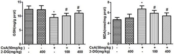
Objective: This study aims to explore the protective effect mechanism of 2-deoxy-D-glucose on nephrotoxicity of cyclosporin A in vivo.

Method: Renal toxicity of SD rats model induced by CsA was established. Serum creatinine, blood urea nitrogen, urine NAG, GSH and MDA were determined and the histopathological changes of rat renal cortex were observed to explore the protective effects of 2-DG on CsA-induced nephrotoxicity.

Results: Serum creatinine, BUN and urinary NAG of rats were significantly changed in experimental groups. Pathological results showed that there was obvious renal tubular injury in model group, however, the renal injury was significantly reduced in pre-treated with 2-DG.

Conclusions: 2-DG had obvious protective effect on nephrotoxicity especially with high dose. This protective effect could be related to the reduction of ROS induced by CsA. However, 2-DG had no effect on the expression of RIP3.

Keywords: 2-deoxy-D-glucose; Cyclosporin A; GSH; MDA; RIP3; SD rats model.


Figures

Figure 1
Figure 1
Effect of 2-DG on CsA induced urine NAG changes in control and treated rats. Values are expressed as mean±SD; n=8 (2 weeks) or n=2 (4 weeks); a: P < 0.05, vs ctrl group; b: P < 0.05, vs CsA group.
Figure 2
Figure 2
Effect of 2-DG on the levels of serum Creatinine of the rats treated with CsA and 2-DG for 2 weeks and 4 weeks. Values are expressed as mean±SD; n=4; *: P < 0.05, vs ctrl group; #: P < 0.05, vs CsA group.
Figure 3
Figure 3
Effect of 2-DG on the levels of serum BUN of the rats treated with CsA and 2-DG for 2 weeks and 4 weeks. Values are expressed as mean±SD; n=4; *: P < 0.05, vs ctrl group; #: P < 0.05, vs CsA group.
Figure 4
Figure 4
Effects of 2-DG on the levels of GSH and MDA in the kidney of the rats treated with CsA for 4 weeks. Values are expressed as mean±SD; n=4; *: P < 0.05, vs ctrl group; #: P < 0.05, vs CsA group.
Figure 5
Figure 5
The expression of RIP3 protein in renal cortex.
Figure 6
Figure 6
Immunohistochemical staining for RIP3 in the renal cortex of rats exposed to CsA and (or) 2-DG (×400). A. Control group; B. 2-DG group; C. CsA group; D. CsA+2-DG(L) group; E. CsA+2-DG(H) group.
Figure 7
Figure 7
The pathological results after being treated with CsA for 2 weeks. A. There was no abnormal renal glomerulus and tubules and no inflammatory cell infiltration in interstitium (control group, HE×200); B. There was no abnormal renal glomerulus and tubules and no inflammatory cell infiltration in interstitium (2-DG group, HE×200); C. There was vacuolar degeneration in some renal tubular epithelia (CsA group, HE×200); D. There was local fibrosis in renal interstitium and acidophilic degeneration in some renal tubular epithelia (CsA group, HE×200); E. There was no abnormal renal glomerulus and tubules and no inflammatory cell infiltration in interstitium (CsA+2-DG low-dosage group, HE×200); F. There was local fibrosis in renal interstitium (yellow arrow) and vacuolar degeneration in some renal tubular epithelia (black arrow) (CsA+2-DG low-dosage group, HE×200); G. There was no abnormal renal glomerulus and tubules and no inflammatory cell infiltration in interstitium (CsA+2-DG high-dosage group, HE×200); H. There was vacuolar degeneration in some renal tubular epithelia (CsA+2-DG high-dosage group, HE×200).
Figure 8
Figure 8
The pathological results after being treated with CsA for 4 weeks. A. There was no abnormal renal glomerulus and tubules and no inflammatory cell infiltration in interstitium (control group, HE×200); B. There was no abnormal renal glomerulus and tubules and no inflammatory cell infiltration in interstitium (2-DG group, HE×200); C. There was vacuolar degeneration in some renal tubular epithelia (black arrow, CsA group, HE×200); D. Renal cysts disappeared (black arrow), glomerular atrophy (red arrow) and regeneration of renal tubular after necrosis (blue arrow) (CsA group, HE×200); E. A small amount of renal tubular epithelial cells shed and formed tube (red arrow), some renal tubular epithelial cells regenerated (yellow arrow) and glomerular congestion (CsA+2-DG low-dosage group, HE×200); F. There was no abnormal renal glomerulus and tubules and no inflammatory cell infiltration in interstitium (CsA+2-DG high-dosage group, HE×200).

References

    1. Laupacis A, Keown PA, Ulan RA, McKenzie N, Stiller CR. Cyclosporin A. a powerful immunosuppressant. Can Med Assoc J. 1982;126:1041–1046. - PMC - PubMed
    1. Hariharan S, Johnson CP, Bresnahan BA, Taranto SE, McIntosh MJ, Stablein D. Improved graft survival after renal transplantation in the United States, 1988 to 1996. N Engl J Med. 2000;342:605–612. - PubMed
    1. Kahan BD. Immunosuppressive therapy. Curr Opin Immunol. 1992;4:553–560. - PubMed
    1. Nakamura T, Nozu K, Iijima K, Yoshikawa N, Moriya Y, Yamamori M, Kako A, Matsuo M, Sakurai A, Okamura N, Ishikawa T, Okumura K, Sakaeda T. Association of cumulative cyclosporine dose with its irreversible nephrotoxicity in Japanese patients with pediatric-onset autoimmune diseases. Biol Pharm Bull. 2007;30:2371–2375. - PubMed
    1. Healy E, Dempsey M, Lally C, Ryan MP. Apoptosis and necrosis: mechanisms of cell death induced by cyclosporine A in a renal proximal tubular cell line. Kidney Int. 1998;54:1955–1966. - PubMed

LinkOut - more resources