. 2014:2014:327452.
doi: 10.1155/2014/327452. Epub 2014 Aug 17.

Antioxidant and anti-inflammatory effects of selected natural compounds contained in a dietary supplement on two human immortalized keratinocyte lines

Affiliations

Antioxidant and anti-inflammatory effects of selected natural compounds contained in a dietary supplement on two human immortalized keratinocyte lines

Elena Fasano et al. Biomed Res Int. 2014.

Abstract

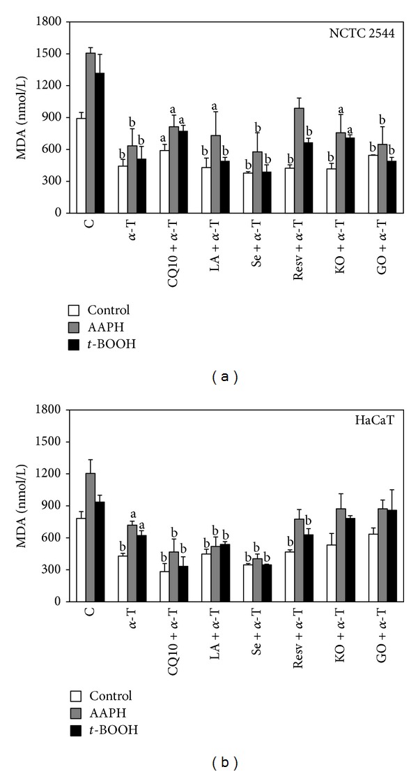
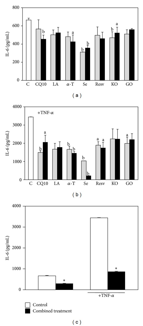
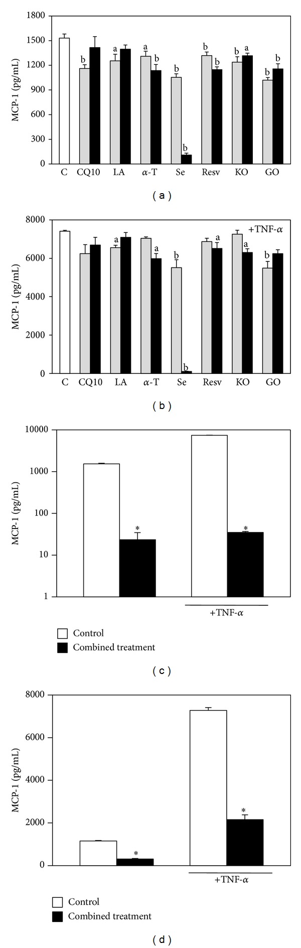
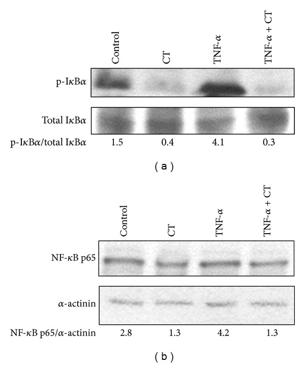
Several advantages may derive from the use of dietary supplements containing multiple natural antioxidants and/or anti-inflammatory agents. At present, however, there is scarce information on the properties and potential of combined supplements. To fill the gap, the antioxidant and anti-inflammatory activities exerted by a combination of seven natural components (coenzyme Q10, krill oil, lipoic acid, resveratrol, grape seed oil, α-tocopherol, and selenium) contained in a dietary supplement used for the prevention of skin disorders were investigated in vitro. Each component was administered, alone or in combination, to human keratinocytes, and the inhibition of Reactive Oxygen Species production and lipid peroxidation as well as the ability to reduce inflammatory cytokine secretion and to modulate Nuclear Factor-κB pathway was evaluated. The combination exhibited high antioxidant activity and in specific conditions the combination's efficiency was higher than that of the most powerful components administered individually. Moreover, the combination showed remarkable anti-inflammatory properties. It reduced more efficiently than each component the secretion of Monocyte Chemoattractant Protein-1, a crucial cytokine for the development of chronic inflammation in skin, and inhibited Nuclear Factor-κB molecular pathway. Overall, our findings suggest that the combined formulation may have the potential to powerfully inhibit oxidative stress and inflammation at skin level.


Figures

Figure 1
Figure 1
Cytokeratin-13 (CK-13) basal expression in NCTC 2544 and HaCaT cells. CK-13-positive cells were evaluated by immunocytochemistry and counted under inverted microscopy. Values represent the means ± SE of CK-13-positive cells counted in 5 and 8 microscopic fields for NCTC 2544 and HaCaT cells, respectively. ∗: significantly different, P < 0.0001, two-tailed unpaired t-test.
Figure 2
Figure 2
Effect of the seven supplement's components on ROS production in keratinocytes in basal conditions. Cells were treated for 24 h with three increasing concentrations of the components administered individually (see Section 2). Data are the means ± SE of a number of determinations ranging from 3 to 8. a: significantly different from control (P < 0.05); b: significantly different from control (P < 0.01), one-way ANOVA followed by Dunnett's test.
Figure 3
Figure 3
Effect of the seven supplement's components on ROS production in keratinocytes exposed to H2O2. Cells were treated for 24 h with three increasing concentrations of the components and then exposed to 100 μM H2O2 for 30 min. The three concentrations used for each component are reported in Section 2. Data are the means ± SE of a number of determinations ranging from 3 to 8. a: significantly different from control (P < 0.05); b: significantly different from control (P < 0.01), one-way ANOVA followed by Dunnett's test.
Figure 4
Figure 4
Effect of the combined treatment with all the seven supplement's components on ROS production in keratinocytes exposed or not to H2O2. Cells were treated simultaneously for 24 h with all the components and then exposed to H2O2 (100 μM, 30 min) or vehicle alone. Each component was used at the highest concentration reported in Section 2. Data are the means ± SE of a number of determinations ranging from 3 to 8. Significance was evaluated by the two-tailed unpaired t-test.
Figure 5
Figure 5
Effect of the combined treatment with all the seven supplement's components on lipid peroxidation in keratinocytes exposed or not to prooxidant agents. Cells were treated simultaneously with all the components for 24 h and then exposed for 1 h to the prooxidant agents AAPH (25 mM) or t-BOOH (0.25 mM) or vehicle alone. Each component was used at the highest concentration reported in Section 2. Lipid peroxidation was evaluated spectrophotometrically as MDA production. Data are the means ± SE of a number of determinations ranging from 3 to 6. ∗: significantly different from respective control (NCTC 2455: CT versus control, P < 0.007; CT +AAPH versus AAPH, P < 0.0003; CT +t-BOOH versus t-BOOH, P < 0.04; HaCaT: CT versus control, P < 0.03; CT +AAPH versus AAPH, P < 0.02; CT +t-BOOH versus t-BOOH, P < 0.03, two-tailed unpaired t-test).
Figure 6
Figure 6
Effect of α-T alone or combined with each of the other supplement's components on lipid peroxidation in keratinocytes exposed or not to prooxidant agents. Cells were treated with α-T alone or combined with each of the other supplement's components for 24 h and then exposed for 1 h to the prooxidant agents AAPH (25 mM) or t-BOOH (0.25 mM) or vehicle alone. Each component was used at the highest concentration reported in Section 2. Lipid peroxidation was evaluated spectrophotometrically as MDA production. Data are the means ± SE of a number of determinations ranging from 3 to 6. a: significantly different from respective control (P < 0.05); b: significantly different from respective control (P < 0.01), one-way ANOVA followed by Dunnett's test.
Figure 7
Figure 7
Effect of the seven supplement's components on HaCaT keratinocyte IL-6 production. Cells were exposed for 18 h to increasing concentrations of the components in the absence (a) or in the presence (b) of TNF-α (20 ng/mL, 6 h pretreatment and further 18 h with the components); the two concentrations used for each component are the lowest and highest concentrations reported in Section 2. Data are the means ± SE of 4 determinations. a: significantly different from control (P < 0.05); b: significantly different from control (P < 0.01), one-way ANOVA followed by Dunnett's test. (c) Cells were treated simultaneously with all the components, both in basal conditions or in the presence of TNF-α (20 ng/mL), following the timing of the previous experiment. Each component was used at the highest concentration reported in Section 2. Data are the means ± SE of 4 determinations. ∗: significantly different from respective control (P < 0.0001, two-tailed unpaired t-test).
Figure 8
Figure 8
Effect of the seven supplement's components on HaCaT keratinocyte MCP-1 production. Cells were exposed to increasing concentrations of the components in the absence (a) or in the presence (b) of TNF-α (20 ng/mL, 6 h pretreatment and further 18 h with the components); the two concentrations used for each component are the lowest and highest concentrations reported in Section 2. Data are the means ± SE of a number of determinations ranging from 4 to 6. a: significantly different from control (P < 0.05); b: significantly different from control (P < 0.01), one-way ANOVA followed by Dunnett's test. (c) Cells were exposed simultaneously to all the components, both in basal conditions or in the presence of TNF-α (20 ng/mL), following the timing of the previous experiment. Each component was used at the highest concentration reported in Section 2. Data are the means ± SE of a number of determinations ranging from 3 to 6. ∗: significantly different from respective control (P < 0.0001, two-tailed unpaired t-test). (d) The cells were exposed simultaneously to all the components excluding Se, both in basal conditions or in the presence of TNF-α (20 ng/mL), following the timing of the previous experiments. Each component was used at the highest concentration reported in Section 2. Data are the means ± SE of a number of determinations ranging from 6 to 8. ∗: significantly different from respective control (P < 0.0001, two-tailed unpaired t-test).
Figure 9
Figure 9
Effect of the combined treatment with all the seven supplement's components on IκBα phosphorylation and p65 expression in HaCaT keratinocytes. Cells were exposed to the combined treatment (CT) in the absence or in the presence of TNF-α (20 ng/mL) for 5 h. (a) A representative Western Blot of three similar experiments with the corresponding p-IκBα/IκBα ratio is shown. (b) A representative Western Blot of three similar experiments with the corresponding p65/α-actinin ratio is shown.

References

    1. Calviello G, Su H, Weylandt KH, Fasano E, Serini S, Cittadini A. Experimental evidence of ω-3 polyunsaturated fatty acid modulation of inflammatory cytokines and bioactive lipid mediators: their potential role in inflammatory, neurodegenerative, and neoplastic diseases. BioMed Research International. 2013;2013:13 pages.743171 - PMC - PubMed
    1. Manfredini S, Vertuani S, Manfredi B, Rossoni G, Calviello G, Palozza P. Novel antioxidant agents deriving from molecular combinations of vitamins C and E analogues: 3,4-dihydroxy-5(R) Bioorganic and Medicinal Chemistry. 2000;8(12):2791–2801. - PubMed
    1. Trombino S, Serini S, di Nicuolo F, et al. Antioxidant effect of ferulic acid in isolated membranes and intact cells: synergistic interactions with alpha-tocopherol, beta-carotene, and ascorbic acid. Journal of Agricultural and Food Chemistry. 2004;52(8):2411–2420. - PubMed
    1. Bjelakovic G, Nikolova D, Gluud C. Antioxidant supplements and mortality. Current Opinion in Clinical Nutrition and Metabolic Care. 2014;17(1):40–44. - PubMed
    1. Speciale A, Chirafisi J, Saija A, Cimino F. Nutritional antioxidants and adaptive cell responses: an update. Current Molecular Medicine. 2011;11(9):770–789. - PubMed

Publication types

MeSH terms