. 2014 Sep 8;9(9):e107001.
doi: 10.1371/journal.pone.0107001. eCollection 2014.

Paracrine effect of mesenchymal stem cells derived from human adipose tissue in bone regeneration

Affiliations

Paracrine effect of mesenchymal stem cells derived from human adipose tissue in bone regeneration

Itali Linero et al. PLoS One. .

Erratum in

Abstract

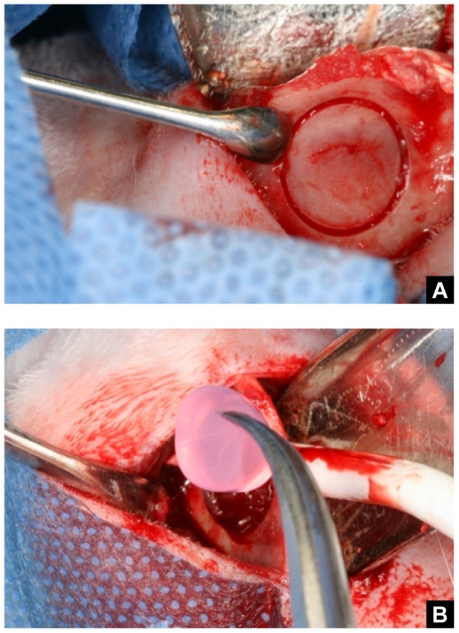
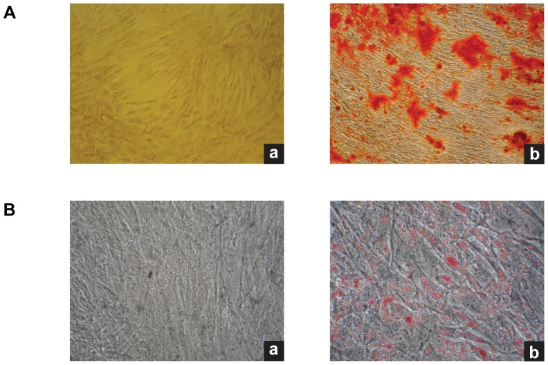
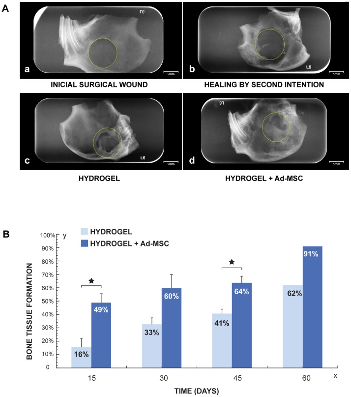
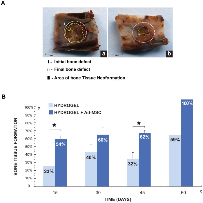
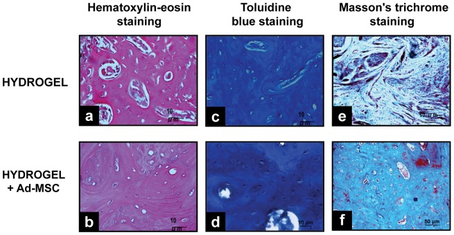
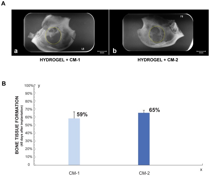
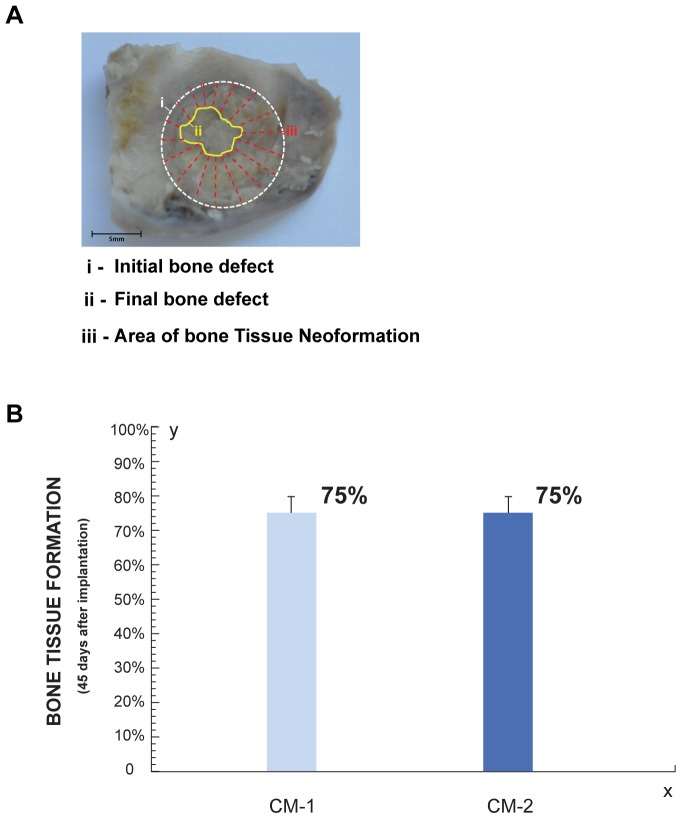
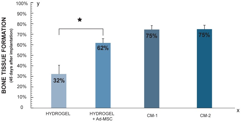
Mesenchymal stem cell (MSC) transplantation has proved to be a promising strategy in cell therapy and regenerative medicine. Although their mechanism of action is not completely clear, it has been suggested that their therapeutic activity may be mediated by a paracrine effect. The main goal of this study was to evaluate by radiographic, morphometric and histological analysis the ability of mesenchymal stem cells derived from human adipose tissue (Ad-MSC) and their conditioned medium (CM), to repair surgical bone lesions using an in vivo model (rabbit mandibles). The results demonstrated that both, Ad-MSC and CM, induce bone regeneration in surgically created lesions in rabbit's jaws, suggesting that Ad-MSC improve the process of bone regeneration mainly by releasing paracrine factors. The evidence of the paracrine effect of MSC on bone regeneration has a major impact on regenerative medicine, and the use of their CM can address some issues and difficulties related to cell transplants. In particular, CM can be easily stored and transported, and is easier to handle by medical personnel during clinical procedures.


Conflict of interest statement

Competing Interests: The authors have declared that no competing interests exist.

Figures

Figure 1
Figure 1. Surgical procedure.
A. Circular demarcation of bone defect. B. Implantation of HBPH.
Figure 2
Figure 2. Ad-MSC characterization: Flow Cytometry.
Sixth passage, 70% confluence Ad-MSC, were labeled with monoclonal antibodies and analyzed by flow cytometry. A. Isotype controls for each of marker. B. Ad-MSC labeled with CD34-FITC, CD45-RPECy5, HLA II-RPE, CD105-PE, CD90-Alexa, HLA I-FITC.
Figure 3
Figure 3. Ad-MSC characterization.
A. Osteogenic differentiation of Ad-MSC. Osteogenic differentiation was evidenced by the detection of calcium deposits with Alizarin Red staining. a. Control Ad-MSCs without osteogenic induction. b. Ad-MSC cultured for 3 weeks in osteogenic differentiation medium. B. Adipogenic differentiation of Ad-MSC. Adipogenic diferentiation was evidenced by the formation of lipid vacuoles after three weeks of cultivation in adipogenic induction medium. a. Control cells without induction. b. Lipid vacuoles staining with oil red O. 10× magnification.
Figure 4
Figure 4. Radiographic Analysis of bone regeneration by implanting HBPHs with Ad-MSC.
A. Radiographic comparison of bone defects at 45 days with different treatments. a. Initial size of surgical wound. b. Healing by second intention. c. Bone defect treated with hydrogel. d. Bone defect treated hydrogel with Ad-MSCs. B. Histogram represent the average of newly formed bone tissue at 15, 30, 45 (n = 4) and 60 (n = 1) days after grafting Hydrogel with Ad-MSC (dark blue) and Hydrogel without Ad-MSC (light blue).
Figure 5
Figure 5. Morphometric Analysis of bone regeneration by implanting HBPHs with Ad-MSC.
A. Surgical specimens 45 days after implantation. a. Hydrogel. b. Hydrogel with Ad-MSC. i, initial bone defect (white circle), ii, final bone defect (yellow line) and iii, new formed bone tissue (red lines). B. Percentage of newly formed bone at 15, 30, 45 (n = 4) and 60 days (n = 1), after application of Hydrogel with or without Ad-MSC.
Figure 6
Figure 6. Histological Analysis of bone regeneration by implanting HBPHs with Ad-MSC.
Bone defects treated with Hydrogel and Hydrogel with Ad-MSCs, 45 days after implantation. a, b, hematoxylin and eosin staining, showing a mild chronic inflammatory response. c, d, blue toluidine staining, evidencing intramembranous ossification. e, f, Masson trichrome staining, showing a better organized bone tissue and increased calcification, where hydrogels with Ad-MSC were implanted (Magnification 10×).
Figure 7
Figure 7. Tracking of implanted Ad-MSC.
Immunohistochemical detection of positive human ß-2 microglobulin Ad-MSC. a, positive control, human skin. b, positive control, human bone. c, negative control, rabbit granulation tissue. Tissue regeneration zone after implantation of blood plasma hydrogel with Ad-MSCs. d. 3 days. e, 6 days. f, 12 days (Magnification 10×).
Figure 8
Figure 8. Radiographic Analysis of bone regeneration by implanting HBPHs with CM.
A Radiographic comparison of bone defects at 45 days with CM. a. Bone defect treated with Hydrogel with CM-1. b. Bone defect treated with Hydrogel with CM-2. The circle represents the initial size of bone defect. B. Histogram shown the percentage of newly formed bone tissue 45 days after implantation of Hydrogel with CM (n = 3).
Figure 9
Figure 9. Morphometric Analysis of bone regeneration by implanting HBPHs with CM.
A. Surgical specimens 45 days after implantation of hydrogel with CM-1. i, initial bone defect (white circle), ii, final bone defect (yellow line) and iii, new formed bone tissue (red lines). Surgical specimens 45 days after implantation. B. Histogram shown the percentage of bone neoformation 45 days after implantation of Hydrogel with CM (n = 3).
Figure 10
Figure 10. Morphometric Analysis of bone regeneration by implanting HBPHs, HBPHs with Ad- MSC and HBPHs with CM.
Percentage of newly formed bone in defects treated with and without Ad-MSC (n = 4) and CM (n = 3). Bone regeneration process improves substantially where hydrogels with Ad-MSC or CM were implanted.
Figure 11
Figure 11. Histological Analysis of bone regeneration by implanting HBPHs with CM.
Histological Analysis of bone defects treated with Hydrogel and Hydrogel with CM, 45 days after implantation. a, b, hematoxylin and eosin staining, showing a mild chronic inflammatory response. c, d, blue toluidine staining, evidencing intramembranous ossification. e, f, Masson trichrome staining, showing a the organized and calcification of bone tissue.

References

    1. Salem HK, Thiemermann C (2010) Mesenchymal stromal cells: current understanding and clinical status. Stem Cells 28: 585–596. - PMC - PubMed
    1. Yamachika E, Tsujigiwa H, Matsubara M, Hirata Y, Kita K, et al. (2012) Basic fibroblast growth factor supports expansion of mouse compact bone-derived mesenchymal stem cells (MSCs) and regeneration of bone from MSC in vivo. J Mol Histol 43: 223–233. - PubMed
    1. Im GI, Shin YW, Lee KB (2005) Do adipose tissue-derived mesenchymal stem cells have the same osteogenic and chondrogenic potential as bone marrow-derived cells? Osteoarthritis Cartilage 13: 845–853. - PubMed
    1. Zuk PA, Zhu M, Ashjian P, De Ugarte DA, Huang JI, et al. (2002) Human adipose tissue is a source of multipotent stem cells. Mol Biol Cell 13: 4279–4295. - PMC - PubMed
    1. Monaco E, Bionaz M, Hollister SJ, Wheeler MB Strategies for regeneration of the bone using porcine adult adipose-derived mesenchymal stem cells. Theriogenology 75: 1381–1399. - PubMed

Publication types

MeSH terms