. 2014 Aug;85(8):798-804.
doi: 10.3357/ASEM.3905.2014.

Optimal 3D culture of primary articular chondrocytes for use in the rotating wall vessel bioreactor

Affiliations

Optimal 3D culture of primary articular chondrocytes for use in the rotating wall vessel bioreactor

Liliana F Mellor et al. Aviat Space Environ Med. 2014 Aug.

Abstract

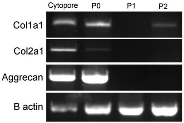
Introduction: Reliable culturing methods for primary articular chondrocytes are essential to study the effects of loading and unloading on joint tissue at the cellular level. Due to the limited proliferation capacity of primary chondrocytes and their tendency to dedifferentiate in conventional culture conditions, long-term culturing conditions of primary chondrocytes can be challenging. The goal of this study was to develop a suspension culturing technique that not only would retain the cellular morphology, but also maintain the gene expression characteristics of primary articular chondrocytes.

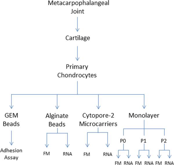
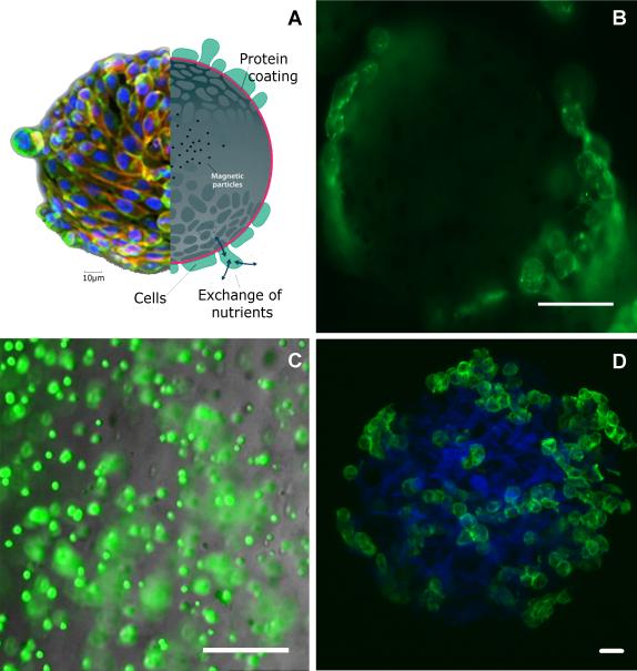
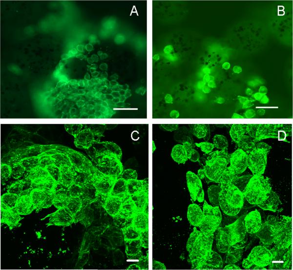
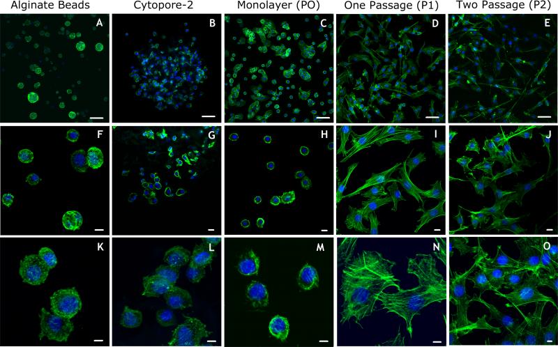
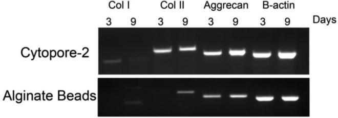
Methods: Three-dimensional culturing methods were compared and optimized for primary articular chondrocytes in the rotating wall vessel bioreactor, which changes the mechanical culture conditions to provide a form of suspension culture optimized for low shear and turbulence. We performed gene expression analysis and morphological characterization of cells cultured in alginate beads, Cytopore-2 microcarriers, primary monolayer culture, and passaged monolayer cultures using reverse transcription-PCR and laser scanning confocal microscopy.

Results: Primary chondrocytes grown on Cytopore-2 microcarriers maintained the phenotypical morphology and gene expression pattern observed in primary bovine articular chondrocytes, and retained these characteristics for up to 9 d.

Discussion: Our results provide a novel and alternative culturing technique for primary chondrocytes suitable for studies that require suspension such as those using the rotating wall vessel bioreactor. In addition, we provide an alternative culturing technique for primary chondrocytes that can impact future mechanistic studies of osteoarthritis progression, treatments for cartilage damage and repair, and cartilage tissue engineering.


Figures

Figure 1
Figure 1. Overview of the experimental design used in this study
Primary chondrocytes were obtained from the metacarpophalangeal joint by aseptic technique and digestion using Pronase and collagenase sequentially. After filtering and washing chondrocytes, cells were counted and cultured either as a monolayer and monitored after successive passages (P0, P1, P2) or on a substrate that supported a 3D cell culture, Cytopore-2, alginate, or Global Eucaryotic Microcarriers (GEM) beads. Cells cultured on different substrates were analyzed for 1) adhesion to specific extracellular matrix molecules, 2) gene expression (RNA), and 3) cytoskeletal organization by fluorescence microscopy (FM).
Figure 2
Figure 2. Three dimensional culturing techniques
The RWV bioreactor requires suspension cultures. A. Global Eukaryotic Microcarriers (GEM), magnetic beads coated with protein to create a substrate for the cells to attach (image adapted from Global Cell Solutions website). B. Fluorescence image of chondrocytes (green=phalloidin bound to the actin cytoskeleton) seeded on GEM beads. C. Epifluorescence image of chondrocytes encapsulated in an alginate bead (green=calcein-AM). D. Confocal image of a Cytopore-2 microcarrier (blue) seeded with chondrocytes (green= phalloidin bound to the actin cytoskeleton).Scale bar dimensions: B. 50μm; C. 100 μm; D. 20 μm.
Figure 3
Figure 3. Chondrocytes detached from protein-coated beads during culture in RWV bioreactor
A. Chondrocytes attached to protein-coated magnetic bead in conventional culturing conditions by epifluorescence microscopy. B. Chondrocytes detached from same beads as shown in A, but under culture conditions using the RWV bioreactor. C. Chondrocytes shown in A at higher magnification. D Chondrocytes shown in B. at higher magnification. Scale bar dimensions: A. and B. 50μm; C. and D. 20 μm.
Figure 4
Figure 4. Primary bovine chondrocyte morphology using different culture techniques
A. Chondrocytes seeded in alginate. B. Chondrocytes seeded on Cytopore-2 microcarriers. C. Primary chondrocytes plated on tissue culture plastic for three days as a monolayer (P0). D. Chondrocytes from C, after trypsin-EDTA treatment and passage onto new tissue culture plastic, grown for three additional days (P1). E. Chondrocytes from D, after an additional trypsin-EDTA treatment and passaged again onto new tissue culture plastic, grown for three additional days (P2). F. and K. Higher magnifications of cells shown in A. G. and L. Higher magnification of cells shown in B. H. and M. Higher magnification of cells shown in C. I. and N. Higher magnification of cells shown in D. J. and O. Higher magnification of cells shown in E. All cells were fixed and stained with AlexaFluor488-phalloidin (green=phalloidin bound to the actin cytoskeleton) to observe changes in the cytoskeletal and cellular morphology. Typical cortical localization of actin and morphology of primary chondrocytes was retained in alginate beads and Cytopore-2 microcarriers as well as chondrocytes prior to trypsin-EDTA treatment and passage (see A, B, and C). Cells became more fibroblast-like in subsequent passages, consistent with dedifferentiation. Scale bar dimensions: A-E, 50 μm; F-J, 10 μm; K-O, 5 μm.
Figure 5
Figure 5. RT-PCR
RNA was extracted from chondrocytes cultured on Cytopore-2 microcarriers or monolayers before passaging with trypsin-EDTA (P0) or after a single (P1) or double passage (P2). Col1a1, Col2a1, aggrecan, and beta actin were detected by RT-PCR. Characteristic gene expression of primary chondrocytes was retained in chondrocytes seeded on Cytopore-2 microcarriers. Chondrocytes grown in a monolayer with no passages (P0) showed a slight decrease in Col2a1 signal while cells in P1 and P2 lost expression of the characteristic markers of chondrocytes.
Figure 6
Figure 6. RT-PCR of chondrocytes grown in alginate beads vs. Cytopore-2 microcarriers for 3 and 9 days
RNA extracted from chondrocytes cultured in alginate beads and Cytopore-2 microcarriers after 3 days and 9 days in culture. Expression of Col1a1, Col2a1, aggrecan, and beta actin were detected by RT-PCR at 3 and 9 days in culture in Cytopore-2 microcarriers and alginate beads.

References

    1. Ab-Rahim S, Selvaratnam L, Raghavendran HR, Kamarul T. Chondrocyte-alginate constructs with or without TGF-beta1 produces superior extracellular matrix expression than monolayer cultures. Mol Cell Biochem. 2013 Apr;376(1-2):11–20. - PubMed
    1. Barzegari A, Saei AA. An update to space biomedical research: tissue engineering in microgravity bioreactors. Bioimpacts. 2012;2(1):23–32. - PMC - PubMed
    1. Connelly JT, Vanderploeg EJ, Levenston ME. The influence of cyclic tension amplitude on chondrocyte matrix synthesis: experimental and finite element analyses. Biorheology. 2004;41(3-4):377–87. - PubMed
    1. Dellacorte C. Isolation of nucleic acids from the sea anemone Condylactis gigantea (Cnidaria: Anthozoa). Tissue Cell. 1994;26(4):613–9. - PubMed
    1. Gefen O, Gabay C, Mumcuoglu M, Engel G, Balaban NQ. Single-cell protein induction dynamics reveals a period of vulnerability to antibiotics in persister bacteria. Proceedings of the National Academy of Sciences of the United States of America. 2008;105(16):6145–9. - PMC - PubMed

Publication types

LinkOut - more resources