. 2014 Sep 29;30(6):645-59.
doi: 10.1016/j.devcel.2014.07.001. Epub 2014 Sep 4.

Local CRH signaling promotes synaptogenesis and circuit integration of adult-born neurons

Affiliations

Local CRH signaling promotes synaptogenesis and circuit integration of adult-born neurons

Isabella Garcia et al. Dev Cell. .

Abstract

Neural activity either enhances or impairs de novo synaptogenesis and circuit integration of neurons, but how this activity is mechanistically relayed in the adult brain is largely unknown. Neuropeptide-expressing interneurons are widespread throughout the brain and are key candidates for conveying neural activity downstream via neuromodulatory pathways that are distinct from classical neurotransmission. With the goal of identifying signaling mechanisms that underlie neuronal circuit integration in the adult brain, we have virally traced local corticotropin-releasing hormone (CRH)-expressing inhibitory interneurons with extensive presynaptic inputs onto new neurons that are continuously integrated into the adult rodent olfactory bulb. Local CRH signaling onto adult-born neurons promotes and/or stabilizes chemical synapses in the olfactory bulb, revealing a neuromodulatory mechanism for continued circuit plasticity, synapse formation, and integration of new neurons in the adult brain.


Figures

Figure 1
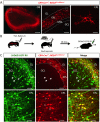
Figure 1. Adult-born granule cells receive presynaptic inputs from external plexiform layer interneurons
(A) Genetic strategy for targeting adult-born neurons for transsynaptic tracing. (B) Dorsal view of mouse brain showing a labeled OB 7 days post infection with SADG-EGFP RV (scale bar 500 μm). (C) Cross-section of (B) (RMS-rostral migratory stream, GCL-granule cell layer, EPL-external plexiform layer, GL-glomerular layer; scale bar 300 μm). (D) High magnification view of (C). tdTomato+/EGFP+ cells (arrows) are newborn neuron ‘source cells’. EGFP+ cells are presynaptic inputs (open arrowheads mark EPL presynaptic inputs, asterisks mark mitral cell inputs; MCL-mitral cell layer; scale bar 80 μm). Inset shows a ‘source cell’ (scale bar 15 μm). (E) EPL presynaptic inputs are PV+ (arrows, scale bar 10 μm), and CRH+ (F) (arrows, arrowheads mark extracellular CRH, scale bar 10 μm). See also Figure S1.
Figure 2
Figure 2. Local CRH+ EPL interneurons make connections onto adult-born granule cells
(A) Genetic lineage of CRH+ neurons in the OB of CRH-Cre+/−; ROSALSL-tdTom+/− mice (GL-glomerular layer, EPL-external plexiform layer, MCL-mitral cell layer, GCL-granule cell layer, scale bars 500, 150, and 25 μm). (B) Experimental scheme to identify presynaptic inputs in CRH lineage traced mice. (C) SADΔG-EGFP RV transsynaptic tracing in CRH-Cre+/−; ROSALSL-tdTom+/− mice (arrows identify newborn neuron ‘source cells’, open arrowheads mark CRH+ presynaptic EPL interneurons, scale bars 150 and 80 μm). See also Figure S2.
Figure 3
Figure 3. Adult-born granule cells dynamically express CRHR1
(A) Semi-quantitative RT-PCR for CRH and CRHR1/2 of whole OB RNA. (B) OB cross-section of CRHR1-EGFP BAC transgenic mice (RMS-rostral migratory stream, GCL-granule cell layer, EPL-external plexiform layer, GL-glomerular layer, scale bar 200 μm). (C) Reporter expression of CRHR1-EGFP; CRH-Cre+/−; ROSALSL-tdTom+/− mice (arrows point to CRHR1+ granule cells, open arrowheads mark CRH+ EPL interneurons, scale bars 60 and 20 μm). (D) Experimental scheme to determine the developmental expression profile of CRHR1 expression in granule cells. (E) CRHR1-expression in newborn neurons 28 days post EdU injection (scale bar 60 μm). (F) Quantification of CRHR1-expression in granule cells (data points represent averages +/− SEM, n=3 animals per time point). (G) CRHR1::EGFP expression in adult-born granule cells (scale bars 100 and 20 μm). See also Figure S3.
Figure 4
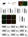
Figure 4. CRH signaling is required for normal levels of adult-born granule cell survival and synaptic protein expression
(A) Experimental scheme to determine cellular proliferation and granule cell survival in CRH mutant alleles (scale bars 100 and 15 μm). (B) Quantification of proliferating cells (BrdU and Ki67 double-positive cells) in the SVZ of control and CRH−/− mice (* p< 0.05 Student’s t-test). (C) Quantification of adult-born granule cell survival in control and CRH−/− mice. (D) Quantification of proliferating cells in the SVZ of CRHR1−/− mice (p>0.05 Student’s t-test). (E) Quantification of adult-born granule cell survival in control and CRHR1−/− mice (*p<0.001 Student’s t-test). (F) Representative images of the GCL of CRHR1+/+ and CRHR1f/f mice that expressed Cre-EGFP or tdTomato in granule cells (scale bar 50 μm). (G) Quantification of the ratio of Cre-EGFP+/tdTom+ granule cells (* p<0.01 Student’s t-test). (H-P) Western blots of the synaptic proteins Synapsin, PSD95, and NR2B of OBs of CRHR1−/−, CRHR1−/−, and CRHR1f/f mice injected with Cre or control viruses (* p<0.05 Student’s t-test). All data points averages +/− SEM, n=4 animals each. See also Figure S4.
Figure 5
Figure 5. Constitutive CRHR-signaling in granule cells promotes synaptogenic changes in the OB
(A) Representative images of granule cells expressing tdTomato or tdTomato and a constitutively-active CRHR1-EGFP fusion construct ((CA)CRHR::EGFP, scale bar 50 μm). (B) Quantification of average granule cell length (p>0.05 Student’s t-test), (C) mean total dendrite length (* p<0.05), and (D) average dendritic branch number in tdTomato and (CA)CRHR+ granule cells (*p<0.01 Student’s t-test). (E) Scholl analysis of the number of intersections in tdTomato or (CA)CRHR+ granule cells (*p<0.05 ANOVA). (F) Quantification of the number of dendritic spines between tdTomato and (CA)CRHR+ granule cells (*p<0.05 ANOVA). N=10 cells each from 3 animals. (G) Representative granule cell morphology reconstructions. (H) Experimental scheme for targeting CRHR1+ OB granule cells for constitutive CRHR1-activation. (I) Expression pattern of AAV-flex-(CA)CRHR::GFP in granule cells (arrows point to (CA)CRHR::GFP+ neurons, scale bars 1000, 100, and 20 μm). (J-O) Western blot analysis of synaptic protein expression of CRHR-Cre+/− OBs injected with either flexed GFP or flexed-(CA)CRHR-GFP AAV (* p<0.05 Student’s t-test, n=4 animals each). All data points averages +/− SEM. See also Figure S5.
Figure 6
Figure 6. Constitutive CRHR-signaling in granule cells promotes synaptic and circuit plasticity in the OB
(A) Representative trace of CRHR1-EGFP+ granule cell before and after CRH application (500 nM CRH). (B) Average mEPSCs before and after CRH. (C-D) Quantification of the mEPSC frequency and amplitude before and after CRH bath application (*p<0.05, n = 13 cells from 3 animals). (E) Representative mEPSC traces of granule cells from CRHR-Cre+/− mice injected with either AAV-flexed EGFP or AAV-flexed-(CA)CRHR::EGFP. (F) Average mEPSCs of EGFP and (CA)CRHR1::EGFP+ granule cells. (G-H) Quantified granule cell mEPSC frequency and amplitude (*p<0.05 Student’s t-test, n = 11 granule cells per group from 3 animals). (I) Representative mIPSC traces of mitral cells from CRHR-Cre+/− mice injected with either AAV-flexed EGFP or AAV-flexed-(CA)CRHR::EGFP. (J) Average mitral cell mIPSCs. (K-L) Average frequency and amplitude of mitral cell mIPSCs in CRHR-Cre+/− OBs in which CRHR-expressing granule cells express either EGFP or (CA)CRHR1::EGFP (* p<0.05 Student’s t-test, n = 13 mitral cells per group from 3 animals). All data points represent mean ±SEM.
Figure 7
Figure 7. Optogenetic activation of CRH+ EPL interneurons induces synaptogenesis in the OB
(A) ChR2 expression pattern of CRH-Cre+/−; ROSALSL-ChR2+/− mice (scale bars 200 and 50 μm). (B-C) Quantification of CRH concentration with hypothalamic or OB optogenetic activation in ROSALSL-ChR2+/− (control) or CRH-Cre+/−; ROSALSL-ChR2+/− mice (*p<0.05 Student’s t-test, n = 3 animals per group). (D) Experimental scheme for in vivo photostimulation of CRH+ EPL interneurons. (E-H) Western blot analysis of synaptic protein expression of ROSALSL-ChR2+/− (control) or CRH-Cre+/−; ROSALSL-ChR2+/− mice (*p<0.05 Student’s t-test, n = 4 animals per group). (I) Model: CRH signaling between local EPL interneurons and granule cells promotes synapse formation and stabilization in the OB. All data points represent mean ±SEM. See also Figure S6.

Comment in

Similar articles

Cited by

References

    1. Abraham NM, Egger V, Shimshek DR, Renden R, Fukunaga I, Sprengel R, Seeburg PH, Klugmann M, Margrie TW, Schaefer AT, et al. Synaptic inhibition in the olfactory bulb accelerates odor discrimination in mice. Neuron. 2010;65:399–411. - PMC - PubMed
    1. Abrous DN, Koehl M, Le Moal M. Adult neurogenesis: from precursors to network and physiology. Physiol Rev. 2005;85:523–569. - PubMed
    1. Alvarez-Buylla A, Temple S. Stem cells in the developing and adult nervous system. J Neurobiol. 1998;36:105–110. - PubMed
    1. Arenkiel BR, Hasegawa H, Yi JJ, Larsen RS, Wallace ML, Philpot BD, Wang F, Ehlers MD. Activity-induced remodeling of olfactory bulb microcircuits revealed by monosynaptic tracing. PLoS One. 2011;6:e29423. - PMC - PubMed
    1. Atasoy D, Aponte Y, Su HH, Sternson SM. A FLEX switch targets Channelrhodopsin-2 to multiple cell types for imaging and long-range circuit mapping. J Neurosci. 2008;28:7025–7030. - PMC - PubMed

Publication types

Substances

LinkOut - more resources