. 2014 Sep 11;8(5):1328-38.
doi: 10.1016/j.celrep.2014.07.062. Epub 2014 Sep 4.

Myeloid malignancies with chromosome 5q deletions acquire a dependency on an intrachromosomal NF-κB gene network

Affiliations

Myeloid malignancies with chromosome 5q deletions acquire a dependency on an intrachromosomal NF-κB gene network

Jing Fang et al. Cell Rep. .

Abstract

Chromosome 5q deletions (del[5q]) are common in high-risk (HR) myelodysplastic syndrome (MDS) and acute myeloid leukemia (AML); however, the gene regulatory networks that sustain these aggressive diseases are unknown. Reduced miR-146a expression in del(5q) HR MDS/AML and miR-146a(-/-) hematopoietic stem/progenitor cells (HSPCs) results in TRAF6/NF-κB activation. Increased survival and proliferation of HSPCs from miR-146a(low) HR MDS/AML is sustained by a neighboring haploid gene, SQSTM1 (p62), expressed from the intact 5q allele. Overexpression of p62 from the intact allele occurs through NF-κB-dependent feedforward signaling mediated by miR-146a deficiency. p62 is necessary for TRAF6-mediated NF-κB signaling, as disrupting the p62-TRAF6 signaling complex results in cell-cycle arrest and apoptosis of MDS/AML cells. Thus, del(5q) HR MDS/AML employs an intrachromosomal gene network involving loss of miR-146a and haploid overexpression of p62 via NF-κB to sustain TRAF6/NF-κB signaling for cell survival and proliferation. Interfering with the p62-TRAF6 signaling complex represents a therapeutic option in miR-146a-deficient and aggressive del(5q) MDS/AML.


Figures

Figure 1
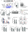
Figure 1. miR-146a deletion mediates HSPC hyperproliferation
(A) Overexpressed genes from low miR-146a-expressing del(5q) MDS/AML patients and miR-146a−/− (KO) marrow cells were compared using ToppGene. (B–C) Cell cycle analysis was performed on LSK and SLAM+ cells from WT (n = 4) and KO (n = 6) mice by Pyronin Y/Hoechst 33342 staining. Shown is a representative plot for LSK (B) and summary for LSK and SLAM+ cells (C). *, P < 0.05. (D) AnnexinV staining was performed on LK cells from WT (n=4) and KO (n = 6) mice. See also Supplemental Figure 1 and Table 1.
Figure 2
Figure 2. Del(5q) MDS/AML are associated with an intrachromosomal NF-κB feed-forward gene network
(A) Gene expression profiling of differentially expressed chr 5q genes (q11-q35) in del(5q) MDS (n = 47) as compared to normal control CD34+ cells (n = 17). (B) GeneConnector functionality in NetWalker was used to build a molecular network of genes that are overexpressed in del(5q) MDS (see Supplemental Table 2 for all genes used in the analysis). Red nodes indicate genes overexpressed in del(5q) MDS. Grey nodes indicate molecularly-connected genes. Grey nodes with red stripes indicate validated miR-146a gene targets. Dotted lines indicate the chromosome position of each gene. (C–D) Enrichment scores for DNA binding motifs in the promoter regions of overexpressed 5q genes (C) and NF-κB node genes (D). Each point represents the enrichment Z-score for a single transcription factor (TF) binding motif. Enriched motifs for NF-κB are indicated with arrows, along with their overall rank amongst the 1867 total human TF motifs analyzed. Horizontal dashed line indicates the (uncorrected) significance threshold corresponding to a P value cutoff of 0.01 (see Table 3). (E) Knockdown of SNCAIP, MAP3K1, PDLIM7, p62, TRAF6, and IRAK1 in HL60 cells was achieved by shRNAs. Solid red histograms represent genes within the “NF-κB node” that reside on chr 5q. Hatched red histograms represent genes within the “NF-κB node” but do not reside on chr 5q (see Figure 2B). Transduced HL60 cells were evaluated for colony formation in methylcellulose following knockdown of each indicated gene. Colonies were scored and normalized to a control shRNA. See also Supplemental Figure 2 and Tables 2–3.
Figure 3
Figure 3. p62 is overexpressed through NF-κB activation and is an essential cofactor in miR-146a−/− HSPC
(A) Schematic of the p62 (SQSTM1) promoter region. UCSC Genome Browser image displays ENCODE consortium data indicating (top to bottom): location of SQSTM1 gene (exons depicted as filled boxes, introns as lines with arrows); location of two NF-κB binding regions, based on ChIP-seq data in B cell lines; location of DNaseI hypersensitive regions (indicative of open chromatin) in HL60 and NB4 cells; various histone marks (all indicative of regulatory regions) in K562 cells. (B) p62 expression in AML marrow cells with high (top 50th percentile) or low (bottom 50th percentile) miR-146a. (C) Immunoblot analysis of p62 in del(5q) AML/MDS patient BM cells and normal CD34+ cells. The miR-146a and p62 locus status is indicated below (N, normal diploid; hap, haploid). (D) qRT-PCR expression of p62 in TF1 leukemic cells transduced with vector or miR-146 decoy and then treated with IL-1β or TNFα. (E) qRT-PCR expression of p62 in HSPC from WT and miR-146−/− mice after treatment with IL-1β at indicated time points. (F) p62 immunoblot analysis on marrow cells from WT and miR-146−/− mice after treated with IL-1β at indicated time points. (G) Del(5q) MDS/AML patients were stratified into miR-146ahigh/p62low and miR-146alow/p62high RNA expression. Overall survival for del(5q) MDS/AML patients based on high miR-146a/low p62 (green, n = 17) and low miR-146a/high p62 (red, n = 8). (H) Cell cycle analysis of WT and miR-146a−/− BM transduced with vector (MSCV-IRES-GFP; MIG) or p62. (I)) Validation of p62 knockdown in WT and miR-146a−/− BM cells by qRT-PCR. (J–K) Cell cycle analysis of WT and miR-146a−/− BM transduced with shCTL or shp62 (J) and summary of replicate experiments (K). (L) WT and miR-146a−/− HSPC (CD45.2) transduced with shCTL or shp62 were mixed with 1×10⁵ WT (CD45.1) marrow cells and then transplanted into lethally-irradiated recipients (n = 8/group). Myeloid and lymphoid proportions in the blood were analyzed 4 weeks post transplantation. *, P < 0.05; #, P < 0.1. See also Supplemental Figure 3.
Figure 4
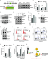
Figure 4. p62 is necessary for MDS/AML cell survival and cell proliferation
(A) p62 knockdown was confirmed by immunoblotting lysates isolated from cells expressing a control or shp62 lentiviral vector. TRAF6 ubiquitination was measured following transduction with shCTL or shp62. (B) TF1 cells cotransduced with the indicated vectors were examined for p65 DNA binding. (C) HL60 cells transduced with shCTL or shp62 were engrafted into NSG mice (n=5–8/group) and monitored for chimerism at the indicated time points. (D) Survival curves for mice receiving shCTL- or shp62-transduced HL60 (top) or MDSL (bottom) cells. (E) AnnexinV staining of the indicated cells was determined following transduction with shCTL or shp62. (F) AnnexinV staining of control CD34+ and two del(5q) AML patient samples (PT-01 and PT-02) was determined following transduction with shCTL or shp62. As a positive control, treatment with an NF-κB inhibitor (BAY, BAY 11-7085) resulted in non-specific cell death. (G) Cell cycle analysis of CD34+ and two patient samples (PT-01 and PT-02) transduced with shCTL or shp62 was determined by Hoechst 33342 incorporation. *, P < 0.05. See also Supplemental Figure 4.
Figure 5
Figure 5. p62 sustains NF-κB activation and leukemic cell function via its TRAF6-binding domain
(A) Schematic of the p62 protein and its domains: PB1, ZZ-type zinc finger domain (ZZ), a TRAF6-binding (TB) domain, and LC3-interacting region (LIR), and a ubiquitin-associated domain (UBA). The amino acid sequence of the TB domain and the schematic of a WT and mutant TB constructs are shown below. (B–C) HL60 expressing MIG, MIG-p62, or MIG-p62ΔTB retroviral vectors were transduced with shCTL or shp62. p62 expression (B) and cell survival (C) was analyzed 4 days post transduction. (D) Expression of empty vector (MSCV-pGK-GFP), TBMut, or TBWT by transient transfection in HEK293 or by transduction in HL60 was confirmed by dot blot immunoassay. (E) HEK293 cells transfected with the indicated constructs were analyzed by co-immunoprecipitation and immunoblotting. (F) NF-κB activation was measured in HEK293 cells transfected with a κB site-luciferase and the indicated vectors (n=3/group). Values represent κB-site firefly over Renilla luciferase. (G) IKKα/β phosphorylation was measured in HL60 cells transduced with TBMut or TBWT by immunoblotting. (H) TRAF6 ubiquitination (TRAF6 IP; Ub IB) was measured in HL60 transduced with TBMut and TBWT. (I) Cell cycle analysis of HL60 and CD34+ cells transduced with TBMut or TBWT was determined by Hoechst 33342 incorporation. (J) AnnexinV staining of HL60 and control CD34+ cells was determined following transduction with TBMut or TBWT. (K) HL60 and CD34+ cells were transduced with TBMut or TBWT and then plated in methylcellulose for progenitor colony formation (n=3/group). Colonies were scored 10–14 days after plating. (L) AnnexinV staining of control CD34+ cells and MDS/AML patient samples (PT-01, PT-02, and PT-03) was determined following transduction with TBMut or TBWT. (M) Model of an intrachromosomal network involving miR-146a/p62/TRAF6/NF-κB in HR del(5q) MDS/AML. miR-146a deletion results in derepression of TRAF6 protein. TRAF6 overexpression results in its autoubiquitination (green circles), which serves to recruit and then activate the NF-κB kinase complex (IKKα/β). NF-κB transcription factors induce p62/SQSTM1 expression from the remaining allele within del(5q). p62 is an important cofactor to sustain NF-κB activation through its TRAF6-binding domain (TB). The p62/TRAF6 signaling complex and subsequent NF-κB activation can be inhibited by expressing the TB motif. *, P < 0.05.

References

    1. Beg AA, Sha WC, Bronson RT, Baltimore D. Constitutive NF-kappa B activation, enhanced granulopoiesis, and neonatal lethality in I kappa B alpha-deficient mice. Genes Dev. 1995;9:2736–2746. - PubMed
    1. Boldin MP, Taganov KD, Rao DS, Yang L, Zhao JL, Kalwani M, Garcia-Flores Y, Luong M, Devrekanli A, Xu J, et al. miR-146a is a significant brake on autoimmunity, myeloproliferation, and cancer in mice. J Exp Med. 2011;208:1189–1201. - PMC - PubMed
    1. Chen FE, Huang DB, Chen YQ, Ghosh G. Crystal structure of p50/p65 heterodimer of transcription factor NF-kappaB bound to DNA. Nature. 1998;391:410–413. - PubMed
    1. Chen J, Bardes EE, Aronow BJ, Jegga AG. ToppGene Suite for gene list enrichment analysis and candidate gene prioritization. Nucleic Acids Res. 2009;37:W305–W311. - PMC - PubMed
    1. Chen L, Wu G, Ji H. hmChIP: a database and web server for exploring publicly available human and mouse ChIP-seq and ChIP-chip data. Bioinformatics. 2011;27:1447–1448. - PMC - PubMed

Publication types

MeSH terms

Associated data