. 2014 Sep 25;8(6):2003-2014.
doi: 10.1016/j.celrep.2014.08.008. Epub 2014 Sep 4.

PTEN interacts with histone H1 and controls chromatin condensation

Affiliations

PTEN interacts with histone H1 and controls chromatin condensation

Zhu Hong Chen et al. Cell Rep. .

Abstract

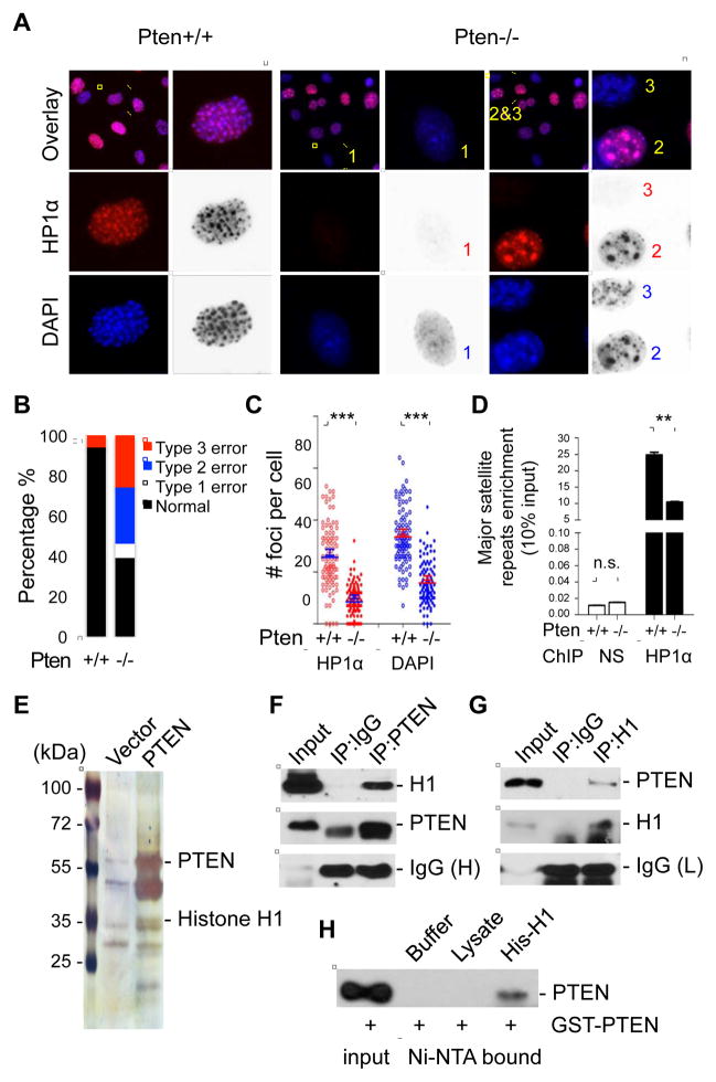
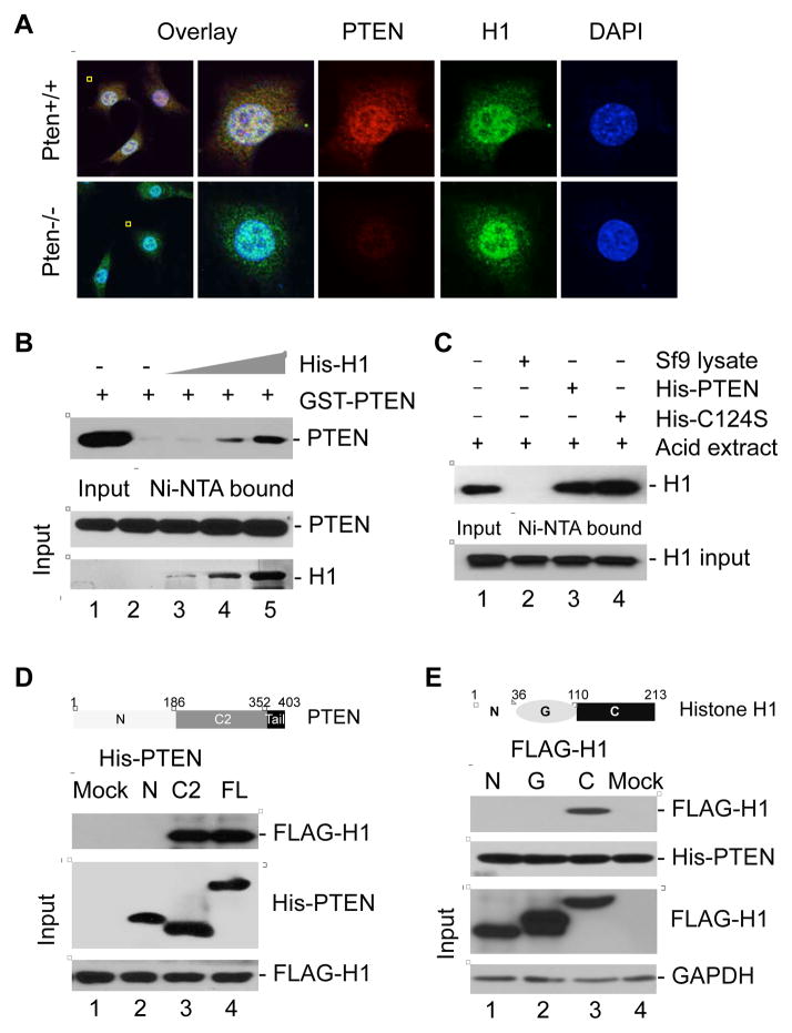
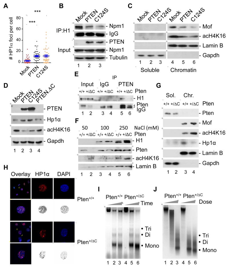
Chromatin organization and dynamics are integral to global gene transcription. Histone modification influences chromatin status and gene expression. PTEN plays multiple roles in tumor suppression, development, and metabolism. Here, we report on the interplay of PTEN, histone H1, and chromatin. We show that loss of PTEN leads to dissociation of histone H1 from chromatin and decondensation of chromatin. PTEN deletion also results in elevation of histone H4 acetylation at lysine 16, an epigenetic marker for chromatin activation. We found that PTEN and histone H1 physically interact through their C-terminal domains. Disruption of the PTEN C terminus promotes the chromatin association of MOF acetyltransferase and induces H4K16 acetylation. Hyperacetylation of H4K16 impairs the association of PTEN with histone H1, which constitutes regulatory feedback that may reduce chromatin stability. Our results demonstrate that PTEN controls chromatin condensation, thus influencing gene expression. We propose that PTEN regulates global gene transcription profiling through histones and chromatin remodeling.


Figures

Figure 1
Figure 1. PTEN regulates HP1α heterochromatic distribution and is physically associated with histone H1
(A) Pten+/+ and Pten−/− MEFs were subjected to immunofluorescent staining for HP1α. DNA was counterstained with DAPI. The selected cell was magnified and foci of HP1α or DAPI are also shown in both color and inverted gray scale. In Pten+/+ cells HP1α exhibited a speckled pattern confined to DAPI-dense heterochromatic regions, while three types of abnormal HP1α distribution were found in the nucleus of Pten−/− cells as indicated. Type 1, complete diffusion of HP1α and DAPI; Type 2, fewer (<10) enlarged foci of HP1α and DAPI; and type 3, HP1α diffusion with relative retention of DAPI foci. (B) Stacked column chart illustrating the proportion of cells with different abnormalities of heterochromatic distribution in Pten+/+ and Pten−/− MEFs. (C) HP1α and DAPI foci were counted in randomly selected Pten+/+ and Pten−/− cells (n=100. ***, p<0.001). (D) ChIP-qPCR analysis of HP1α binding to major satellite repeats. A non-specific antibody (NS) was used as a control. **, p<0.01; n.s., no significance. (E) Identification of histone H1 as a PTEN-associated protein by a FLAG-HA-PTEN pull-down assay. (F and G) Reciprocal immunoprecipitation and immunoblotting of PTEN and histone H1 in Pten+/+ MEFs. (H) In vitro binding assays using purified His-Histone H1 and GST-PTEN proteins. See also Figure S1.
Figure 2
Figure 2. PTEN physically interacts with the C-terminal region of histone H1
(A) Confocal microscopy showing merged signals (yellow) of PTEN (red) with histone H1 (green) in Pten+/+ MEFs. Pten−/− cells were included as a control. (B) GST-tagged PTEN was purified and incubated with increasing doses of purified His-H1.2 for in vitro detection of their direct association. (C) His-tagged wild-type PTEN and a phosphatase-deficient PTEN mutant C124S were purified prior to incubation with an equal amount of acid-extracted native histones for detection of PTEN-bound histone H1. (D) In vitro binding assay with FLAG-tagged full-length histone H1 and His-tagged different domains of PTEN as indicated. The domain structure of PTEN is given above the data. (E) Different domains of histone H1.2 with a FLAG tag were used for in vitro binding assay with His-tagged full length PTEN. The diagram above the data shows different domains of histone H1.2.
Figure 3
Figure 3. PTEN modulates dynamic motility of histone H1 in chromatin and loss of Pten leads to chromatin decondensation
(A) Increasing concentrations of NaCl was used to extract nuclei from Pten+/+ and Pten−/− MEFs, followed by quantification of histone H1 and Npm1 released from nucleosomes. Lamin B was used to confirm equal protein loading in each pair of samples. (B) A sequential cell fractionation procedure was used to separate cytoplasmic, nuclear soluble and chromatin fractions prior to immunoblot analysis of indicated protein molecules in PTEN+/+ and PTEN−/− cells. (C) Co-immunoprecipitation of histone H1 with Hp1α and Npm1 in Pten+/+ and Pten−/− MEFs. (D) FRAP analysis of GFP-histone H1 in Pten+/+ and Pten−/− MEFs (n=15). Bright GFP-H1 regions in the nucleus were selected for photo-bleaching and recovery observation. Data are presented as mean ± SEM. *, p<0.05. (E) Chromatin accessibility assessed by sensitivity to Micrococcal nuclease (MNase) digestion. Nuclei from Pten+/+ and Pten−/− MEFs were prepared and digested with 0.2U/μl MNase for increasing periods of time. The MNase digestion profile is shown with mono-, di- and tri-nucleosomal repeats as indicated. (F) Dose-dependent MNase digestion pattern of the chromatin from Pten+/+ and Pten−/− MEFs. See also Figure S2.
Figure 4
Figure 4. Loss of PTEN induces acetylation of histone H4 at K16 whereas enforced H4K16 acetylation Impairs PTEN-H1 interaction
(A) Evaluation of acH4K16 and acH3K27 levels in low-salt and high-salt extractions from Pten+/+ and Pten−/− MEFs. (B) Chromatin-associated Hp1α, Mof and acH4K16 were evaluated in Pten+/+ and Pten−/− MEFs. Soluble and chromatin fractions were prepared and subjected to Western blotting with antibodies to indicated proteins. (C) Human DLD-1 cells were treated with 2.5 μg/ml TSA for 12 h prior to immunoblot analysis of chromatin-bound H1 and HP1α. (D) The association between PTEN and histone H1 was examined in DLD-1 cells with and without TSA treatment. (E) Pten+/+ MEFs were transfected with wild type histone H4 or an acetylation-mimicking mutant K16Q, followed by PTEN immunoprecipitation and subsequent detection of histone H1. See also Figure S3.
Figure 5
Figure 5. PTEN promotes chromatin association of histone H1 and suppresses H4K16 acetylation in a C-terminus-dependent but phosphatase-independent manner
(A) Pten−/− MEFs transfected with PTEN or a phosphatase-deficient mutant C124S were immunofluorescent stained for HP1α. Numbers of nuclear HP1α foci are summarized in the graph (n=53). ***, p<0.001. (B and C) Pten−/− MEFs transfected with wild-type PTEN and a phosphatase-deficient PTEN mutant (C124S) were subjected to evaluation of H1-NPM1 association (B) and chromatin-bound MOF and acH4K16 (C). (D) HP1α expression and H4K16 acetylation were examined in Pten−/− MEFs transfected with wild-type full length PTEN, the phosphatase-dead C124S mutant and a truncated PTEN with C-terminal deletion, PTEN.ΔC. (E) MEFs isolated from mouse embryo with heterozygous Pten C-terminal deletion (Pten+/ΔC) and a wild-type control embryo (Pten+/+) were used for evaluation of PTEN-H1 interaction by PTEN immunoprecipitation and histone H1 immunoblotting. (F–J) Pten+/+ and Pten+/ΔC MEFs were subjected to salt extraction assay for examination of histone H1 retention and H4K16 acetylation (F), chromatin association of Mof, acH4K16 and Hp1α (G), immunofluorescent staining of HP1α (H), and MNase sensitivity (I and J). See also Figure S4.
Figure 6
Figure 6. Deletion of PTEN or Pten C terminus alters gene expression profiles and impacts the interplay of H1 and acH4K16 occupancy on gene promoters
(A and B) Two pairs of MEFs (Pten+/+ and Pten−/−; Pten+/+ and Pten+/ΔC) were used for microarray analysis. Data were combined and processed in volcano plot (A) and heat map (B) to show significantly altered genes (p< 0.05) in cells lacking Pten or its C terminus. FC, fold change. (C and D) Quantitative PCR (qPCR) analysis of Pten-regulated genes in two cell systems, PTEN+/+ and PTEN−/− DLD-1 cells (C), and Pten+/+ and Pten+/ΔC MEFs (D). (E and F) ChIP-qPCR analysis of non-specific antibody, histone H4, histone H1, and acH4K16 occupancy on the BCL2 and CD44 promoters in PTEN+/+ and PTEN−/− cells. Data are normalized and presented as mean ± SEM. *, p<0.05, **, p<0.01, ***, p<0.001. See also Figures S5 and S6; Tables S1 and S2.

Comment in

References

    1. Akhtar A, Becker PB. Activation of transcription through histone H4 acetylation by MOF, an acetyltransferase essential for dosage compensation in Drosophila. Mol Cell. 2000;5:367–375. - PubMed
    1. Baker SJ. PTEN enters the nuclear age. Cell. 2007;128:25–28. - PubMed
    1. Bannister AJ, Kouzarides T. Regulation of chromatin by histone modifications. Cell Res. 2011;21:381–395. - PMC - PubMed
    1. Beckerman R, Prives C. Transcriptional regulation by p53. Cold Spring Harb Perspect Biol. 2010;2:a000935. - PMC - PubMed
    1. Bednar J, Horowitz RA, Grigoryev SA, Carruthers LM, Hansen JC, Koster AJ, Woodcock CL. Nucleosomes, linker DNA, and linker histone form a unique structural motif that directs the higher-order folding and compaction of chromatin. Proc Natl Acad Sci U S A. 1998;95:14173–14178. - PMC - PubMed

Publication types

LinkOut - more resources