. 2014 Sep 22;211(10):1947-55.
doi: 10.1084/jem.20140860. Epub 2014 Sep 8.

Regulation of proximal T cell receptor signaling and tolerance induction by deubiquitinase Usp9X

Affiliations

Regulation of proximal T cell receptor signaling and tolerance induction by deubiquitinase Usp9X

Edwina Naik et al. J Exp Med. .

Abstract

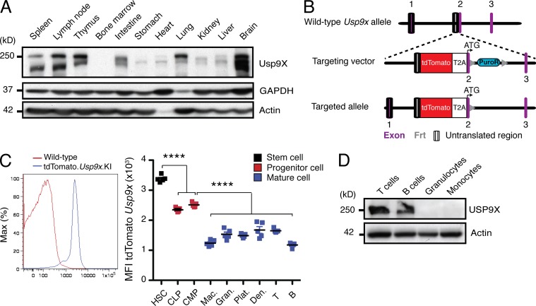
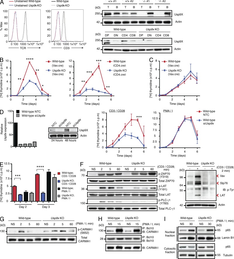
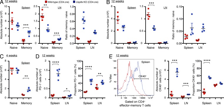
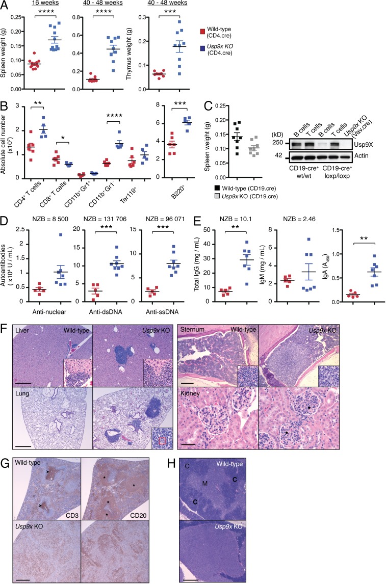
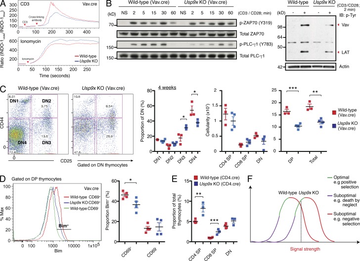
The T cell hyperproliferation and autoimmune phenotypes that manifest in mice lacking E3 ubiquitin ligases such as Cbl, ITCH, or GRAIL highlight the importance of ubiquitination for the maintenance of peripheral T cell tolerance. Less is known, however, about the deubiquitinating enzymes that regulate T cell proliferation and effector function. Here, we define a cell intrinsic role for the deubiquitinase Usp9X during proximal TCR signaling. Usp9X-deficient T cells were hypoproliferative, yet mice with T cell-specific Usp9x deletion had elevated numbers of antigen-experienced T cells and expanded PD-1 and OX40-expressing populations consistent with immune hyperactivity. Aged Usp9x KO mice developed lupus-like autoimmunity and lymphoproliferative disease, indicating that ubiquitin ligases and deubiquitinases maintain the delicate balance between effective immunity and self-tolerance.


Figures

Figure 1.
Figure 1.
Usp9X protein is expressed in lymphocytes. (A) Usp9X protein expression in a panel of adult murine tissues. (B) Organization of the tdTomato.Usp9x knock-in allele. (C) Representative histogram and collated MFI (median fluorescence intensity) of tdTomato.Usp9x mRNA. CMP, common myeloid progenitor; CLP, common lymphoid progenitor; Mac, monocyte/macrophage; Gran, granulocyte; Plat, platelets; Den, dendritic cells; T and B, T and B lymphocytes. (D) Usp9X protein expression in hematopoietic cell subsets. All experiments were conducted a minimum of two independent times with littermate controls on a C57BL/6 background. Each square represents an independent mouse. ****, P ≤ 0.0001 using a two-tailed unpaired Student’s t test.
Figure 2.
Figure 2.
Usp9X regulates proximal TCR signaling events. (A) Representative histograms of TCR and CD3 levels on naive CD4+ T cells purified from CD4.cre mice. The specificity (T vs. B lymphocytes) and kinetics of deletion using CD4.cre was confirmed by Western blot. (B) Naive CD4+ T cell proliferation in response to CD3/CD28. Data represents mean ± SEM of 5–7 independent mice per genotype/time point. (C) Naive CD4+ T cell proliferation in response to PMA/ionomycin. Data represents mean ± SEM of 7–11 independent mice per genotype/time point. (D) Proliferation of wild-type T cells transfected with a nontargeting control (NTC) or Usp9x siRNAs (siUsp9x) that effectively reduced both Usp9x mRNA and protein levels. Data represent mean ± SEM of three independent transfections. (E) CD8+ (CD4.cre) T cell proliferation. Data represents mean ± SEM of 3 independent mice per genotype. (F and G) Naive CD4+ T cells (CD4.cre) were stimulated for the indicated times and proteins were fractionated and probed with the indicated antibodies. N.S., nonstimulated. The induction of tyrosyl-phosphorylated proteins was monitored with a phospho-tyrosine (p-Tyr)-specific antibody. (H) CBM complex formation in naive CD4+ T cells (CD4.cre) was assessed by co-immunoprecipitation of CARMA1 with Bcl10. (I) Nuclear translocation of the p65 subunit of NF-κB was monitored by subcellular fractionation of naive CD4+ T cells. DP, double-positive; DN, double-negative. *, P ≤ 0.05; **, P ≤ 0.01; ***, P ≤ 0.001; and ****, P ≤ 0.0001 of wild-type versus Usp9x KO using a two-tailed unpaired Student’s t test. PMA/I, PMA/ionomycin. c.p.m., counts per minute.
Figure 3.
Figure 3.
Usp9x deficiency expands antigen-experienced, PD-1, and OX40-expressing T cell populations in vivo. The number of naive or effector–memory CD4+ (A and C) or CD8+ (B) T cells in the spleens and LNs of CD4.cre mice. (D) Expansion of PD-1–expressing CD4+ effector–memory populations in Usp9x KO spleens and LNs. (E) Representative histogram depicting OX40 expression on CD4+ effector–memory T cells and the proportion and total number of OX40+ memory T cells. *, P ≤ 0.05; **, P ≤ 0.01; ***, P ≤ 0.001; and ****, P ≤ 0.0001 of wild-type versus Usp9x KO using a two-tailed unpaired Student’s t test.
Figure 4.
Figure 4.
Usp9x KO mice develop spontaneous lupuslike autoimmunity and lymphoproliferative disease. (A) Mass of wild-type or Usp9x KO (CD4.cre) spleens and thymi. (B) Cell subset analysis of 16-wk-old mice. (C) Spleen mass of CD19.cre mice. The specificity of CD19.cre deletion was confirmed by Western blot. (D) Quantitation of serum ANA, double-strand DNA (dsDNA) and single-strand DNA (ssDNA) autoantibody levels by ELISA. (E) IgG, IgM, and IgA levels were quantitated in the sera using ELISA. Where indicated, serum from a New Zealand Black/White (NZB/W) F1 hybrid mouse with proteinuria served as a positive control. (F) Histopathological analysis of organs from 32-wk-old wild-type or Usp9x KO mice. The red box delineates a Mott cell and the asterisks denote areas of expanded mesangium consistent with membranoproliferative glomerulonephritis. Bars: (lung and liver) 500 µm; (sternum) 200 µm; (kidney) 50 µm. (G) CD3 and CD20 expression in the spleens of 48-wk-old mice revealed loss of normal tissue architecture, including periarteriolar lymphoid sheaths (arrowheads) and lymphoid follicles (asterisks). bar, 500 μm. (H) Absence of the cortical/medullary boundary in the thymi of 48-wk-old Usp9x KO mice. C, cortex; M, medulla. Bar, 500 µm. All experiments were conducted a minimum of two independent times with littermate controls on a C576BL/6 background. Each square represents an independent mouse. *, P ≤ 0.05; **, P ≤ 0.01; ***, P ≤ 0.001; and ****, P ≤ 0.0001 of wild-type versus Usp9x KO using a two-tailed unpaired Student’s t test.
Figure 5.
Figure 5.
Defective T cell development in Usp9x KO mice. (A) Thymocyte activation was assessed by the degree of calcium flux elicited by cross-linked anti-CD3 antibody or the calcium ionophore ionomycin. Data are representative of three independent mice per genotype. (B) Western blot analysis confirmed that Usp9x KO thymocytes exhibit similar signaling defects to peripheral T cells. (C) Immature thymic subsets in the Vav.cre strain were enumerated using CD25 and CD44 co-staining to track the transition from DN1 to DN4 before differentiation in to DP thymocytes. (D) Intracellular FACS analysis of the proapoptotic molecule Bim in 4-wk-old Vav.cre mice. (E) Analysis of thymic subsets in 12-wk-old CD4.cre mice. (F) Graphical representation of the selection shift hypothesis. The dashed line denotes a signal strength that in wild-type T cells would result in deletion but in Usp9x KO T cells, results in positive selection. All experiments were conducted a minimum of two independent times with littermate controls on a C576BL/6 background. Each square represents an independent mouse. *, P ≤ 0.05; **, P ≤ 0.01; ***, P ≤ 0.001; and ****, P ≤ 0.0001 of wild-type versus Usp9x KO using a two-tailed unpaired Student’s t test.

Similar articles

Cited by

References

    1. Al-Hakim, A.K., Zagorska A., Chapman L., Deak M., Peggie M., and Alessi D.R.. 2008. Control of AMPK-related kinases by USP9X and atypical Lys(29)/Lys(33)-linked polyubiquitin chains. Biochem. J. 411:249–260 10.1042/BJ20080067 - DOI - PubMed
    1. Alam, S.M., Travers P.J., Wung J.L., Nasholds W., Redpath S., Jameson S.C., and Gascoigne N.R.. 1996. T-cell-receptor affinity and thymocyte positive selection. Nature. 381:616–620 10.1038/381616a0 - DOI - PubMed
    1. Baldwin, T.A., and Hogquist K.A.. 2007. Transcriptional analysis of clonal deletion in vivo. J. Immunol. 179:837–844 10.4049/jimmunol.179.2.837 - DOI - PubMed
    1. Cheng, A.M., Negishi I., Anderson S.J., Chan A.C., Bolen J., Loh D.Y., and Pawson T.. 1997. The Syk and ZAP-70 SH2-containing tyrosine kinases are implicated in pre-T cell receptor signaling. Proc. Natl. Acad. Sci. USA. 94:9797–9801 10.1073/pnas.94.18.9797 - DOI - PMC - PubMed
    1. Croft, M., So T., Duan W., and Soroosh P.. 2009. The significance of OX40 and OX40L to T-cell biology and immune disease. Immunol. Rev. 229:173–191 10.1111/j.1600-065X.2009.00766.x - DOI - PMC - PubMed

MeSH terms