. 2014 Sep 9;130(11 Suppl 1):S60-9.
doi: 10.1161/CIRCULATIONAHA.113.007917.

Cross talk of combined gene and cell therapy in ischemic heart disease: role of exosomal microRNA transfer

Affiliations

Cross talk of combined gene and cell therapy in ischemic heart disease: role of exosomal microRNA transfer

Sang-Ging Ong et al. Circulation. .

Abstract

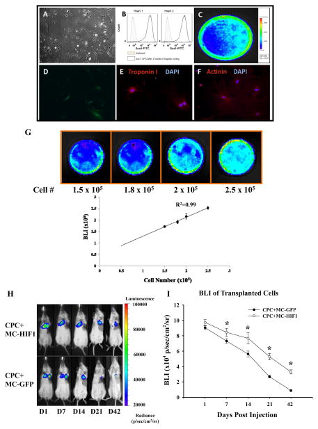
Background: Despite the promise shown by stem cells for restoration of cardiac function after myocardial infarction, the poor survival of transplanted cells has been a major issue. Hypoxia-inducible factor-1 (HIF1) is a transcription factor that mediates adaptive responses to ischemia. Here, we hypothesize that codelivery of cardiac progenitor cells (CPCs) with a nonviral minicircle plasmid carrying HIF1 (MC-HIF1) into the ischemic myocardium can improve the survival of transplanted CPCs.

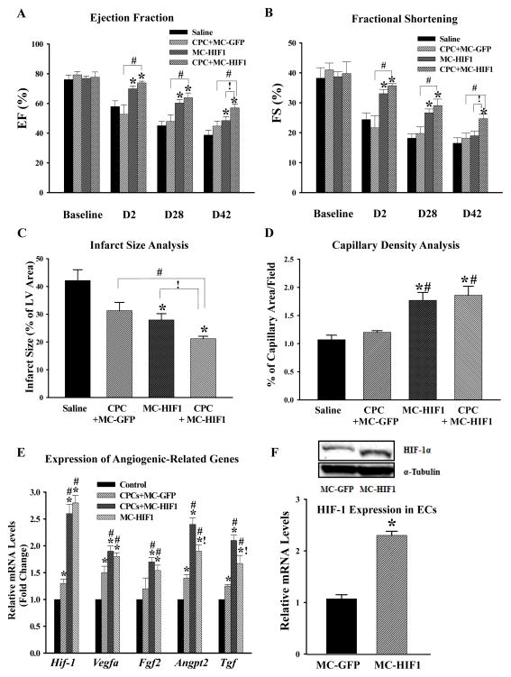
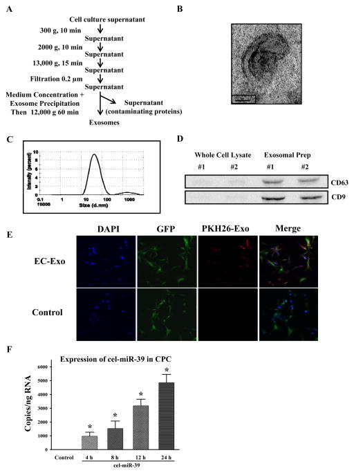
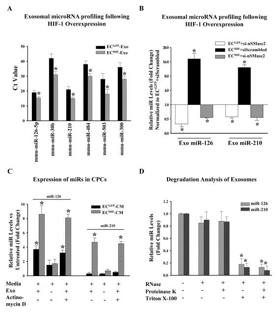
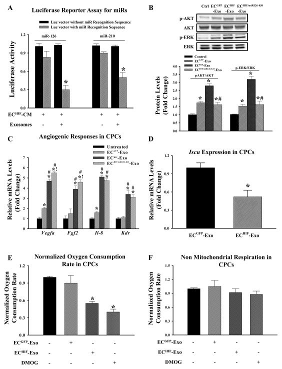
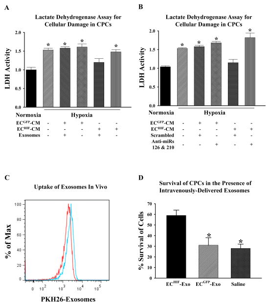
Methods and results: After myocardial infarction, CPCs were codelivered intramyocardially into adult NOD/SCID mice with saline, MC-green fluorescent protein, or MC-HIF1 versus MC-HIF1 alone (n=10 per group). Bioluminescence imaging demonstrated better survival when CPCs were codelivered with MC-HIF1. Importantly, echocardiography showed mice injected with CPCs+MC-HIF1 had the highest ejection fraction 6 weeks after myocardial infarction (57.1±2.6%; P=0.002) followed by MC-HIF1 alone (48.5±2.6%; P=0.04), with no significant protection for CPCs+MC-green fluorescent protein (44.8±3.3%; P=NS) when compared with saline control (38.7±3.2%). In vitro mechanistic studies confirmed that cardiac endothelial cells produced exosomes that were actively internalized by recipient CPCs. Exosomes purified from endothelial cells overexpressing HIF1 had higher contents of miR-126 and miR-210. These microRNAs activated prosurvival kinases and induced a glycolytic switch in recipient CPCs, giving them increased tolerance when subjected to in vitro hypoxic stress. Inhibiting both of these miRs blocked the protective effects of the exosomes.

Conclusions: In summary, HIF1 can be used to modulate the host microenvironment for improving survival of transplanted cells. The exosomal transfer of miRs from host cells to transplanted cells represents a unique mechanism that can be potentially targeted for improving survival of transplanted cells.

Keywords: exosomes; genetic therapy; hypoxia-inducible factor-1; microRNAs; stem cells.


Figures

Figure 1
Figure 1
Co-delivery of hypoxia inducible-factor 1 (HIF-1) driven by minicircle (MC) plasmid promotes survival of transplanted Sca1+ cardiac progenitor cells (CPCs) in the ischemic heart. (A) The morphology of isolated CPCs growing on gelatin coated dish. (B) Flow cytometric analysis of purified Sca1+ CPC population from two preparations. Typical purity of isolation is >95%. CPCs isolated from transgenic mice express robust (C) firefly luciferase (Fluc) and (D) GFP expression. After culturing in cardiac differentiation induction medium, differentiated CPCs stained positively with (E) cardiac Troponin I and (F) α-actinin demonstrating its amenability to lineage commitment. (G) CPCs were seeded into dishes with increasing cell numbers. Assessment of BLI signals showed a robust linear correlation (R2=0.99) between the cell number and Fluc expression, which is crucial for accurate tracking of cell survival by in vivo imaging. (H) Representative BLI of animals injected with CPCs intramyocardially together with either MC-GFP or MC-HIF1 following myocardial infarction (MI) at indicated time points. (I) Quantitative analysis of longitudinal BLI signal demonstrates that CPCs co-delivered with MC-HIF1 had better survival compared to CPCs co-delivered with MC-GFP over a period of 42 days. *P<0.05 (N=10/group).
Figure 2
Figure 2
Combination of cell and gene therapy provides synergistic therapeutic effects following MI. Comparison of (A) ejection fraction and (B) fractional shortening among all 4 groups at indicated time points revealed that CPCs co-delivered with MC-HIF1 had superior therapeutic effects among all groups. *P<0.05 vs. saline group; #P<0.05 vs. CPCs + MC-GFP; !P<0.05 vs. MC-HIF1 (N=10/group). (C) Three days post-MI, mice from each group were sacrificed and hearts were collected for determination of infarct size by tetrazolium chloride staining. *P<0.05 vs. saline group; #P<0.05 vs. CPCs + MC-GFP; !P<0.05 vs. MC-HIF1 (N=6/group). (D) Vascular density in each group was determined by CD31 staining at 7 days post-MI. *P<0.05 vs. saline group; #P<0.05 vs. CPCs + MC-GFP (N=6/group). (E) Areas close to engrafted GFP+ CPCs were laser microdissected to assess levels of angiogenic gene activation. Samples were collected 5 days post-MI. qPCR showed that MC-HIF1 upregulates the expression of angiogenic genes, some of which were further augmented with the presence of CPCs. *P<0.05 vs. control (non-ischemic remote zone of MC-HIF1); #P<0.05 vs. CPCs + MC-GFP; !P<0.05 vs. CPCs + MC-HIF1 (N=5/group). (F) In a separate set of experiments, mice subjected to MI received either MC-GFP or MC-HIF1 only. Three days later, cardiac ECs were isolated from the peri-infarct region, ECs from MC-HIF1 group were found to have higher expression of HIF-1 at both protein (upper panel) and gene levels (lower panel) compared to ECs from MC-GFP group, indicating that cardiac ECs are receptive to MC-HIF1 transfection. *P<0.05 vs. MC-GFP (N=6/group).
Figure 3
Figure 3
Purified exosomes produced by ECs are actively internalized by CPCs in vitro. (A) Purification scheme of exosomes from EC culture supernatant. (B) Cup-shaped morphology of purified ECs’ exosomes assessed by transmission electron microscopy. (C) Dynamic light scattering analysis of purified ECs’ exosomes demonstrating a physical size distribution of 10–110 nm. (D) Immunoblotting revealed an enrichment of exosomal markers CD63 and CD9 in purified exosomal fraction compared to whole EC lysates. Two separate preparations were shown. (E) CPCs were cultured in the presence of EC-derived PKH26-labeled exosomes or absence (control; same volume of PBS labeled similarly) for 6 hours. Exosomes were taken up CPCs as shown by confocal microscopy. (F) ECs were transfected with cel-miR-39 or left untransfected. CPCs were then treated with exosomes isolated from untransfected (control) or cel-miR-39 transfected ECs for the indicated times. *P<0.05 vs. control (N=4/group).
Figure 4
Figure 4
HIF-1 modulates the microRNA (miRs) repertoire in ECs-derived exosomes, which are transferable to CPCs. (A) ECs were transfected with either MC-GFP (control) or MC-HIF1. Following transfection, exosomes were purified from both groups and subjected to miRs profiling. Expression of selected exosomal miRs was increased following MC-HIF1 transfection. *P<0.05 vs ECGFP-Exo (N=4). (B) Based on the profiling results, miR-126 and miR-210 were chosen for detailed analysis. ECGFP and ECHIF were transfected with either scrambled siRNA or siRNA targeting nSMase2. Exosomes were then purified from each group, and the expression of miR-126 and miR-210 were determined by qPCR using Taqman probes, and expressed as relative fold-change normalized to ECGFP transfected with scrambled siRNA. *P<0.05 vs. ECGFP+siScrambled (N=6/group). (C) Recipient CPCs were treated with vehicle or actinomycin D (5 μg/ml). CPCs were then grown in normal growth medium or supplemented with either ECGFP-CM or ECHIF-CM in the absence or presence of exosomes (Exo) as indicated. After 24 hours, the expression of miR-126 and miR-210 in CPCs was determined by qPCR and expressed as fold-change normalized against CPCs grown in normal growth medium. *P<0.05 (N=6). (D) Exosomes were isolated from ECHIF and incubated with the indicated reagents for 45 mins at 37°C before isolation of RNA and measurement of expression levels of miR-126 and miR-210 by qPCR. *P<0.05 vs. untreated (N=4).
Figure 5
Figure 5
Transferred exosomal miRs regulate biological properties of recipient CPCs. (A) CPCs were transfected with a luciferase reporter vector containing either miR-126 or miR-210 recognition sequence or a control luciferase vector lacking the sequence. CPCs were then given ECHIF-CM in the presence or absence of exosomes as indicated. CPCs were then cultured for another 24 hours and luciferase activity was determined and expressed as fold-decrease of cells transfected with the luciferase reporter containing specific miR recognition sequence over the vector lacking the recognition sequence for each culture condition. *P<0.05 (N=4) vs. cells transfected with vector lacking miR recognition sequence. (B) Representative immunoblots and densitometry quantification of indicated proteins in CPCs grown in either normal conditions, or supplemented with ECGFP-Exo, ECHIF-Exo, or ECHIF/miR126-KO-exosomes. *P<0.05 vs. untreated CPCs; #P<0.05 vs. ECHIF (N=4). (C) Expression of angiogenic genes in CPCs grown in each culture condition as indicated was determined by qPCR. *P<0.05 vs. untreated; #P<0.05 vs. ECGFP-Exo; !P<0.05 vs. ECHIF-Exo (N=8). (D) Expression of Iscu, a validated miR-210 target gene, was determined in CPCs supplemented with either ECGFP-Exo or ECHIF-Exo. *P<0.05 (N=4) vs. CPCs supplemented with ECGFP-Exo. CPCs were cultured in conditions as specified for 24 hours. (E) Oxygen consumption and (F) non-mitochondrial respiration were then determined and expressed as fold-change compared to untreated control cells. Dimethyloxalylglycine (DMOG), a known HIF-1 activator was used as positive control for reduced oxygen consumption. *P<0.05 vs. untreated controls (N=6).
Figure 6
Figure 6
Exosomes directly provide CPCs with increased tolerance against ischemic stress both in vitro and in vivo. (A) CPCs were grown in either normal conditions, or supplemented with ECGFP-CM or ECHIF-CM with or without the presence of exosomes as indicated, before being subjected to in vitro hypoxic stress. Lactate dehydrogenase (LDH) was then measured as an indicator of cellular damage and expressed as fold-change compared to untreated cells kept in normoxic conditions. *P<0.05 (N=6) vs. normoxic cells. (B) ECHIF were co-transfected with either scrambled antagomirs or antagomirs targeting miR-126 and miR-210. CPCs were then grown in conditioned medium from each group as indicated before being subjected to LDH assay to assess the importance of miR-126 and miR-210 in ECHIF-CM-mediated protection seen in (A). *P<0.05 vs. normoxic control (N=6). (C) Following MI, CPCs were intramyocardially injected into mice hearts, and a bolus of PKH26-labeled ECs-exosomes was delivered intravenously. After 12 hours, the animals were sacrificed and CPCs were re-isolated from the hearts. Samples were measured by flow cytometry for GFP+ and PKH26+ events. The histogram shows that PKH26-exosomes (blue lines) were detectable in ~10% of GFP+ CPCs (red lines) demonstrating that CPCs are capable of uptaking exosomes in vivo. (D) To determine whether ECHIF-Exo could confer increased tolerance to CPCs in vivo, cells were delivered intramyocardially post-MI into mice concomitantly with intravenous injection of either saline, ECGFP-Exo or ECHIF-Exo. BLI was then performed at day 1 and day 7 post-MI, and survival of CPCs was expressed as a % of signals intensity at day 7 compared to initial signals intensity of day 1. *P<0.05 vs. saline (N=6/group).

Comment in

References

    1. Hausenloy DJ, Erik Botker H, Condorelli G, Ferdinandy P, Garcia-Dorado D, Heusch G, Lecour S, van Laake LW, Madonna R, Ruiz-Meana M, Schulz R, Sluijter JP, Yellon DM, Ovize M. Translating cardioprotection for patient benefit: position paper from the Working Group of Cellular Biology of the Heart of the European Society of Cardiology. Cardiovasc Res. 2013;98:7–27. - PubMed
    1. Liu J, Narsinh KH, Lan F, Wang L, Nguyen PK, Hu S, Lee A, Han L, Gong Y, Huang M, Nag D, Rosenberg J, Chouldechova A, Robbins RC, Wu JC. Early stem cell engraftment predicts late cardiac functional recovery: preclinical insights from molecular imaging. Circ Cardiovasc Imaging. 2012;5:481–90. - PMC - PubMed
    1. Ong SG, Hausenloy DJ. Hypoxia-inducible factor as a therapeutic target for cardioprotection. Pharmacol Ther. 2012;136:69–81. - PubMed
    1. Huang M, Chen Z, Hu S, Jia F, Li Z, Hoyt G, Robbins RC, Kay MA, Wu JC. Novel minicircle vector for gene therapy in murine myocardial infarction. Circulation. 2009;120:S230–7. - PMC - PubMed
    1. Hu S, Huang M, Nguyen PK, Gong Y, Li Z, Jia F, Lan F, Liu J, Nag D, Robbins RC, Wu JC. Novel microRNA prosurvival cocktail for improving engraftment and function of cardiac progenitor cell transplantation. Circulation. 2011;124:S27–34. - PMC - PubMed

Publication types

MeSH terms