. 2014;42(17):11203-17.
doi: 10.1093/nar/gku822. Epub 2014 Sep 8.

Direct binding of the Alu binding protein dimer SRP9/14 to 40S ribosomal subunits promotes stress granule formation and is regulated by Alu RNA

Affiliations

Direct binding of the Alu binding protein dimer SRP9/14 to 40S ribosomal subunits promotes stress granule formation and is regulated by Alu RNA

A Berger et al. Nucleic Acids Res. 2014.

Abstract

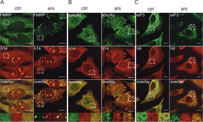
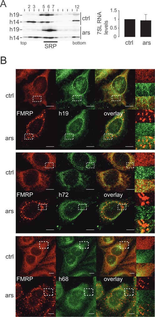
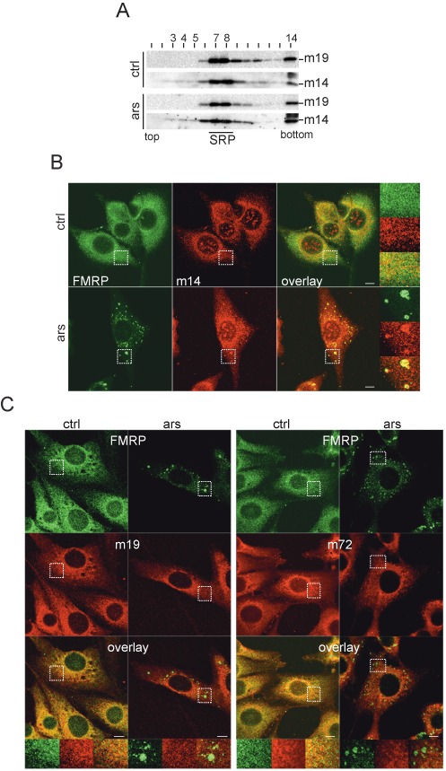
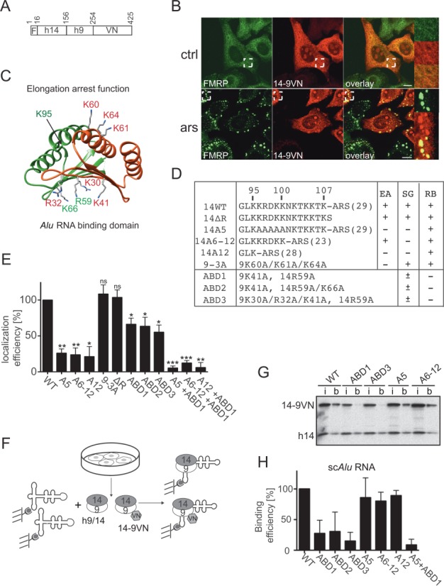
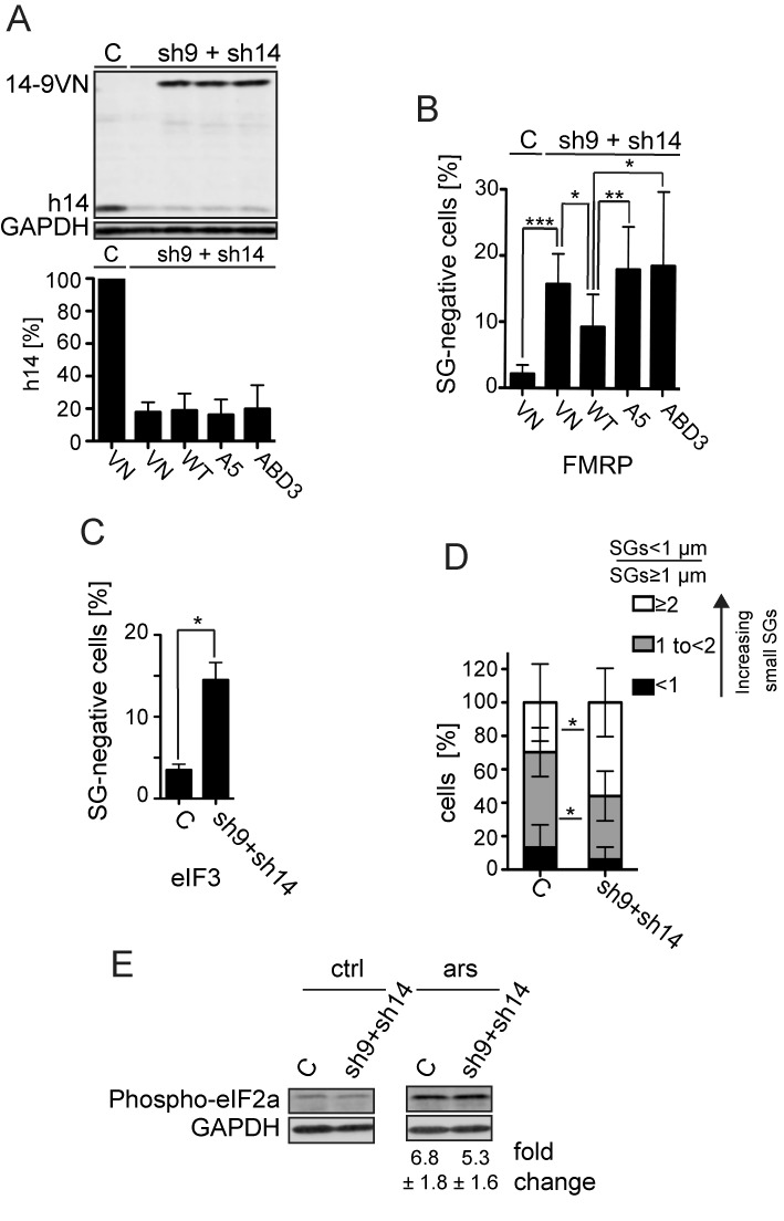
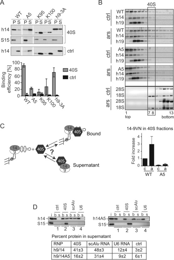
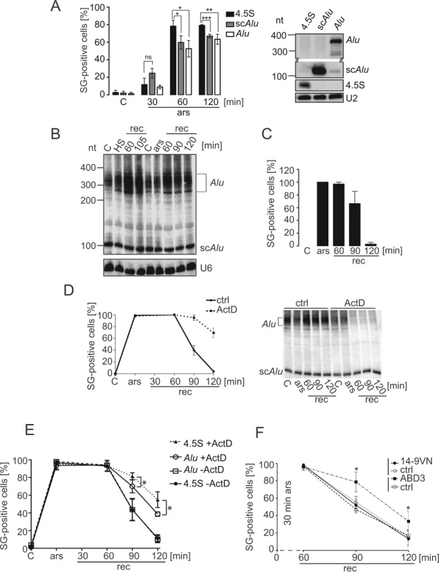
Stress granules (SGs) are formed in response to stress, contain mRNAs, 40S ribosomal subunits, initiation factors, RNA-binding and signaling proteins, and promote cell survival. Our study describes a novel function of the protein heterodimer SRP9/14 and Alu RNA in SG formation and disassembly. In human cells, SRP9/14 exists assembled into SRP, bound to Alu RNA and as a free protein. SRP9/14, but not SRP, localizes to SGs following arsenite or hippuristanol treatment. Depletion of the protein decreases SG size and the number of SG-positive cells. Localization and function of SRP9/14 in SGs depend primarily on its ability to bind directly to the 40S subunit. Binding of SRP9/14 to 40S and Alu RNA is mutually exclusive indicating that the protein alone is bound to 40S in SGs and that Alu RNA might competitively regulate 40S binding. Indeed, by changing the effective Alu RNA concentration in the cell or by expressing an Alu RNA binding-defective protein we were able to influence SG formation and disassembly. Our findings suggest a model in which SRP9/14 binding to 40S promotes SG formation whereas the increase in cytoplasmic Alu RNA following stress promotes disassembly of SGs by disengaging SRP9/14 from 40S.


Figures

Figure 1.
Figure 1.
SGs contain SRP9/14. Immunofluorescence images of HeLa cells stained with antibodies against human SRP14 (h14), SRP9 (h9) and against different SG markers. ars: cells treated with 500 μM sodium arsenite for 30 min; ctrl: untreated cells. (A) Antibodies against h14 and FMRP. (B) Anti-h14 antibodies and in situ hybridization of mRNAs with oligo(dT). (C) Anti-h9 and anti-eIF3 antibodies. Anti-h9 antibodies failed to stain nucleoli. (A–C) Images were captured at the SP2 laser scanning confocal microscope (63x/1.4 numerical aperture, PlanApo). Areas denoted by rectangles are shown at higher magnification. Scale bars: 10 μm.
Figure 2.
Figure 2.
The SRP complex remains intact during stress and does not localize to SGs. (A) Velocity sedimentation fractionations of HeLa cell postnuclear supernatants on 12–30% glycerol gradients. Left panel: Western blots of gradient fractions using anti-h14 and anti-h19 antibodies. Right panel: quantification of 7SL RNA in HeLa cell extracts by qRT-PCR. Levels were standardized to GAPDH mRNA and normalized to control cells. Error bars are shown as SD, n = 3. (B) Immunofluorescence images of HeLa cells stained with antibodies against h19 and FMRP (upper panel) against h72 and FMRP (middle panel) and against h68 and FMRP (lower panel). Images were captured using a 63x lens on LSM-710 Laser scanning microscope. Areas denoted by rectangles are shown at higher magnification. Scale bars: 10 μm. ars: 500 μM sodium arsenite for 30 min; ctrl: untreated cells. h19: human SRP19; h72: human SRP72; h68: human SRP68
Figure 3.
Figure 3.
SRP9/14 localizes to SGs following stress in murine cells. (A) Velocity sedimentation fractionation of NIH 3T3 postnuclear supernatants on 12–30% glycerol gradients. Western blots of gradient fractions using anti-h14 and anti-h19 antibodies. (B) Double immunofluorescence staining of NIH 3T3 cells with anti-h14 and anti-FMRP antibodies. (C) Double immunofluorescence staining of NIH 3T3 cells with anti-h19 (left panel) and anti-h72 (right panel) antibodies together with anti-FMRP antibodies. Images were captured using a 63x lens on LSM-710 laser scanning microscope. Areas denoted by rectangles are shown at higher magnification. Scale bars: 10 μm. m14: murine SRP14; m19: murine SRP19; m72: murine SRP72. ars: 500 μM sodium arsenite for 1h; ctrl: untreated cells.
Figure 4.
Figure 4.
Functional determinants for SG localization in SRP9/14. (A) Schematic representation of the 14-9VN fusion protein. F: flag epitope: VN: 173 N-terminal amino acid residues of the Venus protein. Numbering refers to amino acid residues in 14-9VN. (B) Double immunofluorescence staining of HeLa cells with anti-GFP and anti-FMRP antibodies. ars: 500 μM sodium arsenite for 30 min; ctrl: untreated cells. Images were captured using a 63x lens on the LSM-710 laser scanning microscope. Areas denoted by rectangles are shown at higher magnification. Scale bars: 10 μm. (C) Structure of the protein dimer. h9: red, h14: green. Mutated amino acids are highlighted. The structure of the protein sequence following K95 in h14 could not be solved in the SRP-Alu-h9/14 complex (32). (D) Description of the mutations in the reporter protein 14-9VN. Amino acids are numbered according to the human sequences. ARS: alanine-rich sequence; brackets: number of amino acid residues; EA: elongation arrest activity; SG: SG localization; RB: Alu RNA binding activity. ABD1–3: proteins with mutations in the Alu RNA Binding Domain. (E) Efficiency of SG localization of the mutated proteins. Wild-type (WT) and mutated reporter proteins were expressed in HeLa Kyoto cells. Double immunofluorescence staining of cells with antibodies against GFP and FMRP revealed the fusion proteins and SGs, respectively. The presence of the reporter protein in SGs was counted in 100 transfected and SG-positive cells. In 74 ± 10% (n = 8) of these cells, 14-9VN was present in SGs. Localization efficiencies of the mutated proteins were normalized to 14-9VN, which was arbitrarily set to 100%. Error bars are shown as SD, n = 3. (F) Schematic representation of the RNA binding assay. Synthetic biotinylated scAlu RNA immobilized on magnetic streptavidin beads was incubated with postnuclear supernatants of HeLa Kyoto cells expressing the fusion proteins. (G) Equivalent aliquots of the input (i) and scAlu RNA-bound (b) protein fractions were analyzed by Western blot. Additional Western blots of RNA binding experiments are shown in Supplementary Figure S4. (H) Quantification of the RNA binding efficiencies of all fusion proteins normalized to the WT, which was set to 100%. Error bars are shown as SD, n ≥ 2. *P ≤ 0.05, **P ≤ 0.01, ***P ≤ 0.001.
Figure 5.
Figure 5.
Effect of the SRP9/14 knockdown on SG formation. HeLa cells were transfected simultaneously with plasmids expressing shRNAs and the reporter proteins as indicated. After 24 h, cells were selected with puromycin (3 μg/ml) for 24 h and harvested following 30 min of sodium arsenite (500 μM) treatment at 72 h posttransfection. (A) Western blot (upper panel) and quantification standardized to GAPDH (lower panel) of the h9/14 knockdown. C: shLuc RNA; VN: venus protein; WT: 14-9VN; A5: 14-9VNA5; ABD3: 14-9VNABD3. Values were normalized to C, which was set to 100%. Error bars are shown as SD, n = 7. (B and C) Number of cells without SGs in knockdown cells. Arsenite-treated cells from (A) were subjected to immunofluorescence staining using anti-FMRP antibodies (B) or anti-eIF3 antibodies (C) and the number of cells devoid of SGs was counted in a sample of 100 cells. Error bars are shown as SD, n = 7 (B) or n = 2 (C). Unpaired one-tailed t-test. (D) Cells were categorized into three groups according to the ratio = SGs < 1 μm of diameter/SGs ≥ 1 μm of diameter. The three categories <1, 1 to <2 and ≥2 represent cells with an increasing number of small SGs. Error bars are shown as SD, n = 5 (E) Phosphorylation of eIF2α in response to stress in SRP9/14 knockdown cells. Western blots of cell lysates of arsenite-treated (ars) and untreated (ctrl) cells using anti-phospho-eIF2α antibodies. Phosphorylated protein levels were standardized to GAPDH. Numbers indicate the fold increases of phosphorylated eIF2α in response to arsenite treatment. The difference was not significant between the samples. Error bars are shown as SD; n = 4. *P≤ 0.05, **P ≤ 0.01, ***P ≤ 0.001.
Figure 6.
Figure 6.
SRP9/14 binds to 40S subunits and cannot bind Alu RNA and 40S subunits simultaneously. (A) Binding of WT and mutated h9/14 to 40S. Equal amounts (100 nM) of purified recombinant proteins and purified rabbit 40S ribosomal subunits were incubated in a buffer containing 150 mM potassium acetate and 1.5 mM magnesium acetate. 40S-bound and free proteins were separated on sucrose cushions. Pellet (P) and supernatant (S) fractions were analyzed by Western blots (upper panel) and the results quantified (lower panel). K100 and K95 proteins were truncated at positions 100 and 95 in h14, respectively (see Figure 4D) (9,10). Ctrl: without 40S. Error bars are shown as SD, n ≥ 2. (B) Velocity sedimentation fractionation on 10–40% sucrose gradients of postnuclear supernatants obtained from HEK 293T cells expressing 14-9VN and 14-9VNA5. Western blots of gradient fractions with anti-h14 and anti-h19 antibodies. Bottom panels: agarose gels of RNA samples stained with ethidium bromide. ars: 500 μM sodium arsenite for 40 min; ctrl: untreated cells. Lower panel: relative amounts of the proteins 14-9VN and 14-9VNA5 present in fractions 7–8 as compared to untreated cells expressing 14-9VN (upper panel). Error bars are shown as SD, n ≥ 3. (C) Schematic representation of the protein transfer assay (PTA). Purified scAlu RNP is captured on magnetic streptavidin beads and purified 40S added in a buffer containing 150 mM potassium acetate and 5 mM magnesium acetate. Immobilized Alu RNP (b) is separated from the supernatant (s) after 5 min. (D) Western blots of the fractions from PTA experiments using anti-S15 and anti-h14 antibodies. Left panel: WT h9/14. Right panel: h9/14A5. Lane 1: mock control; lane 2: 40S; lane 3: nonbiotinylated scAlu RNA; lane 4: nonbiotinylated U6 RNA. (i): aliquots of h9/14 and 40S equivalent to the amounts loaded on the beads. Lower panel: quantification of the results.
Figure 7.
Figure 7.
Alu RNA expression affects SG formation and disassembly. (A) Quantification of SG formation upon Alu RNA expression. HeLa cells expressing the flag-VN protein as a reporter for transfection, together with either 4.5S, scAlu or Alu RNA were incubated with 50 μM sodium arsenite for the indicated time periods and processed for double-immunofluorescence staining with anti-flag antibodies to identify transfected cells and with anti-eIF3 antibodies as a marker of SGs. Left panel: quantification. Error bars are shown as SD, n = 3. Right panel: Northern blot showing the expression of the 4.5S, scAlu and Alu RNAs. Expression levels of scAlu and Alu RNAs were significantly increased as compared to the endogenous levels, which were hardly detectable. (B) Expression of Alu RNAs during stress recovery. HeLa Kyoto cells were heat-shocked or arsenite-treated and allowed to recover for the times indicated. RNA samples were displayed on 6% denaturing polyacrylamide gel followed by Northern blot analysis. HS: heated 45°C for 30 min; ars: 500 μM sodium arsenite for 30 min; rec: recovery. (C) Quantification of SG-positive cells during recovery. SGs were revealed with anti-FMRP antibodies in arsenite-treated HeLa Kyoto cells. Error bars are shown as SD, n = 3. (D) Time course of SG-positive cells during recovery with and without actinomycin D. Stress: 30 min of 500 μM sodium arsenite. Left panel: quantification of SG-positive cells during recovery (rec) using FMRP antibodies. Right panel: Northern blot of RNA samples. ActD: 8 μM actinomycin D; ctrl: without actinomycin D. Error bars are shown as SD, n = 3. (E) Arsenite-treated HeLa cells expressing Alu or 4.5S RNA were allowed to recover with and without actinomycin D and the number of SGs counted as indicated in (A) at the time periods indicated. Error bars are shown as SD, n ≥ 3. (F) Arsenite-treated HeLa Kyoto cells expressing 14-9VN or 14-9VNABD3 were allowed to recover for the indicated time periods and processed for immunofluorescence staining using anti-FMRP antibodies. SG-positive cells were counted in 100 cells expressing the fusion proteins. In each sample, untransfected SG-positive cells were also quantified (ctrl). Error bars are shown as SD, n ≥ 2. *P ≤ 0.05, **P ≤ 0.01, ***P ≤ 0.001.

References

    1. Holcik M., Sonenberg N. Translational control in stress and apoptosis. Nat. Rev. Mol. Cell Biol. 2005;6:318–327. - PubMed
    1. Buchan J.R., Parker R. Eukaryotic stress granules: the ins and outs of translation. Mol. Cell. 2009;36:932–941. - PMC - PubMed
    1. Gilks N., Kedersha N., Ayodele M., Shen L., Stoecklin G., Dember L.M., Anderson P. Stress granule assembly is mediated by prion-like aggregation of TIA-1. Mol. Biol. Cell. 2004;15:5383–5398. - PMC - PubMed
    1. Tourriere H., Chebli K., Zekri L., Courselaud B., Blanchard J.M., Bertrand E., Tazi J. The RasGAP-associated endoribonuclease G3BP assembles stress granules. J. Cell Biol. 2003;160:823–831. - PMC - PubMed
    1. Zhang J., Okabe K., Tani T., Funatsu T. Dynamic association-dissociation and harboring of endogenous mRNAs in stress granules. J. Cell Sci. 2011;124:4087–4095. - PubMed

Publication types