. 2014 Oct 29;42(19):12138-54.
doi: 10.1093/nar/gku815. Epub 2014 Sep 8.

Snapshots of pre-rRNA structural flexibility reveal eukaryotic 40S assembly dynamics at nucleotide resolution

Affiliations

Snapshots of pre-rRNA structural flexibility reveal eukaryotic 40S assembly dynamics at nucleotide resolution

Ralph D Hector et al. Nucleic Acids Res. .

Abstract

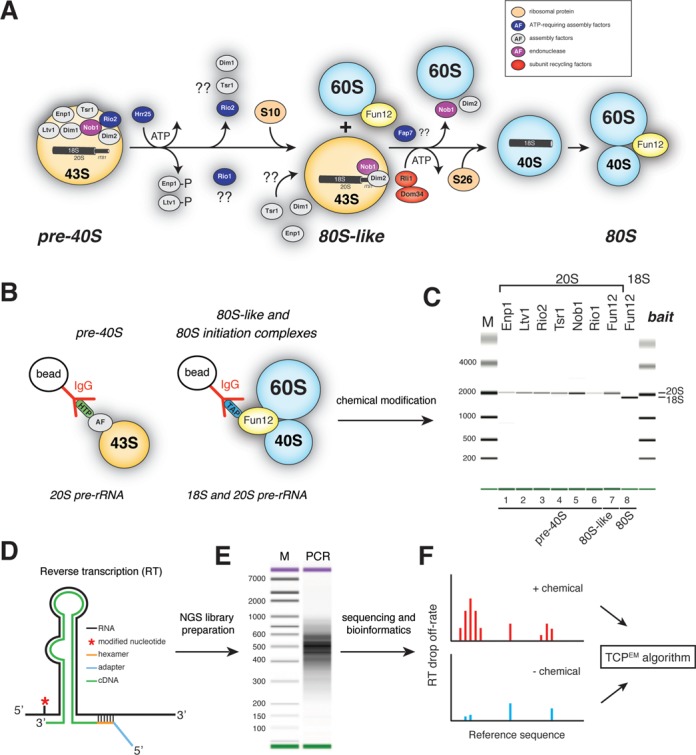
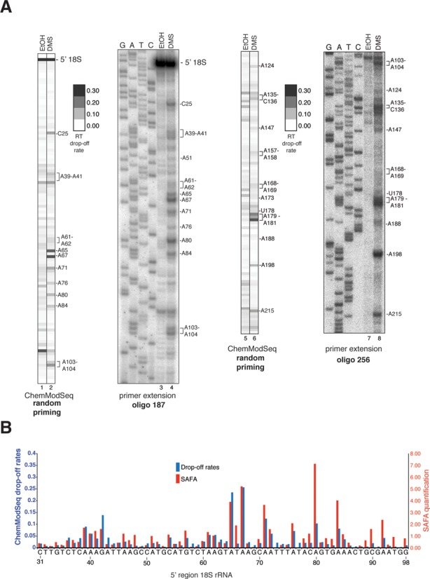
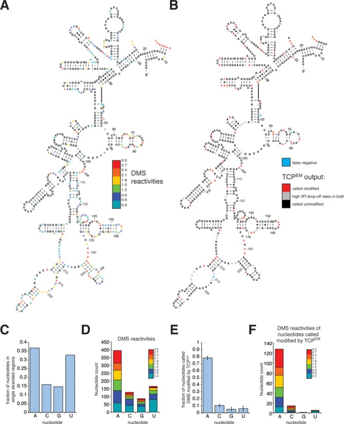
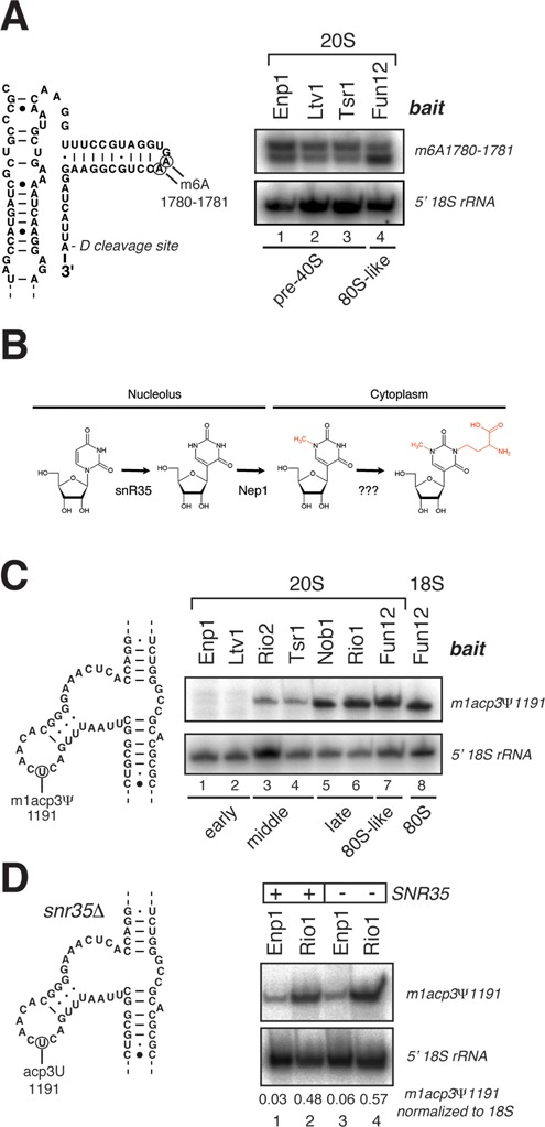
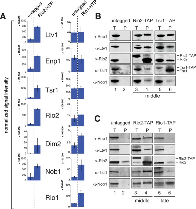
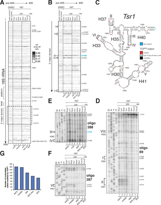
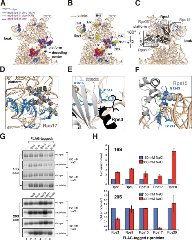
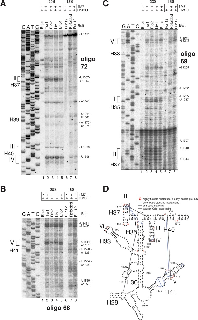
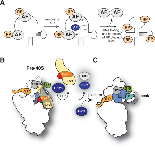
Ribosome assembly in eukaryotes involves the activity of hundreds of assembly factors that direct the hierarchical assembly of ribosomal proteins and numerous ribosomal RNA folding steps. However, detailed insights into the function of assembly factors and ribosomal RNA folding events are lacking. To address this, we have developed ChemModSeq, a method that combines structure probing, high-throughput sequencing and statistical modeling, to quantitatively measure RNA structural rearrangements during the assembly of macromolecular complexes. By applying ChemModSeq to purified 40S assembly intermediates we obtained nucleotide-resolution maps of ribosomal RNA flexibility revealing structurally distinct assembly intermediates and mechanistic insights into assembly dynamics not readily observed in cryo-electron microscopy reconstructions. We show that RNA restructuring events coincide with the release of assembly factors and predict that completion of the head domain is required before the Rio1 kinase enters the assembly pathway. Collectively, our results suggest that 40S assembly factors regulate the timely incorporation of ribosomal proteins by delaying specific folding steps in the 3' major domain of the 20S pre-ribosomal RNA.


Figures

Figure 1.
Figure 1.
40S subunit maturation in Saccharomyces cerevisiae. (A) Schematic representation of the cytoplasmic 40S synthesis pathway. (B and C) Pre-40S particles at different stages of assembly are purified using strains expressing TAP-or HTP-tagged assembly factors. Particles are chemically modified (B), RNA is extracted and gel purified 20S and 18S rRNAs are analyzed on a Bioanalyzer (C). (D and E) cDNA libraries, generated by random priming, were PCR amplified and (E) sequenced on a HiSeq. (F) Chemically modified samples were compared to solvent treated samples using a two-channel Poisson expectation maximization algorithm (TCMPEM) to identify nucleotides that had the highest likelihood of being modified.
Figure 2.
Figure 2.
ChemModSeq accurately and quantitatively measures RNA secondary structure. (A) Primer extension results and ChemModSeq drop-off rates for DMS modified and unmodified 18S rRNA, focusing on the first ∼100 nucleotides. To compare ChemModSeq with primer extension data, drop-off rates were converted to gray scale heat maps in which each block represents a single nucleotide. The darker the color of the block, the higher the drop-off rate. Oligos used for primer extensions are indicated in bold (see Supplementary Table S4). The positions of the modified nucleotides are indicated on the right side of each panel. (B) ChemModSeq data agree well with primer extension results. Direct comparison of the primer extension results shown in (A), quantified using SAFA (34) (red bars) and ChemModSeq RT drop-off rates (blue bars) for the same region (nucleotides 31–98).
Figure 3.
Figure 3.
ChemModSeq accurately and quantitatively measures RNA secondary structure. (A and B) DMS reactivities (A) and TCPEM outputs (B) for nucleotides in the 18S rRNA depicted in the secondary structure of the 5′ region of the yeast 18S rRNA. DMS reactivities were calculated as described (31) and capped at 3. Red colored letters in (B) indicate nucleotides called modified by the TCPEM algorithm. Blue letters in (B) indicate nucleotides visually identified as modified by DMS in primer extension reactions (see Figure 2A) but not called modified by the TCPEM algorithm. (C and D) Distribution of nucleotides located in single-stranded regions in the 18S rRNA (C) and the distribution of nucleotides with DMS reactivities higher than zero (D). (E and F) Fraction of nucleotides called DMS-modified by the TCPEM algorithm (E; average and standard deviations from three independent experiments) and the DMS reactivities of these nucleotides (F). DMS reactivities were calculated by summing drop-off counts from three independent experiments.
Figure 4.
Figure 4.
Pre-40S complexes can be classified into early, middle and late particles. (A) Detection of cytoplasmic methylation at A1780 and A1781 by primer extension in 20S pre-rRNAs. As loading controls, we performed primer extensions to detect the A1 cleavage site at the 5′ end of the pre-rRNAs. (B) Hypermodification of U1191 in the 18S rRNA. U1191 is first pseudouridinylated and N1-methylated by snR35 and Nep1 in the nucleolus, respectively, and then aminocarboxypropyl (acp)-modified in the cytoplasm by an unknown enzyme. (C) Detection of U1191 acp modification in 20S and 18S rRNAs by primer extension. Baits used to purify the individual pre-rRNA species are indicated above the panel. Loading controls were as in (A). (D) Same as in (C) but compared to strains lacking the SNR35 gene.
Figure 5.
Figure 5.
Rio1 associated pre-40S complexes contain fewer 40S assembly factors. (A) HTP-tagged Rio2 and Rio1 were affinity purified and associated proteins were quantified by label-free LC-MS. Shown are the averages and standard deviations from two (Rio2) or three (Rio1) replicate experiments. The y-axis indicates the relative abundance of the proteins in the purified complexes. The number of peptides used to calculate these abundances is listed in Supplementary Table S1. No Rio1 peptides were found in the Rio2 affinity purification. (B and C) Western blot analysis of protein composition of middle (Rio2, Tsr1) and late (Rio1) pre-40S particles purified by Tandem Affinity Purification (TAP) using TAP-tagged strains.
Figure 6.
Figure 6.
A digital snapshot of pre-rRNA secondary structures. (A and B) ChemModSeq and primer extension results obtained using rRNAs extracted from in vitro 1M7 modified particles. Heat maps of average RT drop-off rates in 18S coding sequences (n ≥ 2) (A) or the 3′ major domain (B) of purified rRNAs. The positions of the modified nucleotides are indicated on the right side of each panel. (C) Early-middle and late pre-40S particles are structurally distinct. The 3′ major domain secondary structure represents the ChemModSeq results for the middle (Tsr1) particle. Red nucleotides were indicated as modified by the TCPEM algorithm. Blue nucleotides were visibly modified by 1M7 but not called modified by the TCPEM algorithm (false negative). (DF) Representative primer extension reactions for the 3′ major domain. Roman numbers indicate sites where differences in SHAPE reactivity were observed between pre-40S particles. Oligonucleotides used for primer extensions are indicated in bold. The positions of the modified nucleotides are indicated on the right side of each panel. (G) Pre-rRNAs in early and middle pre-40S complexes adopt more flexible conformations compared to late particles. Plotted are the average number of nucleotides called modified by the TCPEM algorithm (y-axis) for early, middle, late, 80S-like and mature 80S translation initiation complexes (x-axis).
Figure 7.
Figure 7.
Ribosomal protein rRNA binding sites are highly flexible in early and middle pre-40S complexes. (A and B) Nucleotides in the head domain reactive to SHAPE chemicals (A) cluster near assembly factor UV cross-linking sites (B). (CF) SHAPE-modified nucleotides (blue) in the head domain coincide with Rps3, Rps15, Rps17 and Rps20 binding sites. Relevant nucleotide positions are indicated. Green dots highlight the 2′-OH. (G and H) Ribosomal proteins Rps3 and Rps17 form salt-labile interactions with pre-40S complexes. Immunoprecipitations were performed using strains expressing FLAG-tagged r-proteins (indicated above the panels). Co-precipitated 20S was detected by northern hybridization with oligo 004, 18S rRNA was detected by ethidium bromide staining (EtBr). Input indicates 1% of total RNA extracted from cell lysates. (H) Quantification of results shown in (G). Error bars indicate standard deviations obtained from two biological replicates.
Figure 8.
Figure 8.
Ribosome assembly factors are required to maintain an open head domain conformation. (AC) Comparison of primer extension data obtained from 1M7 modified early (Ltv1, Enp1), middle (Tsr1, Rio2) pre-40S complexes (lanes 2–5), mature 40S subunits (Fun12) (lane 8) and in vitro refolded 18S rRNA (lane 7, Refolded). Unmodified 20S and 18S rRNAs were used as control samples (lanes 1 and 6). The positions of the modified nucleotides are indicated on the right side of each panel. (D) Overview of TCPEM output generated from in vitro refolded 18S rRNA ChemModSeq data. Shown are the results for the 3′ major domain. Yellow letters indicate nucleotides that the algorithm predicted were most likely 1M7 modified.
Figure 9.
Figure 9.
A model for structural rearrangements during late stages of 40S subunit maturation. (A) Proposed model for the function of ribosome assembly factors in 40S assembly. Binding of assembly factors (AF) to RNA is required to maintain a more flexible conformation. Their presence allows the assembly of ribosomal proteins (RP) but prevent that these RPs adopt their final conformation. At some point during the maturation pathway, the system receives a signal that certain ribosome assembly/RNA folding steps have been completed and AFs are no longer needed. Their release could be triggered through phosphorylation (such as Hrr25-dependent phosphorylation of Enp1 and Ltv1). This allows completion of RNA folding steps and binding of late RPs. (B and C) Model for rearrangements that take place in the head domain during late stages of 40S assembly. In early and middle pre-40S complexes, binding of Ltv1 and Enp1 to the head domain is required to maintain a more flexible conformation in the 3′ major domain. Rps3, Rps17, Rps20 and Rps17 assemble into pre-ribosomes but do not adopt their final conformation. Hrr25 phosphorylation of Ltv1 and Enp1 triggers their release, allowing late RNA folding steps and assembly of head domain r-proteins to be completed. We propose that Rio2 and Tsr1 are released shortly afterward (B) and Rio1 enters the assembly pathway. The exact function of Rio1, however, remains unclear.

References

    1. Henras A.K., Soudet J., Gerus M., Lebaron S., Caizergues-Ferrer M., Mougin A., Henry Y. The post-transcriptional steps of eukaryotic ribosome biogenesis. Cell. Mol. Life Sci. 2008;65:2334–2359. - PMC - PubMed
    1. Lafontaine D., Delcour J., Glasser A.L., Desgres J., Vandenhaute J. The DIM1 gene responsible for the conserved m6(2)Am6(2)A dimethylation in the 3′-terminal loop of 18 S rRNA is essential in yeast. J. Mol. Biol. 1994;241:492–497. - PubMed
    1. Brand R.C., Klootwijk J., Planta R.J., Maden B.E. Biosynthesis of a hypermodified nucleotide in Saccharomyces carlsbergensis 17S and HeLa-cell 18S ribosomal ribonucleic acid. Biochem. J. 1978;169:71–77. - PMC - PubMed
    1. Liang X.H., Liu Q., Fournier M.J. Loss of rRNA modifications in the decoding center of the ribosome impairs translation and strongly delays pre-rRNA processing. RNA. 2009;15:1716–1728. - PMC - PubMed
    1. Granneman S., Petfalski E., Swiatkowska A., Tollervey D. Cracking pre-40S ribosomal subunit structure by systematic analyses of RNA-protein cross-linking. EMBO J. 2010;29:2026–2036. - PMC - PubMed

Publication types

MeSH terms

Associated data