. 2014;42(17):11233-45.
doi: 10.1093/nar/gku789. Epub 2014 Sep 8.

The spontaneous replication error and the mismatch discrimination mechanisms of human DNA polymerase β

Affiliations

The spontaneous replication error and the mismatch discrimination mechanisms of human DNA polymerase β

Myong-Chul Koag et al. Nucleic Acids Res. 2014.

Abstract

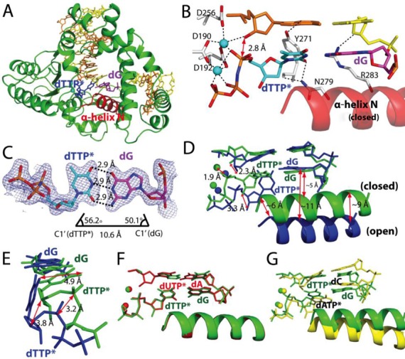
To provide molecular-level insights into the spontaneous replication error and the mismatch discrimination mechanisms of human DNA polymerase β (polβ), we report four crystal structures of polβ complexed with dG•dTTP and dA•dCTP mismatches in the presence of Mg2+ or Mn2+. The Mg(2+)-bound ground-state structures show that the dA•dCTP-Mg2+ complex adopts an 'intermediate' protein conformation while the dG•dTTP-Mg2+ complex adopts an open protein conformation. The Mn(2+)-bound 'pre-chemistry-state' structures show that the dA•dCTP-Mn2+ complex is structurally very similar to the dA•dCTP-Mg2+ complex, whereas the dG•dTTP-Mn2+ complex undergoes a large-scale conformational change to adopt a Watson-Crick-like dG•dTTP base pair and a closed protein conformation. These structural differences, together with our molecular dynamics simulation studies, suggest that polβ increases replication fidelity via a two-stage mismatch discrimination mechanism, where one is in the ground state and the other in the closed conformation state. In the closed conformation state, polβ appears to allow only a Watson-Crick-like conformation for purine•pyrimidine base pairs, thereby discriminating the mismatched base pairs based on their ability to form the Watson-Crick-like conformation. Overall, the present studies provide new insights into the spontaneous replication error and the replication fidelity mechanisms of polβ.


Figures

Figure 1.
Figure 1.
Base pairings of G•T and A•C mismatches. (A) The formation of G•T base pair via wobble, Watson–Crick and pseudo-Watson–Crick base pairings. Base pairings involving enolized or ionized thymine are not shown. (B) The formation of A•C base pair via wobble and Watson–Crick base pairings. Watson–Crick base pair involving the imino tautomer of adenine is not shown.
Figure 2.
Figure 2.
Ternary structure of polβ with dG•dTTP* mismatch and the active-site Mg2+ (PDB ID 4PGQ). (A) Overall structure of polβ with the templating dG paired with an incoming nonhydrolyzable dTMPNPP (dTTP*) in the presence of Mg2+. The template strand is shown in yellow, and the primer and downstream strands are shown in orange. The templating dG is shown in magenta, and the incoming nucleotide is shown in blue. The α-helix N, shown in red, adopts an open conformation. The DNA sequence used for the crystallographic studies is shown. (B) Close-up view of the active site of the dG•dTTP*-Mg2+ ternary structure. The O4 of the incoming dTTP* forms an H-bond with N1 of the templating dG. Tyr271 is H-bonded to N2 of the templating dG. The three catalytic aspartic acid residues and the minor groove recognition amino acids (Asn279 and Arg283) are indicated. A 2FoFc map contoured at 1σ around dTTP* and the templating dG. (C) Close-up view of the metal ion-binding site. Note that Asp256 is not coordinated to the catalytic metal ion, but is coordinated to a water molecule, which is in turn coordinated to the catalytic Mg2+. (D) The H-bonding interactions of the templating dG with Tyr271 and the incoming dTTP*.
Figure 3.
Figure 3.
Ternary structure of polβ with dG•dTTP* mismatch and the active-site Mn2+ (PDB ID 4PGX). (A) Overall structure of the dG•dTTP*-Mn2+ ternary structure. Polβ adopts a closed conformation, and dTTP* and dG form a coplanar base pair conformation. (B) Close-up view of the active site of the dG•dTTP*-Mn2+ ternary structure. (C) Base-pairing properties of dTTP* and dG. (D) Comparison of the dG•dTTP*-Mn2+ ternary structure (green) with the dG•dTTP*-Mg2+ ternary structure (blue). (E) Comparison of the dG•dTTP* base pairs in the Mn2+ (green) and the Mg2+ (blue) complexes. (F) Comparison of the dG•dTTP*-Mn2+ structure (green) with the published dA•dUTP*-Mn2+ structure (PDB ID 2FMS, shown in red). (G) Comparison of the dG•dTTP*-Mn2+ structure (green) with the published dC•dATP*-Mn2+ structure (PDB ID 3C2L, yellow).
Figure 4.
Figure 4.
Ternary structures of polβ with dA•dCTP* mismatch (PDB ID 4PHA and 4PHD). (A) Overall structure of the active site of the polβ-dA•dCTP*-Mg2+ complex. (B) Comparison of the lyase domains of the dA•dCTP*-Mg2+ (white) and dC•dATP*-Mn2+ (blue) structures. (C) Close-up view of the dA•dCTP*-Mg2+ structure. Key H-bonding interactions are indicated as dotted lines. (D) Comparison of the dA•dCTP*-Mg2+ (blue) and dG•dTTP*-Mg2+ (green) structures. (E) Comparison of the dA•dCTP*-Mg2+ (blue) and dG•dTTP*-Mn2+ (pale cyan) structures. (F) Comparison of the dA•dCTP*-Mg2+ (blue) and dC•dATP*-Mn2+ (yellow) structures. (G) Close-up view of the active site of the dA•dCTP*-Mn2+ structure. A 2FoFc map contoured at 1σ around dCTP* and the templating dA. Key H-bonding interactions are indicated as dotted lines. (H) Comparison of the dA•dCTP*-Mg2+ (blue) and the dA•dCTP*-Mn2+ (magenta) structures.
Figure 5.
Figure 5.
Average RMSDs of the thumb subdomain of polβ and their fluctuations. In the left panel, RMSDs are compared for the closed conformation simulations, and in the right panel, for the open conformation simulations. For each coordinate during 150 ns MD simulation, RMSDs are computed relative to the open (red) and to the closed (black) reference conformation, respectively, in which the polβ-dG•dTTP*-Mn2+ ternary structure (for the closed conformation, PDB ID 4PGX) and the polβ-dG•dTTP*-Mg2+ ternary structure (for the open conformation, PDB ID 4PGQ) are used as their corresponding reference structures. ‘Apo’ denotes the single-nulcleotide gapped binary complex with the templating dG.
Figure 6.
Figure 6.
In the lower panel, the RMSDs of the thumb subdomain relative to the closed conformations are shown. The RMSD for the dG•dCTPInt system shows that the structure approaches the state of the closed conformation around 125 ns. In the upper panel, the RMSDs relative to the open conformation are presented for the dG•dCTPInt (black) and dG•dCTPopen-2Mg2+ (green) systems, respectively, showing that the ‘intermediate’ system deviates from the open conformation.
Figure 7.
Figure 7.
Ground-state and ‘pre-chemistry-state’ structures of polβ ternary complexes. The Mg2+-bound (left panels) and the Mn2+-bound (right panels) structures most likely represent the ground-state and the ‘pre-chemistry-state’ structures of the polβ ternary complexes, respectively. (A) The dG•dTTP*-Mg2+ structure. Tyr271 and dTTP form two H-bonds with the templating dG. (B) The dG•dTTP*-Mn2+ structure. Substituting Mn2+ for Mg2+ triggers an open-to-closed conformational change of the protein and a staggered-to-Watson–Crick conformational change of the dG•dTTP base pair. (C) The dA•dCTP*-Mg2+ structure. dA•dCTP* forms a non-coplanar conformation. (D) The dA•dCTP*-Mn2+ structure. The Mn2+ substitution does not induce a conformational change of the protein and the base pair. (E) Published polβ structure with dG•dCTP* and the active-site Mg2+ (PDB ID 1BPY).

References

    1. Halliday J.A., Glickman B.W. Mechanisms of spontaneous mutation in DNA repair-proficient Escherichia coli. Mutat. Res. 1991;250:55–71. - PubMed
    1. Bacolla A., Cooper D.N., Vasquez K.M. Mechanisms of base substitution mutagenesis in cancer genomes. Genes. 2014;5:108–146. - PMC - PubMed
    1. Keohavong P., Thilly W.G. Fidelity of DNA polymerases in DNA amplification. Proc. Natl Acad. Sci. U.S.A. 1989;86:9253–9257. - PMC - PubMed
    1. Bebenek K., Joyce C.M., Fitzgerald M.P., Kunkel T.A. The fidelity of DNA synthesis catalyzed by derivatives of Escherichia coli DNA polymerase I. J. Biol. Chem. 1990;265:13878–13887. - PubMed
    1. Lee H.R., Johnson K.A. Fidelity of the human mitochondrial DNA polymerase. J. Biol. Chem. 2006;281:36236–36240. - PubMed

Publication types