. 2014;42(17):11056-70.
doi: 10.1093/nar/gku752. Epub 2014 Sep 8.

Characterization of three mycobacterial DinB (DNA polymerase IV) paralogs highlights DinB2 as naturally adept at ribonucleotide incorporation

Affiliations

Characterization of three mycobacterial DinB (DNA polymerase IV) paralogs highlights DinB2 as naturally adept at ribonucleotide incorporation

Heather Ordonez et al. Nucleic Acids Res. 2014.

Abstract

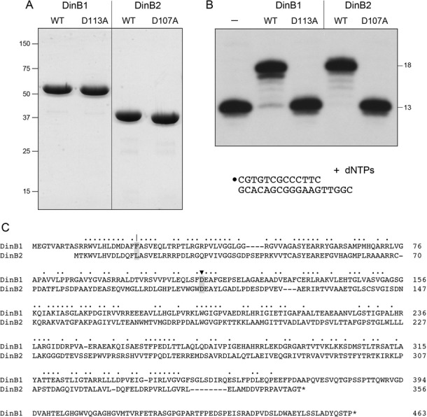
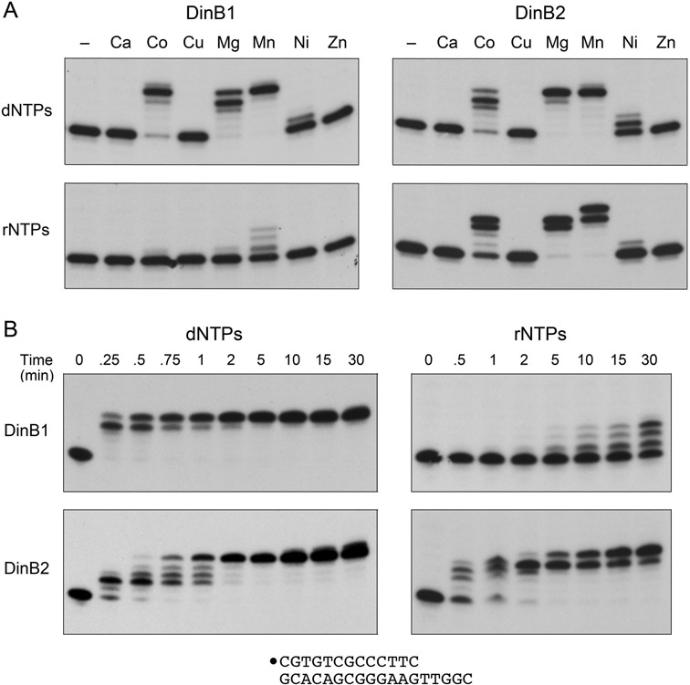
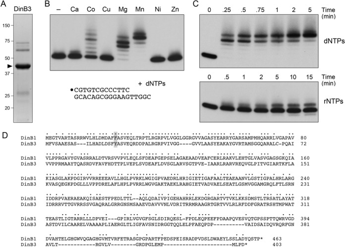
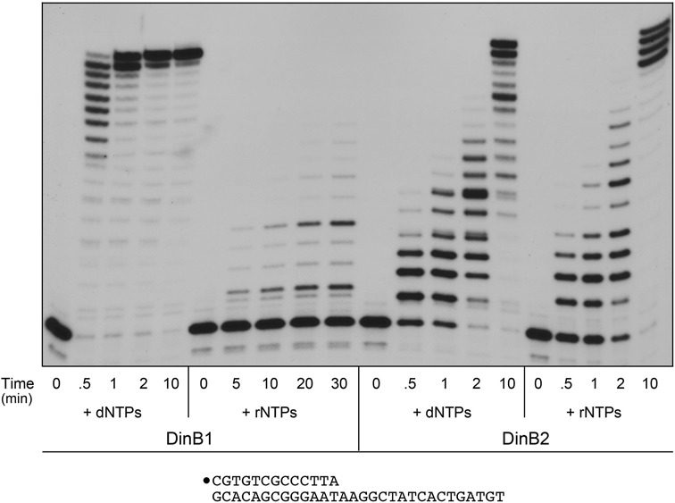
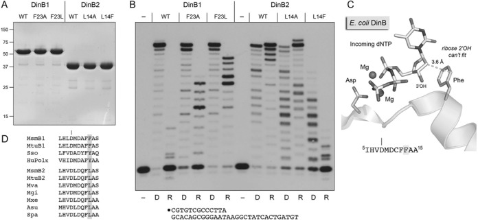
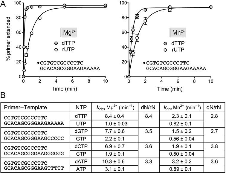
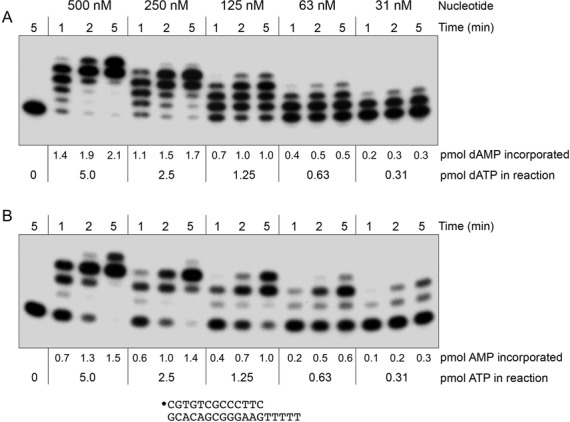
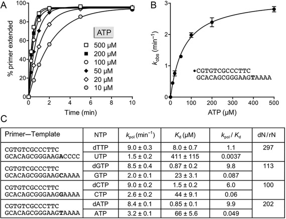
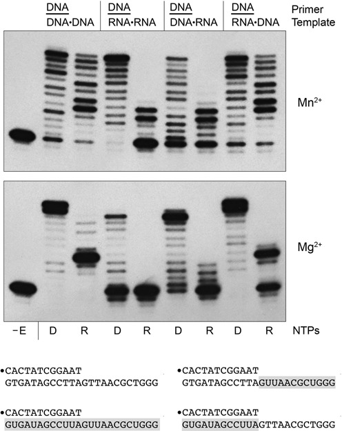
This study unveils Mycobacterium smegmatis DinB2 as the founder of a clade of Y-family DNA polymerase that is naturally adept at incorporating ribonucleotides by virtue of a leucine in lieu of a canonical aromatic steric gate. DinB2 efficiently scavenges limiting dNTP and rNTP substrates in the presence of manganese. DinB2's sugar selectivity factor, gauged by rates of manganese-dependent dNMP versus rNMP addition, is 2.7- to 3.8-fold. DinB2 embeds ribonucleotides during DNA synthesis when rCTP and dCTP are at equimolar concentration. DinB2 can incorporate at least 16 consecutive ribonucleotides. In magnesium, DinB2 has a 26- to 78-fold lower affinity for rNTPs than dNTPs, but only a 2.6- to 6-fold differential in rates of deoxy versus ribo addition (kpol). Two other M. smegmatis Y-family polymerases, DinB1 and DinB3, are characterized here as template-dependent DNA polymerases that discriminate strongly against ribonucleotides, a property that, in the case of DinB1, correlates with its aromatic steric gate side chain. We speculate that the unique ability of DinB2 to utilize rNTPs might allow for DNA repair with a 'ribo patch' when dNTPs are limiting. Phylogenetic analysis reveals DinB2-like polymerases, with leucine, isoleucine or valine steric gates, in many taxa of the phylum Actinobacteria.


Figures

Figure 1.
Figure 1.
DinB1 and DinB2 are DNA polymerases. (A) Purification. Aliquots (5 μg) of recombinant wild-type DinB1, DinB1-D113A, wild-type DinB2 and DinB2-D107A were analyzed by SDS-PAGE. The Coomassie Blue-stained gel is shown. The positions and sizes (kDa) of marker polypeptides are indicated on the left. (B) DNA polymerase reaction mixtures (10 μl) containing 10 mM Tris-HCl, pH 7.5, 5 mM MnCl2, 125 μM each of dATP, dGTP, dCTP and dTTP (dNTPs), 100 nM 5′ 32P-labeled 13-mer/18-mer primer–template (depicted at bottom, with the 5′ 32P label denoted by •) and 500 nM of the DinB protein were incubated at 37°C for 15 min. The reaction products were analyzed by urea-PAGE and visualized by autoradiography. (C) DinB1 and DinB2 primary structures. The amino acid sequences of the MsmDinB1 and MsmDinB2 proteins are aligned. Positions of side chain identity/similarity are denoted by dots. Gaps in the alignments are denoted by dashes. The metal-binding aspartate residues Asp113 and Asp107 that were mutated in (A) and (B) are shaded and denoted by ▾. The steric gate residues are shaded and denoted by |.
Figure 2.
Figure 2.
Characterization of DinB1 and DinB2 polymerase activities. (A) Divalent cation specificity. Polymerase reaction mixtures (10 μl) containing 10 mM Tris-HCl, pH 7.5, 100 nM 5′ 32P-labeled 13-mer/18-mer primer–template, 125 μM each of dATP, dGTP, dCTP and dTTP (dNTPs, top panels) or ATP, GTP, CTP and UTP (rNTPs, bottom panels) as specified, 1 μM DinB1 (left panels) or DinB2 (right panels) as indicated, and either no added metal cofactor (lanes –) or 5 mM of the indicated divalent cation (as the chloride salt) were incubated at 37°C for 15 min. (B) Kinetic profiles. Polymerase reaction mixtures containing 10 mM Tris-HCl, pH 7.5, 5 mM MnCl2, 100 nM 5′ 32P-labeled 13-mer/18-mer primer–template, 125 μM dNTP (left panels) or rNTPs (right panels), and 1 μM DinB1 (top panels) or DinB2 (bottom panels) were incubated at 37°C. Aliquots (10 μl) were withdrawn at the times specified and quenched immediately with EDTA/formamide. The reaction products were analyzed by urea-PAGE and visualized by autoradiography.
Figure 3.
Figure 3.
Characterization of DinB3 polymerase activity. (A) Purification. An aliquot (5 μg) of recombinant DinB3 was analyzed by SDS-PAGE. The Coomassie Blue-stained gel is shown. The positions and sizes (kDa) of marker polypeptides are indicated on the left. The DinB3 polypeptide is denoted by ▸. (B) Metal specificity. Polymerase reaction mixtures (10 μl) containing 10 mM Tris-HCl, pH 7.5, 100 nM 5′ 32P-labeled 13-mer/18-mer primer–template, 125 μM each of dATP, dGTP, dCTP and dTTP (dNTPs), 1 μM DinB3, and either no added metal cofactor (lane –) or 5 mM of the indicated divalent cation (as the chloride salt) were incubated at 37°C for 15 min. (C) Kinetic profile. Polymerase reaction mixtures containing 10 mM Tris-HCl, pH 7.5, 5 mM MnCl2, 100 nM 5′ 32P-labeled 13-mer/18-mer primer–template, 125 μM dNTPs (top panel) or rNTPs (bottom panel) and 1 μM DinB3 were incubated at 37°C. Aliquots (10 μl) were withdrawn at the times specified and quenched immediately with EDTA/formamide. The reaction products were analyzed by urea-PAGE and visualized by autoradiography. (D) DinB3 primary structure. The amino acid sequences of the MsmDinB1 and MsmDinB3 proteins are aligned. Positions of side chain identity/similarity are denoted by dots. Gaps in the alignments are denoted by dashes. The steric gate residues are shaded.
Figure 4.
Figure 4.
Longer tracts of templated synthesis by DinB1 and DinB2. Polymerase reaction mixtures containing 10 mM Tris-HCl, pH 7.5, 5 mM MnCl2, 50 nM 5′ 32P-labeled 13-mer/30-mer primer–template (depicted at bottom, with the 5′ 32P label denoted by •), dNTPs (125 μM each) or rNTPs (125 μM each) as specified, and 1 μM DinB1 or DinB2 were incubated at 37°C. Aliquots (10 μl) were withdrawn at the times specified and quenched immediately with EDTA/formamide. The reaction products were analyzed by urea-PAGE and visualized by autoradiography.
Figure 5.
Figure 5.
DinB2 can embed ribonucleotides during DNA synthesis. Polymerase reaction mixtures (20 μl) containing 10 mM Tris-HCl, pH 7.5, 5 mM MnCl2, 50 nM 5′ 32P-labeled 13-mer/30-mer primer–template, either 125 μM each of dATP, dGTP, dTTP and dCTP (dAGTC), 125 μM each of ATP, GTP, UTP and CTP (rAGUC), 125 μM each of dATP, dGTP, dTTP and rCTP (dAGT/rC), or 125 μM each of dATP, dGTP, dTTP plus 63 μM each of dCTP and rCTP (dAGTC/rC), and either 1 μM DinB2 or no enzyme (–E) were incubated for 20 min at 37°C. The reactions were quenched by adjustment to 20 mM EDTA. Aliquots (10 μl) of each sample were then adjusted to either 0.25 M NaOH (denoted by +) or 0.25 M NaCl (denoted by −) and incubated at 25°C for 16 h. The NaOH samples were then neutralized with 0.25 M HCl. The reaction products were analyzed by urea-PAGE and visualized by autoradiography. The three alkaline hydrolysis products indicative of rCMP incorporation at +3, +4 and +13 during DNA synthesis in the presence of all four dNTPs are indicated by •. The gel was scanned with a Fujix BAS2500 imager and the extents of rCMP incorporation at +3, +4 and +13 sites were calculated as described in the Results section.
Figure 6.
Figure 6.
A single amino acid polymorphism governs ribonucleotide utilization. (A) Purification. Aliquots (5 μg) of the indicated DinB1 and DinB2 proteins were analyzed by SDS-PAGE. The Coomassie Blue-stained gel is shown. The positions and sizes (kDa) of marker polypeptides are indicated on the left. (B) Polymerase reaction mixtures (10 μl) containing 10 mM Tris-HCl, pH 7.5, 5 mM MnCl2, 50 nM 5′ 32P-labeled 13-mer/30-mer primer–template, dNTPs (125 μM each; lanes D) or rNTPs (125 μM each; lanes R) as specified and 1 μM of the indicated DinB proteins were incubated for 10 min at 37°C. The reaction products were analyzed by urea-PAGE and visualized by autoradiography. (C) Structure of E. coli DinB in complex with two magnesium ions and an incoming dNTP (from pdb 4IR1). The N-terminal DinB segment containing the first metal-binding aspartate and the steric gate phenylalanine is shown. The amino acid sequence is specified at bottom. (D) Distinct clades of DinB homologs with aromatic versus aliphatic steric gates. The amino acid sequences in the vicinity of the metal-binding aspartate and steric gate residues are aligned for exemplary DinB/PolIV homologs that either have an aromatic steric gate side chain (top grouping) or a leucine instead (bottom grouping). The aromatic gate group includes M. smegmatis DinB1, M. tuberculosis DinB1, Sulfolobus sulfataricus Dpo4 and human Pol kappa. The leucine gate clade includes DinB2-like proteins from Actinobacteria species M. smegmatis, M. tuberculosis, M. vanbaalenii, M. gilvum, M. xenopi, Amycolicicoccus subflavus and Saccharomonospora paurometabolica.
Figure 7.
Figure 7.
Kinetics of templated dNMP and rNMP incorporation. (A) Polymerase reaction mixtures contained 10 mM Tris-HCl, pH 7.5, 50 nM 5′ 32P-labeled 13-mer/18-mer primer–template with a homo-oligo-dA tail, and either 5 mM MnCl2 and 500 μM dTTP or rUTP (left panel) or 1 mM MnCl2 and 100 μM dTTP or rUTP (right panel). Aliquots (10 μl) were withdrawn at the times specified and quenched immediately with EDTA/formamide. The reaction products were analyzed by urea-PAGE and the percent of primer strand extended by one or more nucleotides was quantified by scanning the gel with a Fujix BAS2500 imager. The % primer extension is plotted as a function of reaction time. Each datum in the graphs is the average of three separate experiments ±SEM. The rate constants (kobs ± SE) for the first step of dTTP and UTP addition to the primer–template were obtained by non-linear regression curve fitting of the data to a one-phase association function in Prism and are shown in panel (B). A series of 5′ 32P-labeled 13-mer/18-mer primer–templates with identical duplex segments and either homo-oligo-dC, homo-oligo-dG or homo-oligo-dT template tails was used to assay the kinetics of G, C and A nucleotide addition to the primer strand, with magnesium or manganese as the metal cofactor. The rate constants are compiled in panel (B). The ratios of the rate of the first deoxynucleotide and ribonucleotide addition steps are indicated in the columns dN/rN.
Figure 8.
Figure 8.
DinB2 scavenges nucleotides in the presence of manganese. Reaction mixtures containing 10 mM Tris-HCl, pH 7.5, 1 mM MnCl2, 50 nM 5′ 32P-labeled 13-mer/18-mer primer–template with a homo-oligothymidylate tail, 1 μM of DinB2, and either 500 nM, 250 nM, 125 nM, 63 nM or 31 nM dATP (top panel) or ATP (bottom panel) were incubated at 37°C. Aliquots (10 μl) were withdrawn at 1, 2 and 5 min. The reaction products were analyzed by urea-PAGE and visualized by autoradiography. The input amounts (pmol) of dATP or ATP substrates in the 10 μl aliquots of reaction mixture are indicated below the autoradiograms. The gel was scanned with a Fujix BAS2500 imager and the pmol of dAMP/AMP incorporation into the primer strand were calculated as described in the Results section; the values are shown below each lane in the gels.
Figure 9.
Figure 9.
DinB2 polymerization rates and affinities for individual nucleotide substrates with magnesium as metal cofactor. Polymerase reaction mixtures containing 10 mM Tris-HCl, pH 7.5, 5 mM MgCl2, 50 nM 5′ 32P-labeled 13-mer/18-mer primer–template with dT as the first template nucleobase (shown in panel B), 1 μM DinB2 and the indicated concentrations of ATP were incubated at 37°C. Aliquots (10 μl) were withdrawn at times specified and quenched with EDTA/formamide. The products were analyzed by urea-PAGE and quantified by scanning the gels. (A) The % of primer extended is plotted as a function of reaction time for each concentration of ATP substrate. The data were fit by non-linear regression in Prism to a one-phase exponential. (B) The kobs values calculated in Prism are plotted as a function of ATP concentration; each datum in the graph is the average of two separate experiments. The data were fit by non-linear regression to a single binding function, from which the Kd for nucleotide and turnover number (kpol) at saturating nucleotide were derived. (C) A series of 5′ 32P-labeled 13-mer/18-mer primer–templates with identical duplex segments and either dA, dC, dG or dT as the first template nucleobase was used to assay the kinetics of the indicated nucleotide addition to the primer strand, as a function of dNTP/rNTP nucleotide concentration, with magnesium as the metal cofactor. The data were analyzed as in panels (A) and (B). The kpol and Kd values and kpol/Kd ratio (catalytic efficiency) for each dNTP and rNTP substrate are indicated. The ratios of the catalytic efficiencies for deoxynucleotide and ribonucleotide substrates are indicated in the column dN/rN.
Figure 10.
Figure 10.
DinB2 can synthesize DNA using an RNA template. Polymerase reaction mixtures (10 μl) containing 10 mM Tris-HCl, pH 7.5, 50 nM 5′ 32P-labeled 12-mer/24-mer primer–template as specified, either 1 mM MnCl2 and 100 μM of each dNTP or rNTP (top panel) or 5 mM MgCl2 and 125 μM of each dNTP or rNTP (bottom panel), and 1 μM DinB2 were incubated at 37°C for 15 min. The reaction products were analyzed by urea-PAGE and visualized by autoradiography. The dNTP (D) or rNTP (R) substrates are indicated below the lanes. DinB was omitted from the control reaction in lane –E. The primer–templates are depicted at bottom with RNA in the template strand shaded gray.

Similar articles

Cited by

References

    1. Gong C., Martins A., Bongiorno P., Glickman M., Shuman S. Biochemical and genetic analysis of the four DNA ligases of mycobacteria. J. Biol. Chem. 2004;279:20594–20606. - PubMed
    1. Gong C., Bongiorno P., Martins A., Stephanou N.C., Zhu H., Shuman S., Glickman M. S. Mechanism of non-homologous end joining in mycobacteria: a low-fidelity repair system driven by Ku, ligase D and ligase C. Nat. Struct. Mol. Biol. 2005;12:304–312. - PubMed
    1. Akey D., Martins A., Aniukwu J., Glickman M.S., Shuman S., Berger J.M. Crystal structure and nonhomologous end joining function of the ligase domain of Mycobacterium DNA ligase D. J. Biol. Chem. 2006;281:13412–13423. - PubMed
    1. Sinha K.M., Stephanou N.C., Gao F., Glickman M.S., Shuman S. Mycobacterial UvrD1 is a Ku-dependent DNA helicase that plays a role in multiple DNA repair events, including double-strand break repair. J. Biol. Chem. 2007;282:15114–15125. - PubMed
    1. Sinha K.M., Glickman M.S., Shuman S. Mutational analysis of Mycobacterium UvrD1 identifies functional groups required for ATP hydrolysis, DNA unwinding, and chemomechanical coupling. Biochemistry. 2009;48:4019–4030. - PMC - PubMed

Publication types

MeSH terms