. 2014 Nov:76:159-68.
doi: 10.1016/j.yjmcc.2014.08.021. Epub 2014 Sep 6.

Connexin40 abnormalities and atrial fibrillation in the human heart

Affiliations

Connexin40 abnormalities and atrial fibrillation in the human heart

Joanna Gemel et al. J Mol Cell Cardiol. 2014 Nov.

Abstract

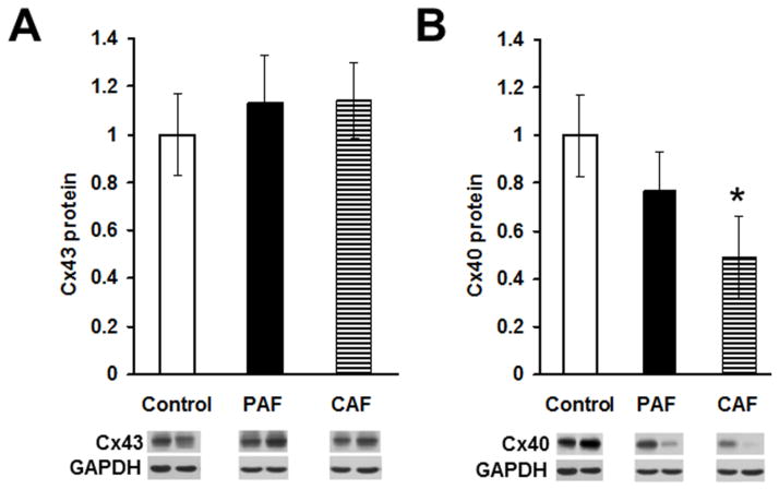
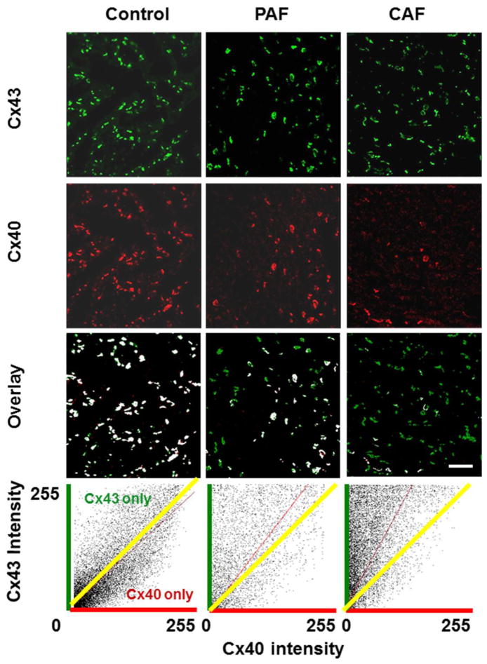
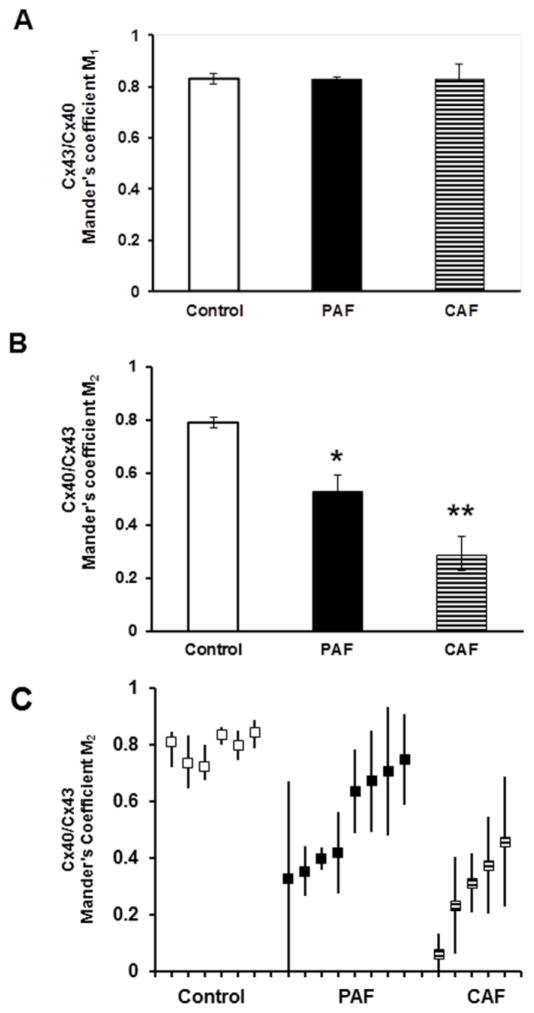
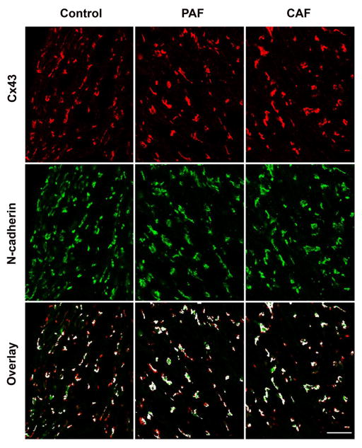
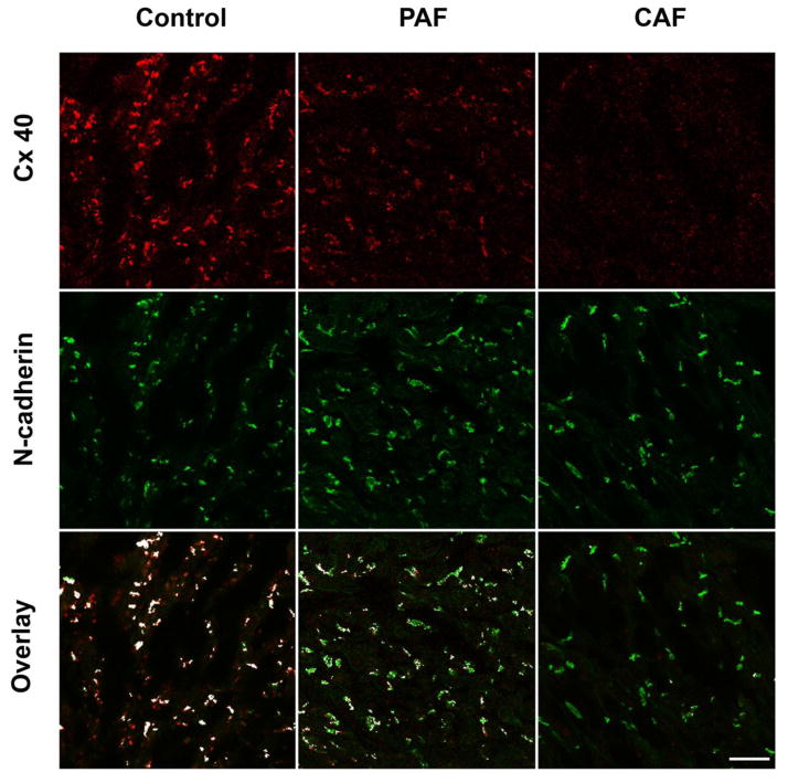
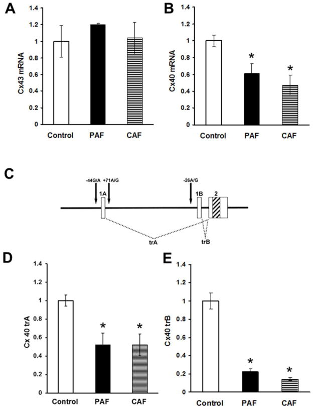
Normal atrial conduction requires similar abundances and homogeneous/overlapping distributions of two connexins (Cx40 and Cx43). The remodeling of myocyte connections and altered electrical conduction associated with atrial fibrillation (AF) likely involves perturbations of these connexins. We conducted a comprehensive series of experiments to examine the abundances and distributions of Cx40 and Cx43 in the atria of AF patients. Atrial appendage tissues were obtained from patients with lone AF (paroxysmal or chronic) or normal controls. Connexins were localized by double label immunofluorescence confocal microscopy, and their overlap was quantified. Connexin proteins and mRNAs were quantified by immunoblotting and qRT-PCR. PCR amplified genomic DNA was sequenced to screen for connexin gene mutations or polymorphisms. Immunoblotting showed reductions of Cx40 protein (to 77% or 49% of control values in samples from patients with paroxysmal and chronic AF, respectively), but no significant changes of Cx43 protein levels in samples from AF patients. The extent of Cx43 immunostaining and its distribution relative to N-cadherin were preserved in the AF patient samples. Although there was variability of Cx40 staining among paroxysmal AF patients, all had some fields with substantial Cx40 heterogeneity and reduced overlap with Cx43. Cx40 immunostaining was severely reduced in all chronic AF patients. qRT-PCR showed no change in Cx43 mRNA levels, but reductions in total Cx40 mRNA (to <50%) and Cx40 transcripts A (to ~50%) and B (to <25%) as compared to controls. No Cx40 coding region mutations were identified. The frequency of promoter polymorphisms did not differ between AF patient samples and controls. Our data suggest that reduced Cx40 levels and heterogeneity of its distribution (relative to Cx43) are common in AF. Multiple mechanisms likely lead to reductions of functional Cx40 in atrial gap junctions and contribute to the pathogenesis of AF in different patients.

Keywords: Arrhythmia; Atrial fibrillation; Connexin; Connexin40; Gap junction.


Figures

Figure 1
Figure 1. Levels of Cx40 (but not Cx43) are reduced in atrial homogenates of patients with atrial fibrillation
Homogenates were prepared from atrial appendage samples from control hearts or from patients with paroxysmal (PAF) or chronic (CAF) atrial fibrillation, and Cx43 (A) and Cx40 (B) were detected by immunoblotting. Bands at the bottom are representative blots from two control samples and from two patients with PAF or CAF. Gels were loaded with equal amounts of total protein and were also blotted with antibodies directed against GAPDH (as a loading control). Graphs show the amounts (mean ± SEM) of immunoreactive connexin in each group determined by densitometry (adjusted for GAPDH densitometry and normalized to the mean control values). While the abundance of Cx43 did not differ significantly between control and disease groups, the mean level of Cx40 in the CAF group (but not the PAF group) was significantly less than in controls (*, p < 0.05, Student’s t test).
Figure 2
Figure 2. Immunolocalization of Cx43 and Cx40 shows reductions of Cx40 in sections from hearts of patients with atrial fibrillation
Representative photomicrographs are shown for sections from atrial appendage samples from control hearts (left panels) or from patients with paroxysmal (PAF, center panels) or chronic (CAF, right panels) atrial fibrillation that were studied by double label immunofluorescence for Cx43 (green, top row) and Cx40 (red, second row). The third row shows the overlay between Cx43 and Cx40; spots containing both connexins are shown in white, while those containing only Cx43 are green and those containing only Cx40 are red. While the abundance of Cx43 appears similar in all three samples, Cx40 is diminished in PAF and severely reduced in CAF. This results in fewer white spots and many “green only” spots in the overlay images for PAF and CAF. The bottom panels show cytofluorogram scatter plots of the intensities of all pixels; Cx43 and Cx40 intensities are quantified according to a 0 – 255 gray scale. The yellow line shows the theoretical predicted mean for a perfect overlap of the distribution and intensities of Cx40 and Cx43. The red lines show the determined distributions, and illustrate the shift towards Cx43 predominance in the atrial fibrillation samples and the selective loss of Cx40. Bar, 42 μm.
Figure 3
Figure 3. Co-localization coefficients show that Cx40 was reduced relative to Cx43 in atria of patients with atrial fibrillation
The distribution of Cx43 and Cx40 and their overlap were determined by immunofluorescence (as illustrated in Fig. 2) for multiple sections from each control and diseased sample. For each section the overlap was quantified by calculating the Mander’s coefficients as described in Methods. A coefficient of 1.0 indicates perfect overlap while a coefficient of 0 indicates no overlap. Variables were defined so that Mander’s coefficient M1 is an expression of the overlap of Cx40 on Cx43 (Cx40 spots that also contain Cx43) and Mander’s coefficient M2 is an expression of the overlap of Cx43 on Cx40 (Cx43 spots that also contain Cx40). (A. B) The graphs show the Mander’s M1 (A) and M2 (B) coefficients (mean ± SEM) for groups of control (n = 6), PAF (n = 8), and CAF (n = 5) samples. Mean M1 values did not show any differences between control and diseased specimens. However, mean M2 values were reduced in PAF samples as compared to controls (*, p < 0.05, ANOVA) and reduced in CAF samples as compared to either controls or PAF samples (**, p < 0.05, ANOVA). These data quantify the selective loss of Cx40 from gap junctions in the atria of patients with paroxysmal and chronic atrial fibrillation. (C) The graph shows the mean M2 coefficient (solid squares) for each control, PAF or CAF sample analyzed. The bars represent the range of different M2 values determined from images of different sections in the same individual. Thus, there was a wide variation between AF patients (especially those with PAF) and within many individual AF patients.
Figure 4
Figure 4. Immunolocalization of Cx43 vs. N-cadherin does not differ between control and AF patients
Representative photomicrographs are shown for sections from atrial appendage samples from control hearts (left panels) or from patients with paroxysmal (PAF, center panels) or chronic (CAF, right panels) atrial fibrillation that were studied by double label immunofluorescence for Cx43 (red, top row) and N-cadherin (green, second row). The third row shows the overlay between Cx43 and N-cadherin; spots containing both proteins are shown in white, while those containing only Cx43 are red and those containing only N-cadherin are green. The abundance of Cx43 and N-cadherin appears similar in all three samples. Bar, 40 μm.
Figure 5
Figure 5. Immunolocalization of Cx40 and N-cadherin shows reductions of Cx40 in sections from hearts of patients with atrial fibrillation
Representative photomicrographs are shown for sections of atrial appendage samples from control hearts (left panels) or from patients with paroxysmal (PAF, center panels) or chronic (CAF, right panels) atrial fibrillation that were studied by double label immunofluorescence for Cx40 (red, top row) and N-cadherin (green, second row). The third row shows the overlay between Cx40 and N-cadherin; spots containing both connexins are shown in white, while those containing only N-cadherin are green and those containing only Cx40 are red. While the abundance of N-cadherin appears similar in all three samples, Cx40 is diminished in PAF and CAF. This results in fewer white spots and many “green only” spots in the overlay images for PAF and CAF. Bar, 40 μm.
Figure 6
Figure 6. Levels of Cx40 (but not Cx43) mRNA are reduced in atrial homogenates of patients with atrial fibrillation
Total RNA was prepared from atrial homogenates of control hearts or patients with paroxysmal (PAF) or chronic (CAF) atrial fibrillation. Total mRNAs for Cx43 (A) and Cx40 (B) were quantified by qRT-PCR. Graphs show the values normalized to the average values in control samples (mean ± SEM). (A) Total levels of Cx43 mRNA did not differ among groups. (B) Total levels of Cx40 mRNA were significantly reduced in both PAF and CAF groups. (C) A diagram illustrates the structure of the Cx40 gene and the mRNAs generated from it. The gene contains two alternate first exons (1A and 1B) a single second exon (2) indicated by boxes with the coding region indicated by diagonal stripes. Differential exon usage leads to the production of two different transcripts (trA and trB). The two transcriptional start sites are flanked by polymorphic variants (−44G/A and +71A/G for A and −26A/G for B) that are indicated by arrows. (D, E) Levels of both Cx40 trA (panel D) and trB (panel E) were significantly reduced in both PAF and CAF groups (*, p < 0.05 as compared to controls).

References

    1. Allessie MA, Boyden PA, Camm AJ, kleber AG, Lab MJ, Legato MJ, et al. Pathophysiology and prevention of atrial fibrillation. Circulation. 2001;103:769–77. - PubMed
    1. Iwasaki YK, Nishida K, Kato T, Nattel S. Atrial fibrillation pathophysiology: implications for management. Circulation. 2011;124:2264–74. - PubMed
    1. Spach MS, Starmer CF. Altering the topology of gap junctions: a major therapeutic target for atrial fibrillation. Cardiovasc Res. 1995;30:337–44. - PubMed
    1. Saffitz JE, Kanter HL, Green KG, Tolley TK, Beyer EC. Tissue-specific determinants of anisotropic conduction velocity in canine atrial and ventricular myocardium. Circ Res. 1994;74:1065–70. - PubMed
    1. Lin X, Gemel J, Glass A, Zemlin CW, Beyer EC, Veenstra RD. Connexin40 and connexin43 determine gating properties of atrial gap junction channels. J Mol Cell Cardiol. 2010;48:238–45. - PMC - PubMed

Publication types