. 2014 Oct 23;21(10):1318-1329.
doi: 10.1016/j.chembiol.2014.06.010. Epub 2014 Sep 4.

Maresin-like lipid mediators are produced by leukocytes and platelets and rescue reparative function of diabetes-impaired macrophages

Affiliations

Maresin-like lipid mediators are produced by leukocytes and platelets and rescue reparative function of diabetes-impaired macrophages

Song Hong et al. Chem Biol. .

Abstract

Nonhealing diabetic wounds are associated with impaired macrophage (Mf) function. Leukocytes and platelets (PLT) play crucial roles in wound healing by poorly understood mechanisms. Here we report the identification and characterization of the maresin-like(L) mediators 14,22-dihydroxy-docosa-4Z,7Z,10Z,12E,16Z,19Z-hexaenoic acids, 14S,22-diHDHA (maresin-L1), and 14R,22-diHDHA (maresin-L2) that are produced by leukocytes and PLT and involved in wound healing. We show that 12-lipoxygenase-initiated 14S-hydroxylation or cytochrome P450 catalyzed 14R-hydroxylation and P450-initiated ω(22)-hydroxylation are required for maresin-L biosynthesis. Maresin-L treatment restores reparative functions of diabetic Mfs, suggesting that maresin-Ls act as autocrine/paracrine factors responsible for, at least in part, the reparative functions of leukocytes and PLT in wounds. Additionally, maresin-L ameliorates Mf inflammatory activation and has the potential to suppress the chronic inflammation in diabetic wounds caused by activation of Mfs. These findings provide initial insights into maresin-L biosynthesis and mechanism of action and potentially offer a therapeutic option for better treatment of diabetic wounds.


Figures

Figure 1
Figure 1. Structure Identification and Elucidation of Novel 14,22-dihydroxy-4,7,10,12,16,19-Docosahexaenoic acids, Produced from DHA by Human Leukocytes with or without Platelets
Human blood leukocytes (3 × 10⁶ monocytes + 3 × 10⁷ neutrophils + 3 × 10⁶ lymphocytes) were incubated with or without 3 × 10⁷ platelets in 3 μM DHA or DHA-21,21,22,22,22-d5, then stimulated by wound healing relevant conditions. Leukocytes were also incubated with 3 μM 14S-HDHA or 14R-HDHA and then stimulated. The incubations were analyzed by aR chiral LC-UV-MS (AD-RH column). MS/MS and UV (inset) spectra of aR chiral LC-MS/MS chromatographic peaks demonstrate the structures (right). Diagnostic MS/MS ions are interpreted in the insets. Representative results are shown (n = 3). (A) MS/MS, UV spectra of 14,22-diHDHA for peak II in panel C. (B) MS/MS, UV spectra of DHA-d5-derived 14,22-diHDHA-d4 corresponding to peak II in (C). (C) Chromatograms of aR chiral LC-MS/MS, for 14R,22-diHDHA and 14S,22-diHDHA generated from 14R-HDHA (top) and 14S-HDHA (middle), respectively, by leukocytes; or for both 14,22-diHDHAs generated from DHA (bottom) by leukocytes with or without platelets.
Figure 2
Figure 2. Intermediates 22-HDHA, 14R-HDHA, and 14S-HDHA were Generated from DHA by Human Blood Leukocytes plus Platelets or Platelets
Human blood platelets (3 × 10⁷) or [leukocytes + platelets] (3 × 10⁶ monocytes + 3 × 10⁶ neutrophils + 3 × 10⁶ lymphocytes + 3 × 10⁷ platelets) were incubated with 3 μM DHA or DHA-21,21,22,22,22-d5, then stimulated as in Figure 1. The incubations were analyzed by aR chiral LC-UV-MS (IA column). n = 3. (A) 22-HDHA was identified from [leukocytes + platelets] by its MS/MS spectrum of the aR chiral LCMS/MS chromatographic peak in (B). (B) 22-HDHA produced by [leukocytes + platelets], shown by its aR chiral LC-MS/MS chromatogram. (C) MS/MS spectrum of 22-HDHA-d4 generated from DHA-21,21,22,22,22-d5. (D) 14S- and 14R-HDHA produced by platelets shown by aR chiral LC-MS/MS chromatogram.
Figure 3
Figure 3. Identification of Pathways in the Biosynthesis of 14S,22-diHDHA and 14R,22-diHDHA: Inhibition of Cytochrome P450 in Leukocytes and Platelets Diminished Production of 14S,22-diHDHA 14R,22-diHDHA, and 22-HDHA, Knockout of L12-LO Diminished 14S,22-diHDHA Production by Macrophages. (see also Figure S1, S2 and Table S1)
Human blood cells (3 × 10⁶ monocytes + 3 × 10⁶ neutrophils + 3 × 10⁶ lymphocytes + 3 × 10⁸ platelets) were incubated in 3 μM DHA, then stimulated as in Figure 1 except that P450 inhibitor 17-ODYA or vehicle alone was added, 10 min before DHA was supplied, and added again at the same amount when stimulating. Macrophages (Mfs, 3 × 10⁶) isolated from L12-LO–/– or wildtype control mice were incubated in 3 μM DHA and stimulated. Incubations were analyzed via aR chiral LC-UV-MS with an IA column. Representative results are shown (n = 3). (A) Inhibition of P450 diminished the formation of 14S,22-diHDHA, 14R,22-diHDHA, and the intermediate or biosynthetic markers 14R-HDHA and 22-HDHA, but not 14S-HDHA. (B) Knockout of L12-LO diminished Mf formation of 14S,22-diHDHA, and its biosynthetic intermediate and marker 14S-HDHA.
Figure 4
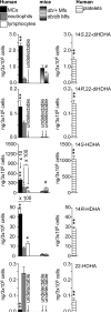
Figure 4. Differential Activities of Human Monocytes, Neutrophils, Lymphocytes, or Platelets; or Macrophages of Diabetic db/db or non-diabetic db/+ mice in Producing 14S,22-diHDA, 14R,22-diHDHA, and The Biosynthetic Intermediates or Markers 22-HDHA, 14S-HDHA, and 14R-HDHA. (See also Figure S2 and Table S1, Figure S3)
Human monocytes (MCs) (3 × 10⁶), neutrophils (3 × 10⁶), lymphocytes (3 × 10⁶), or platelets (3 × 10⁸); or macrophages (Mfs) of db/db or db/+ mice were incubated in 3 μM DHA, then stimulated as in Figure 1. The incubations were analyzed using aR chiral LC-UV-MS. For Mfs of db/db or db/+ mice, the media also contained 25 mM glucose. Results are mean ± SEM (n = 4), * p < 0.05, ** p < 0.01, versus neutrophils. # p < 0.05 versus db/+ Mfs.
Figure 5
Figure 5. Maresin-L (14,22-diHDHA) Treatment Restored Diabetes-impaired db/db Macrophage Functions in Promoting Wound Healing and in Recruiting MSCs
Macrophages (Mfs) were isolated from diabetic db/db or non-diabetic control db/+ mice. Scratch wounds were generated in the monolayers of fibroblasts and epithelial cells in dishes, which were then treated, for 34 h and 24 h, respectively, with conditioned medium (CM) from db/db or db/+ Mfs cultured without or with maresin-Ls (14,22-diHDHAs) (0, 10, or 50 nM). Wound healing was % of cell densities recovered in the original wound zone relative to control without CM. MSC transmigration was started with adding murine MSCs into the upper chambers of transwells, and adding CM of db/db or db/+ Mfs cultured without or with 14,22-diHDHAs (0, 10, or 50 nM) into the lower chamber. The transmigration was expressed as % of migrated MSCs relative to control without CM. Results are means ± SEM (n = 4), * p < 0.01 compared to CM of db/db Mfs without maresin-L treatment. Quantitative results are on the left; representative microphotographs are in the middle. The cell morphologies are on the right. (A) Scratch wound healing of fibroblasts in CM of Mfs. (B) Scratch wound healing of epithelial cells in CM of Mfs. (C) Transmigration of MSCs toward CM of Mfs.
Figure 6
Figure 6. Maresin-L (14,22-diHDHA) Treatment Increased db/db Macrophage Expression of HGF but Decreased db/db Macrophage Production of TNFα and TXB2 under Hypoxia and High Glucose Condition Relevant to Diabetic Wounds
Macrophages (Mfs) isolated from diabetic db/db or non-diabetic control db/+ mice were cultured in medium containing 0, 10, or 50 nM maresin-Ls, and 25 mM glucose under hypoxia (95% N2, 5%CO2, 24 h). For comparison, Mfs isolated from non-diabetic db/+ mice were cultured in medium without maresin-Ls and with normal glucose (5 mM) under hypoxia (95% N2, 5%CO2, 24 h). The Mf-conditioned media were analyzed for HGF, TNFα, and TXB2 by ELISA. Results are means ± SEM (n = 3); * p < 0.05, ** p < 0.01 compared to CM of db/db Mfs without maresin-L treatment. # p < 0.05 compared to CM of db/+ Mfs without maresin-L treatment and with normal glucose. (A) HGF. (B) TNFα. (C) TXB2.
Figure 7
Figure 7. A Scheme Outlining the Tentative Biosynthetic Pathways and Bioactions of ω-Hydroxy Maresin-like Docosahexaenoic Mediators Generated by Leukocytes and Platelets. (See also Figure S4)
DHA is transformed by leukocytes and platelets via h15-LO-1 or L12-LO (12/15-LO) and P12-LO, respectively, to 14S-hydroperoxy-DHA (14S-HpDHA), which is reduced to 14S-HDHA. These cells further convert 14S-HDHA to 14S,22-diHDHA (maresin-L1) through P450 catalyzed ϖ(22)-oxidation. Alternatively, by P450 activities of the cells, DHA is first transformed to 14S-HDHA, 14R-HDHA, and 22-HDHA. Then 14R-HDHA is converted by P450 to 14R,22-diHDHA (maresin-L2). The double-bond geometries of 14S (or R)-HDHA are conserved after being converted to maresin-Ls based on our results and reported analogous data for eicosanoids. This conservation also applies for the transformation of DHA to 22-HDHA. Maresin-Ls represent autacrine/paracrine factors that are produced by monocytes, macrophages, neutrophils, and/or platelets. They act on macrophages, in the promotion of wound healing and MSC transmigration.

References

    1. Bannon P, Wood S, Restivo T, Campbell L, Hardman MJ, Mace KA. Diabetes induces stable intrinsic changes to myeloid cells that contribute to chronic inflammation during wound healing in mice. Disease models & mechanisms. 2013;6:1434–1447. - PMC - PubMed
    1. Biswas SK, Mantovani A. Orchestration of metabolism by macrophages. Cell metabolism. 2012;15:432–437. - PubMed
    1. Brem H, Tomic-Canic M. Cellular and molecular basis of wound healing in diabetes. The Journal of clinical investigation. 2007;117:1219–1222. - PMC - PubMed
    1. Capdevila JH, Holla VR, Faick JR. Cytochrome P450 and the Metabolism and Bioactivation of Arachidonic Acid and Eicosanoids. In: Paul R.O.d.M., editor. Cytochrome P450: Structure, Mechanism, and Biochemistry, 3e. Kluwer Academic/Plenum Publishers; New York: 2005.
    1. Chiang N, Takano T, Clish CB, Petasis NA, Tai HH, Serhan CN. Aspirin-triggered 15-epi-lipoxin A4 (ATL) generation by human leukocytes and murine peritonitis exudates: development of a specific 15-epi-LXA4 ELISA. The Journal of pharmacology and experimental therapeutics. 1998;287:779–790. - PubMed

Publication types

MeSH terms

Substances

LinkOut - more resources