

Progressive interatrial block and supraventricular arrhythmias
- PMID: 25201217
- PMCID: PMC6931540
- DOI: 10.1111/anec.12208
Progressive interatrial block and supraventricular arrhythmias
Abstract
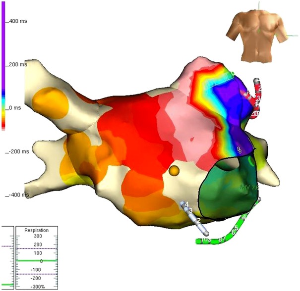
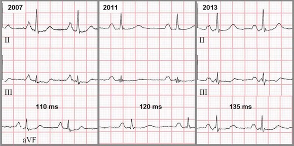
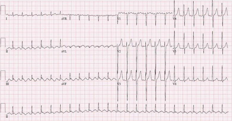
Interatrial conduction disorders are frequent in patients with structural heart diseases, including hypertension, coronary disease, and hypertrophic cardiomyopathy, and they are strongly associated with atrial tachyarrhythmias, especially atrial fibrillation and flutter. Conduction delays lead to dispersion of refractory periods and participate in initiating and maintaining reentry circuits, facilitating atrial arrhythmias. In this case, the changing pattern over time is a manifestation of progressive atrial remodeling and conduction delay. The terminal negative component of the P wave in the inferior leads suggests block of the electrical impulse in the Bachman bundle zone, with retrograde activation of the left atria via muscular connections at the coronary sinus. This has been reproduced in experimental models and confirmed by endocardial mapping. Physicians should be aware of the association between advanced interatrial block and development of atrial arrhythmias as its recognition could prompt early and aggressive antiarrhythmic treatment.
Keywords: atrial fibrillation; atrial flutter; interatrial block.
© 2014 Wiley Periodicals, Inc.
Figures
Similar articles
-
[Interatrial block as anatomical-electrical substrate for supraventricular arrhythmias: Bayés syndrome].Arch Cardiol Mex. 2014 Jan-Mar;84(1):32-40. doi: 10.1016/j.acmx.2013.10.004. Epub 2014 Feb 13. Arch Cardiol Mex. 2014. PMID: 24529591 Review. Spanish.
-
Bayés' syndrome: the association between interatrial block and supraventricular arrhythmias.Expert Rev Cardiovasc Ther. 2015 May;13(5):541-50. doi: 10.1586/14779072.2015.1037283. Expert Rev Cardiovasc Ther. 2015. PMID: 25907617 Review.
-
Advanced interatrial block as a substrate of supraventricular tachyarrhythmias: a well recognized syndrome.J Electrocardiol. 2015 Mar-Apr;48(2):135-40. doi: 10.1016/j.jelectrocard.2014.12.015. Epub 2015 Jan 9. J Electrocardiol. 2015. PMID: 25637273 Review.
-
Interatrial conduction block and retrograde activation of the left atrium and paroxysmal supraventricular tachyarrhythmia.Eur Heart J. 1988 Oct;9(10):1112-8. doi: 10.1093/oxfordjournals.eurheartj.a062407. Eur Heart J. 1988. PMID: 3208776
-
Assessment of atrial conduction time in patients with polycystic ovary syndrome.J Interv Card Electrophysiol. 2014 Nov;41(2):137-43. doi: 10.1007/s10840-014-9925-8. Epub 2014 Jul 9. J Interv Card Electrophysiol. 2014. PMID: 25005453
Cited by
-
Progressive interatrial block associated with atrial fibrillation in a patient with hypertrophic cardiomyopathy.Ann Noninvasive Electrocardiol. 2017 May;22(3):e12403. doi: 10.1111/anec.12403. Epub 2016 Sep 11. Ann Noninvasive Electrocardiol. 2017. PMID: 27615799 Free PMC article.
-
The P-wave morphology: what does it tell us?Herzschrittmacherther Elektrophysiol. 2015 Sep;26(3):192-9. doi: 10.1007/s00399-015-0385-3. Herzschrittmacherther Elektrophysiol. 2015. PMID: 26264481
-
Intra- and interatrial conduction abnormalities: hemodynamic and arrhythmic significance.J Interv Card Electrophysiol. 2018 Aug;52(3):293-302. doi: 10.1007/s10840-018-0413-4. Epub 2018 Aug 20. J Interv Card Electrophysiol. 2018. PMID: 30128800 Review.
-
Bayés' syndrome: Time to consider early anticoagulation?North Clin Istanb. 2018 May 23;5(4):370-378. doi: 10.14744/nci.2017.60251. eCollection 2018. North Clin Istanb. 2018. PMID: 30815636 Free PMC article. Review.
-
Value of Interatrial Block for the Prediction of Silent Ischemic Brain Lesions.J Atr Fibrillation. 2018 Oct 31;11(3):2037. doi: 10.4022/jafib.2037. eCollection 2018 Oct-Nov. J Atr Fibrillation. 2018. PMID: 31139269 Free PMC article.
References
-
- Bayés de Luna A, Platonov P, Cosio FG, et al. Interatrial blocks. A separate entity from left atrial enlargement: a consensus report. J Electrocardiol 2012;45:445–451. - PubMed
-
- Ariyarajah V, Spodick DH. Progression of partial to advanced interatrial block. J Electrocardiol 2006;39(2):177–179 - PubMed
-
- Bayés de Luna A, Cladellas M, Oter R, et al. Interatrial conduction block and retrograde activation of the left atrium and paroxysmal supraventricular tachyarrhythmia. Eur Heart J 1988;9:1112–1118. - PubMed
-
- Conde D, Baranchuk A. Interatrial block as anatomical‐electrical substrate for supraventricular arrhythmias: Bayes’ syndrome. Arch Mex Cardiol 2014;84(1):32–40. - PubMed
-
- Caldwell J, Koppikar S, Barake W, et al. Prolonged P‐wave duration is associated with atrial fibrillation recurrence after successful pulmonary vein isolation for paroxysmal atrial fibrillation. J Interv Card Electrophysiol 2014;39(2):131–138. - PubMed
Publication types
MeSH terms
LinkOut - more resources
Full Text Sources
Other Literature Sources
Medical
