. 2015 Apr;135(4):1019-1030.e8.
doi: 10.1016/j.jaci.2014.07.036. Epub 2014 Sep 4.

Autocrine hemokinin-1 functions as an endogenous adjuvant for IgE-mediated mast cell inflammatory responses

Affiliations

Autocrine hemokinin-1 functions as an endogenous adjuvant for IgE-mediated mast cell inflammatory responses

Tina L Sumpter et al. J Allergy Clin Immunol. 2015 Apr.

Abstract

Background: Efficient development of atopic diseases requires interactions between allergen and adjuvant to initiate and amplify the underlying inflammatory responses. Substance P (SP) and hemokinin-1 (HK-1) are neuropeptides that signal through the neurokinin-1 receptor (NK1R) to promote inflammation. Mast cells initiate the symptoms and tissue effects of atopic disorders, secreting TNF and IL-6 after FcεRI cross-linking by antigen-IgE complexes (FcεRI-activated mast cells [FcεRI-MCs]). Additionally, MCs express the NK1R, suggesting an adjuvant role for NK1R agonists in FcεRI-MC-mediated pathologies; however, in-depth research addressing this relevant aspect of MC biology is lacking.

Objective: We sought to investigate the effect of NK1R signaling and the individual roles of SP and HK-1 as potential adjuvants for FcεRI-MC-mediated allergic disorders.

Methods: Bone marrow-derived mast cells (BMMCs) from C57BL/6 wild-type (WT) or NK1R(-/-) mice were used to investigate the effects of NK1R signaling on FcεRI-MCs. BMMCs generated from Tac1(-/-) mice or after culture with Tac4 small interfering RNA were used to address the adjuvancy of SP and HK-1. WT, NK1R(-/-), and c-Kit(W-sh/W-sh) mice reconstituted with WT or NK1R(-/-) BMMCs were used to evaluate NK1R signaling on FcεRI-MC-mediated passive local and systemic anaphylaxis and on airway inflammation.

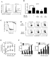
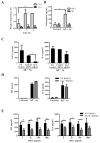
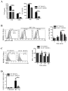
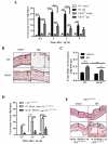
Results: FcεRI-activated MCs upregulated NK1R and HK-1 transcripts and protein synthesis, without modifying SP expression. In a positive signaling loop HK-1 promoted TNF and IL-6 secretion by MC degranulation and protein synthesis, the latter through the phosphoinositide 3-kinase/Akt/nuclear factor κB pathways. In vivo NK1R signaling was necessary for the development of passive local and systemic anaphylaxis and airway inflammation.

Conclusions: FcεRI stimulation of MCs promotes autocrine secretion of HK-1, which signals through NK1R to provide adjuvancy for efficient development of FcεRI-MC-mediated disorders.

Keywords: FcεRI; Hemokinin-1; IL-6; IgE; TNF; airway inflammation; mast cells; neurokinin-1 receptor; passive anaphylaxis; substance P.


Figures

FIG 1
FIG 1. Role of NK1R in FcεRI-BMMCs
A, Expression of NK1R in control (filled histogram) and FcεRI-BMMCs (open histogram) [loaded with IgE (SPE-07, 1.0 μg/ml, 1 h then cross-linked with Ag (DNP-HSA, 200 ng/ml) for 18 h]. Mean ± 1 SD of the percent positive from 3 experiments. B, Signaling pathways involved in NK1R transcript synthesis in FcεRI-BMMCs 2 hours after FcεRI ligation with Ag in the presence of inhibitors specific for the indicated pathways. Means + 1 SD from 3 experiments. C, Calcium flux in WT (black line) or NK1R−/− (gray line) BMMCs loaded with Ig and pulsed with Ag or ionomycin at the indicated time. One representative of 3 experiments. D–E, Degranulation of WT and NK1R−/− FcεRI-BMMCs, 90 min following FcεRI ligation with Ag. D, A representative flow plot from three experiments; values are means ± 1 SD of the percentage of degranulating BMMCs either loaded with IgE (1.0 μg/ml, 1 h) then cross-linked with Ag (200 ng/ml, 90 min) or activated with compound (c) 48/80 (1.0 μg/ml). E, Data points depict the mean ± 1 SD of the percentage of degranulating BMMCs from three experiments. F, TNF and IL-6 release by FcεRI-BMMCs, 18h Ag. Mean + 1 SD of duplicate values from a representative of three experiments. *p<0.05 **p<0.01, ***p<0.001, ****p<0.0001.
FIG 2
FIG 2. Hemokinin-1 is required for maximal TNF-α secretion from FcεRI BMMCs
A, Quantification of Tac4 or Tac1 mRNAs in WT FcεRI-BMMCs. Means + 1 SD from two experiments. B, HK-1 and SP levels secreted by WT FcεRI-BMMCs 18 h after Ag cross-linking. Means + 1 SD from three experiments. C, TNF and IL-6 secretion by Tac4-silenced WT FceRI-BMMCs, or D, Tac1−/− BMMCs. Means + 1 SD from two experiments. E, TNF and IL-6 secreted by WT or NK1R−/− FcεRI-BMMCs in which exogenous HK-1 was added at the time of Ag. Means + 1 SD from three independent experiments. Cytokines were measured in supernatants 18h after Ag cross-linking. *p<0.05, **p<0.01, ***p<0.001.
FIG 3
FIG 3. NK1R signaling potentiates PI3K/Akt/NFκB activation in FcεRI-BMMCs
A, TNF and IL-6 released by BMMCs. Mean + 1 SD from three experiments. B, Left, histograms comparing intracellular phospho-(p)Ser473Akt in WT FcεRI- (black line) and NK1R−/− BMMCs (gray line) loaded with IgE and stimulated with Ag for 10 min as detected by flow cytometry. Filled histograms are Ig isotype controls. Right, depicts the mean ± 1 SD of the MFI for the kinetics of pSer473-Akt detected by intracellular flow cytometry in WT or NK1R−/− FcεRI-BMMCs. from 3 independent experiments. C, Left, IκB-α expression in FcεRI-BMMCs, 2h after Ag cross-linking. Right, kinetics of IκB-α degradation in FcεRI-BMMCs expressed as the percentage of the MFI of untreated controls. Means ± 1 SD from 3 independent experiments. D, Reporter assays of NFκB activity in FcεRI-BMMCs transfected with pLuc-NFκB, measured after 18h of FcεRI ligation. Mean + 1 SD of quadruplicates from a representative of 3 experiments. *p<0.05, **p<0.01.
FIG 4
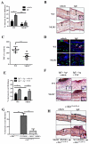
FIG 4. NK1R signaling amplifies the early phase of PCA
PCA was induced by injecting IgE (20 ng/ear, i.d.). 24h later, Ag (DNP-HSA, 100 μg, i.v.). was administered. A, Increases in ear thickness following Ag injection. Means + 1 SEM of 13 mice from three independent experiments. B, Histology of skin sections comparing edema (arrows) in IgE-sensitized or control ears of WT and NK1R−/− mice, 2h after Ag challenge. 200x. C, Evan's Blue extravasion from ear tissue, 2h after Ag challenge. Means + 1 SEM from 2 experiments. D–E, c-KitW-sh/W-sh mice were reconstituted with WT or NK1R−/− BMMCs, (1 × 10⁶/ear, i.d) 8 weeks prior to the PCA induction. D, Depicts increases in ear thickness following induction of PCA. Mean + 1 SEM from 4–5 mice per group. E, Histology of skin sections comparing edema, 2h after sensitization. 200x. **p<0.01, ***p<0.005, ****p<0.001.
Fig. 5
Fig. 5. NK1R signaling is required for the TNF dependent development of late phase PCA
A, Increase in ear thickness and B, histology of mouse ears 24h following Ag challenge in late PCA. C, TNF levels from the IgE-sensitized ears depicted as percent control ears. Means ± 1 SEM of two experiments. C, Immunofluorescence microscopy of ears of WT mice 24 h following the induction of PCA, showing c-Kit+ MCs (green) containing TNF-α (red). 200x. D, Increase in ear thickness in IgE sensitized WT or NK1R−/− mice injected with TNF-α (40 U/ear). Means + 1 SEM of 5 mice/experimental group. E, Histology illustrating the severity of the inflammatory infiltrate in ear dermis and epidermis. H&E, 200x. Inset shows PMN leukocytes, 1000x. G–H, Late phase PCA was evaluated in c-KitW-sh/W-sh mice reconstituted (i.d) with either WT or NK1R−/− BMMCs. G, Increases in ear thickness or H, histology of skin sections, 24h after PCA induction. 200x and inset 500x. *p<0.05, **p<0.01 ***p<0.001.
FIG 6
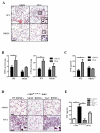
FIG 6. The NK1R is required for onset of MC-dependent airway inflammation
To induce MC-dependent chronic asthma, mice were treated with either vehicle or OVA (10 μg, i.p.) on days 0, 2, 4, 6, 8, 10, and 12 then challenged (200 μg, i.n.) on days 40, 43, and 46. A, Inflammatory infiltrate in OVA-sensitized/challenged lungs or vehicle controls mice at day 47. H&E, 200x. Insets show PMN leukocytes including eosinophils and neutrophils. 500x. Bar=20 μm. B, Quantification of EPO and MPO in lungs. Means + 1 SEM of 5 mice/group are illustrated. C, TNF concentration in BAL from WT or NK1R−/− mice. D, Histology of KitW-sh/W-sh lungs from mice reconstituted with WT, NK1R−/− or Tac1−/− BMMCs 8 weeks prior to induction of airway inflammation. H&E, 200x. Inset shows eosinophils in the lung of KitW-sh/W-sh reconstituted with WT BMMCs, 1000x. E, TNF in BALs of c-KitW-sh/W-sh mice reconstituted with WT, NK1R−/−, or Tac1−/− BMMCs prior to induction of experimental airway inflammation. Means ± SEM of 3 mice/group. *p<0.05, **p<0.01.

Similar articles

Cited by

References

    1. Chiu IM, von Hehn CA, Woolf CJ. Neurogenic inflammation and the peripheral nervous system in host defense and immunopathology. Nat Neuro. 2012;15:1063–7. - PMC - PubMed
    1. Douglas SD, Leeman SE. Neurokinin-1 receptor: functional significance in the immune system in reference to selected infections and inflammation. Ann N Y Acad Sci. 2011;1217:83–95. - PMC - PubMed
    1. Mathers AR, Tckacheva OA, Janelsins BM, Shufesky WJ, Morelli AE, Larregina AT. In vivo signaling through the neurokinin 1 receptor favors transgene expression by Langerhans cells and promotes the generation of Th1- and Tc1-biased immune responses. J Immunol. 2007;178:7006–17. - PubMed
    1. Janelsins BM, Mathers AR, Tkacheva OA, Erdos G, Shufesky WJ, Morelli AE, et al. Proinflammatory tachykinins that signal through the neurokinin 1 receptor promote survival of dendritic cells and potent cellular immunity. Blood. 2009;113:3017–26. - PMC - PubMed
    1. Janelsins BM, Sumpter TL, Tkacheva OA, Rojas-Canales DM, Erdos G, Mathers AR, et al. Neurokinin-1 receptor agonists bias therapeutic dendritic cells to induce type-1 immunity by licensing host dendritic cells to produce IL-12. Blood. 2013 - PMC - PubMed

Publication types

MeSH terms