Review
. 2014 Dec:29:17-25.
doi: 10.1016/j.sbi.2014.08.011. Epub 2014 Sep 15.

Structural mechanisms in NLR inflammasome signaling

Affiliations
Review

Structural mechanisms in NLR inflammasome signaling

Bernhard C Lechtenberg et al. Curr Opin Struct Biol. 2014 Dec.

Abstract

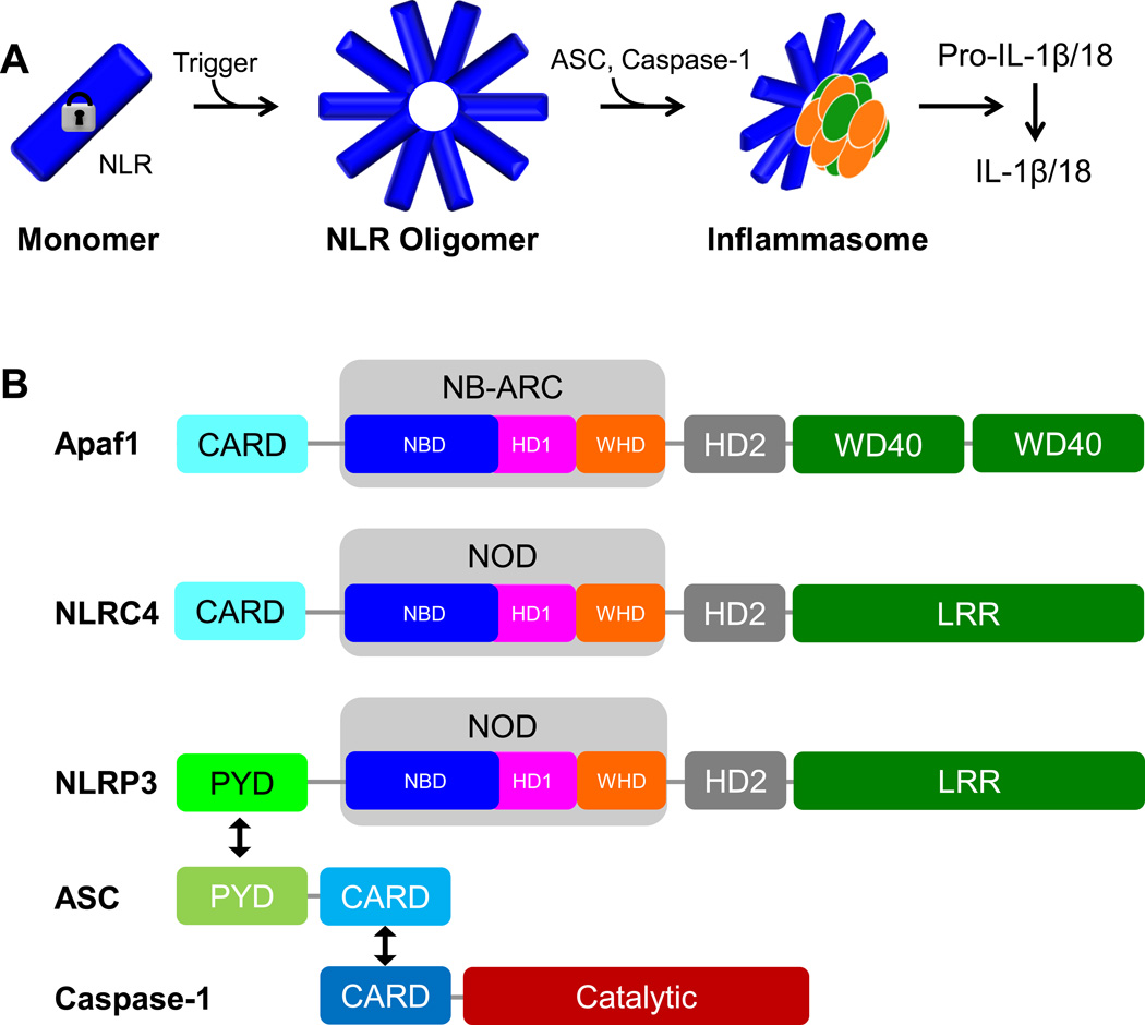
Members of the NOD-like receptor (NLR) family mediate the innate immune response to a wide range of pathogens, tissue damage and other cellular stresses. They achieve modulation of these signals by forming oligomeric signaling platforms, which in analogy to the apoptosome are predicted to adopt a defined oligomeric architecture and will here be referred to as NLR oligomers. Once formed, oligomers of the NLR proteins NLRP3 or NLRC4 'recruit' the adaptor protein ASC and the effector caspase-1, whereby NLRC4 can also directly interact with caspase-1. This results in large multi-protein assemblies, termed inflammasomes. Ultimately, the formation of these inflammasomes leads to the activation of caspase-1, which then processes the cytokines IL-1β and IL-18 triggering the immune response. Here we review new insights into NLR structure and implications on NLR oligomer formation as well as the nature of multi-protein inflammasomes. Of note, so dubbed 'canonical inflammasomes' can also be triggered by the NLR NLRP1b and the non-NLR protein AIM2, however the most detailed mechanistic information at hand pertains to NLRC4 while NLRP3 represents the quintessential inflammasome trigger. Thus these two NLRs are mainly used as examples in this article.


Figures

Figure 1
Figure 1. Inflammasome signaling
A. Illustration of inflammasome formation. Upon a trigger a monomeric autoinhibited NLR (blue) becomes activated and oligomerizes to form a defined oligomer. Recruitment of ASC (orange) and caspase-1 (green) results in formation of a large signaling particle (inflammasome) which leads to the activation of caspase-1 and processing of Pro-IL1β or IL-18. B. Domain organization of Apaf-1, NLRC4, NLRP3, ASC and caspase-1. Black arrows symbolize homotypical PYD/PYD and CARD/CARD interactions. Abbreviations: Apaf-1: Apoptotic protease-activating factor 1; NLRC4: NLR family CARD domain-containing protein 4; NLRP3: Nucleotide-binding domain and leucine-rich repeat pyrin domain 3; ASC: Apoptosis-associated speck-like protein containing a CARD; CARD: Caspase activation and recruitment domain; PYD: Pyrin domain; NOD: Nucleotide-binding oligomerization domain; NB-ARC: nucleotide-binding, APAF-1, R proteins, and CED-4; NBD: Nucleotide-binding domain; HD: Helical domain; WHD: winged-helix domain; WD40: WD40 domain; LRR: Leucine-rich repeat domain.
Figure 2
Figure 2. Auto-inhibited NLRC4 and Apaf-1
A. Structures of monomeric NLRC4 and Apaf-1. Displayed are the crystal structures of mouse NLRC4 (delta CARD; PDBid: 4KXF [15]; left) and mouse Apaf-1 (PDBid: 3SFZ [17]; with CARD modeled based on the structure of human Apaf-1ΔWD40, PDBid: 1Z6T [14]) in similar orientation (overlay of NBD and HD1). Domains are colored analogous to Figure 1b with the relative positions of NBD, HD1 and HD2 emphasized by blue, magenta and grey spheroids respectively. The position of the bound ADP molecule is indicated by a red sphere. B. Winged helix domains of NLRC4 and Apaf-1 exhibit different shapes. Close-up onto the respective WHDs with the critical histidines coordinating the phosphate moieties of the bound ADP depicted (colored “rainbow” - N to C-terminus: blue to red). C. HD2 of NCLRC4 and Apaf-1 adopt different tertiary fold. Display analogous to panel B depicting the different shapes of the respective HD2 domains.
Figure 3
Figure 3. The LRR lock – internal LRR domain interactions; PYD/CARD structures
A. The NLRC4 LRR lock. Interaction of NLRC4 LRR (green) with HD2 (grey, position of phospho-Ser553 is indicated). Close up on the interaction pattern of HD2 pSer553 with LRR residues. His693 and Glu720 from the LRR are colored in orange (PDBid: 4KXF [15]). B. NLRX1: LRR-LRRCT interaction. In NLRX1 the LRR is internally bound by a helix domain C-terminal to the LRR domain (LRRCT) endemic to NLRX1. LRRCT interacts with the LRR similar to HD2 in NLRC4 thus overriding the internal LRR lock in NLRX1 (PDBid: 3UN9 [22]). C/D. Structures of inflammasome PYD/CARD domains - potential conformational flexibility. C. Structures of NLRP3 PYD (PDBid: 3QF2 [40]), ASC full length (PDBid: 2KN6 [37]) and ICEBERG (PDBid: 1DGN [39]; CARD-only relative of caspase-1 CARD for which no structure is available) as indicated. D. Possible conformational flexibility in the death domain fold – “Open conformations”. NLRP14 PYD (grey, PDBid: 4N1J [44]), NOD1 CARD (magenta, from a domain swapped dimer in the crystal structure; PDBid: 2NZ7 [43]). Two examples for death domains showing an open conformation due to a helix-5/helix-6 fusion captured in non-swapped crystal structures (grey: MALT1 death domain (DD), PDBid: 2G7R; orange: Fas DD, PDBid 3EZQ [51]). Helix 6 is indicated in red. For NOD1 CARD and Fas DD the closed forms are also displayed (analogous color code, PDBids: 2B1W [52] and 1DDF [41], respectively); no structures of closed NLRP14 PYD, MALT1 DD exist.
Figure 4
Figure 4. Model of NLRC4/NLRP3 activation and inflammasome formation – endogenous NLRP3 inflammasomes
A. Autoinhibited NLRC4/NLRP3. Prior sensing of a trigger NLRC4/NLRP3 exist as autoinhibited monomers. Displayed is the structure of NLRC4 (PDBid: 4KXF [15]) with the PYD (NLRP3) or CARD (NLRC4) schematically depicted (light green). B. Schematic of formed NLRP3 oligomer poised for inflammasome formation. Binding of PAMPs/DAMPs (red star) and exchange of bound ADP with ATP leads to the open NLR conformation and formation of the NLR oligomer. A NLRP3 oligomer is depicted schematically (based on the architecture of apoptosomes) with domain color code analogous to Figure 1b. The oligomer is formed through a ring-like interaction of neighboring NBD domains and a secondary ring formed by HD1 and WHD interactions (six molecules are displayed, yet the exact number of molecules in the oligomers is not unambiguously known – numbers of 5–11 have been proposed for NLR oligomers [23,53]). The NLRP3 PYD is oligomerized and poised to “recruit and seed” ASC and caspase-1 molecules (dark green: ASC PYD, light blue: ASC CARD, dark blue: Caspase-1 CARD with catalytic domain indicated in brown). C. Schematic of the NLRP3/ASC/caspase-1 inflammasome. The NLR oligomers have seeded an ASC/Caspase-1 network that is structurally formed through both PYD and CARD interactions leading to the formation of a large multi-protein particle. Illustrated is a potentially heterogeneous nature of the particle that resembles more a signaling entity than a highly defined structural entity. Ultimately caspase-1 catalytic domains are brought into close proximity leading to their activation (illustrated by a yellow star). D. Endogenous NLRP3/ASC inflammasomes. Image depicting endogenous inflammasomes (red) reproduced with generous permission from Akira et al. [49] (Copyright 2013 John Wiley & Sons Ltd). As outlined in Akira et al. [49], the image depicts the assembly of ASC and NLRP3 on microtubules after stimulation with nigericin. Mouse bone marrow-derived macrophages were stimulated with nigericin and fixed. Samples were subjected to proximity ligation assay followed by fluorescence staining with Alexa488-labelled anti-α-tubulin antibody (green) and Hoechst dye (DNA, blue). Endogenous proximity of NLRP3 with ASC (red) on microtubules was observed by super resolution structured illumination microscopy. Highlights: We discuss recent advances in understanding inflammasome formation and regulation. We compare the recent structure of NLRC4 to Apaf-1 and apoptosomes. NLRC4 autoinhibition and NLR oligomerization are discussed. The nature of NLRC4- and NLRP3/ASC/Caspase-1 inflammasome assemblies is discussed. We discuss role and nature of PYD and CARD domains in inflammasomes.

References

    1. Lamkanfi M, Dixit VM, et al. Mechanisms and Functions of Inflammasomes. Cell. 2014;157:1013–1022. - PubMed
    1. Ting JP, Lovering RC, Alnemri ES, Bertin J, Boss JM, Davis BK, Flavell RA, Girardin SE, Godzik A, Harton JA, et al. The NLR gene family: a standard nomenclature. Immunity. 2008;28:285–287. - PMC - PubMed
    1. Schroder K, Tschopp J. The inflammasomes. Cell. 2010;140:821–832. - PubMed
    1. Kersse K, Bertrand MJ, Lamkanfi M, Vandenabeele P. NOD-like receptors and the innate immune system: coping with danger, damage and death. Cytokine Growth Factor Rev. 2011;22:257–276. - PubMed
    1. Chen GY, Nunez G. Sterile inflammation: sensing and reacting to damage. Nat Rev Immunol. 2010;10:826–837. - PMC - PubMed

Publication types

MeSH terms

LinkOut - more resources