. 2014 Sep 18;55(6):891-903.
doi: 10.1016/j.molcel.2014.08.006. Epub 2014 Sep 4.

IRAK4 dimerization and trans-autophosphorylation are induced by Myddosome assembly

Affiliations

IRAK4 dimerization and trans-autophosphorylation are induced by Myddosome assembly

Ryan Ferrao et al. Mol Cell. .

Abstract

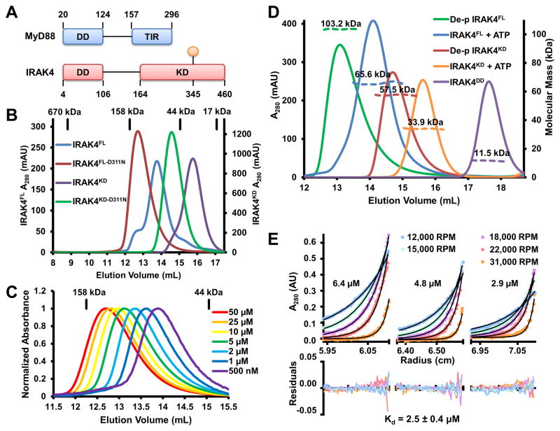
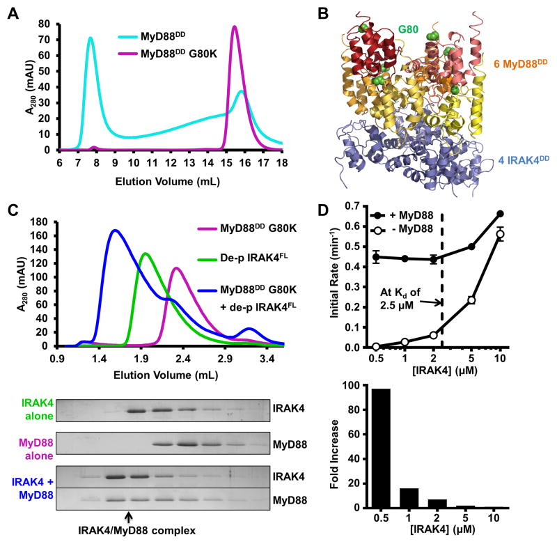
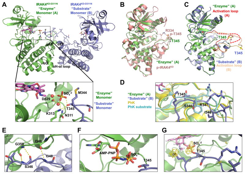
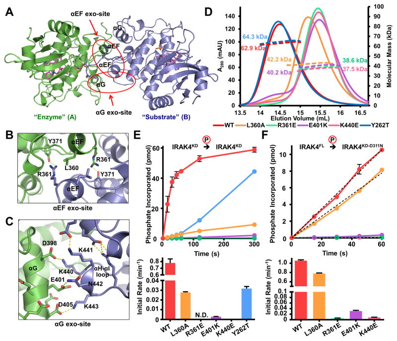
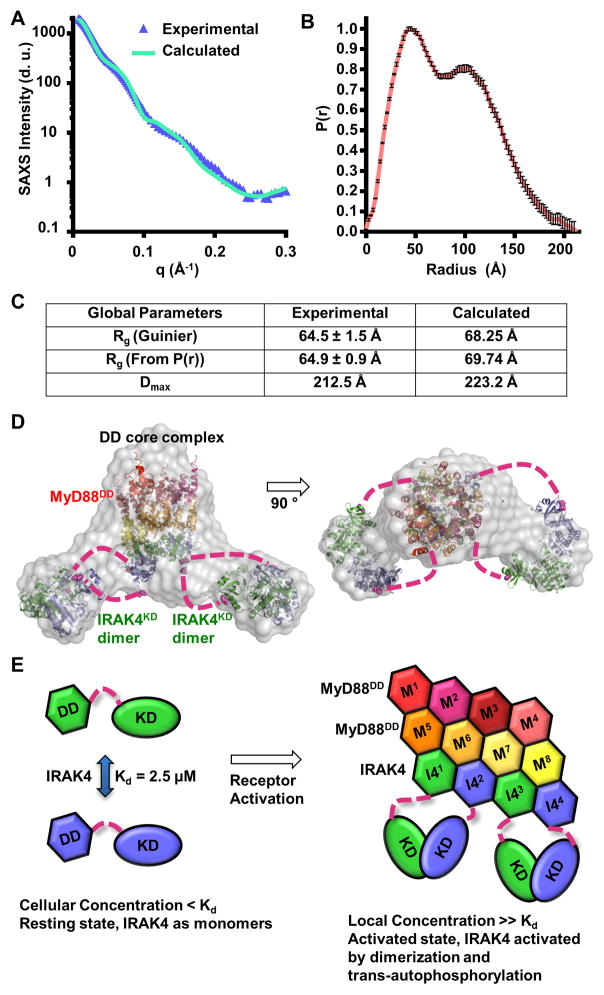
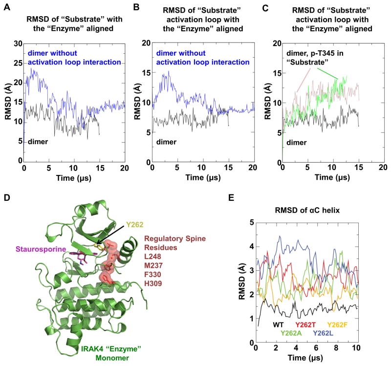
Trans-autophosphorylation is among the most prevalent means of protein kinase activation, yet its molecular basis is poorly defined. In Toll-like receptor and interleukin-1 receptor signaling pathways, the kinase IRAK4 is recruited to the membrane-proximal adaptor MyD88 through death domain (DD) interactions, forming the oligomeric Myddosome and mediating NF-κB activation. Here we show that unphosphorylated IRAK4 dimerizes in solution with a KD of 2.5 μM and that Myddosome assembly greatly enhances IRAK4 kinase domain (KD) autophosphorylation at sub-KD concentrations. The crystal structure of the unphosphorylated IRAK4(KD) dimer captures a conformation that appears to represent the actual trans-autophosphorylation reaction, with the activation loop phosphosite of one IRAK4 monomer precisely positioned for phosphotransfer by its partner. We show that dimerization is crucial for IRAK4 autophosphorylation in vitro and ligand-dependent signaling in cells. These studies identify a mechanism for oligomerization-driven allosteric autoactivation of IRAK4 that may be general to other kinases activated by autophosphorylation.


Figures

Figure 1
Figure 1. Unphosphorylated IRAK4 is Dimeric
(A) Domain organization of IRAK4 and MyD88. Approximate domain boundaries are labeled with residue numbers and the prototypical phosphorylation site at T345 is shown in orange. (B) Size-exclusion chromatograms of full-length and kinase domain of IRAK4 (IRAK4FL and IRAK4KD), and their catalytically inactive forms (IRAK4FL-D311N and IRAK4KD-D311N). Elution volumes of protein standards are indicated above. (C) Normalized size-exclusion chromatograms of IRAK4FL-D311N at different concentrations. (D) Size-exclusion chromatograms (solid lines) and molecular mass as measured by multi-angle light scattering (MALS) (dotted lines) of dephosphorylated (de-p) IRAK4FL and IRAK4KD, IRAK4FL and IRAK4KD after ATP/Mg2+ incubation, and the IRAK4 death domain (IRAK4DD). (E) Sedimentation equilibrium analytical ultracentrifugation of IRAK4FL-D311N at 6.4 μM, 4.8 μM, and 2.9 μM. Samples were run at five different speeds shown in revolutions per minute (RPM). Solid black lines correspond to the fitting of a monomer-dimer self-association model, which produced a dimerization Kd of 2.5 ± 0.4 μM. Residuals are plotted below. See also Figure S1.
Figure 2
Figure 2. MyD88 Enhances IRAK4 Trans-autophosphorylation
(A) Size-exclusion chromatograms of MyD88DD (cyan) and MyD88DD with a solubilizing mutation (G80K, purple). (B) Structure of the Myddosome DD complex containing MyD88DD (warm colors) and IRAK4DD (blue). Positions of MyD88 residue G80 are shown as green spheres. (C) Size-exclusion chromatograms of MyD88DD G80K (purple), dephosphorylated IRAK4FL (green), and the IRAK4/MyD88 complex (blue) formed in vitro (top). SDS-PAGE of the fractions is shown with Coomassie blue staining (below). Black arrow indicates co-migration of IRAK4 and MyD88 in a high molecular weight complex. (D) Autophosphorylation rates of dephosphorylated IRAK4FL at different concentrations, with (black circle) and without (open circle) pre-incubation with MyD88DD G80K. Dotted black line indicates the Kd of IRAK4 dimerization. Data represent mean ± SEM. See also Figure S2.
Figure 3
Figure 3. Asymmetric Enzyme-Substrate Embrace in the IRAK4 Dimer Structure
(A) Ribbon diagram of IRAK4KD-D311N dimer structure, shown in green for the “enzyme” monomer A and blue for the “substrate” monomer B. The pan-kinase inhibitor staurosporine (magenta), sulfate ion (red and yellow), “enzyme” monomer catalytic residues, and “substrate” monomer activation loop side chains are shown as sticks. Detailed interactions at the active site of the “enzyme” monomer are shown (inset). Hydrogen bonds are shown as dotted yellow lines. T345 is the prototypical phosphorylation residue. (B) Superposition of IRAK4KD-D311N “enzyme” monomer (green) with phosphorylated IRAK4KD structure (p-IRAK4KD, PDB: 2NRY) (pink). (C) Superposition of IRAK4KD-D311N “enzyme” monomer A (green) with “substrate” monomer B (blue). Monomer A activation loop (red) and monomer B activation loop (peach) adopt distinct conformations. Side chains of T345 are represented as yellow sticks to highlight differences in conformation. (D) Superposition of IRAK4KD-D311N “enzyme” monomer with phosphorylase kinase (PhK) (yellow) bound to a peptide substrate (teal) (PDB: 2PHK). Side chains of “substrate” monomer and PhK peptide are shown as sticks. Labels correspond to IRAK4KD-D311N “substrate” monomer residues. (E) Anti-parallel hydrogen bonding interactions between IRAK4KD-D311N “enzyme” monomer (green) and “substrate” monomer (blue). (F) Modeled ATP analogue AMP-PNP in the active site of IRAK4KD-D311N “enzyme” monomer based on superposition with p-IRAK4KD/AMP-PNP complex structure (PDB: 2OID). AMP-PNP, sulfate ion and T345 of the “substrate” monomer are shown as sticks. (G) Sulfur anomalous difference Fourier of IRAK4KD-D311N structure contoured at 2.5σ (yellow). See also Table 1 and Figure S3.
Figure 4
Figure 4. Symmetrical Exo-site Interactions in IRAK4 Dimerization and Autophosphorylation
(A) Ribbon diagram of the IRAK4KD-D311N dimer viewed down the approximate dimer axis, colored as in Figure 3A. Positions of the αG and αEF helices are indicated. (B) Detailed interactions at the αEF exo-site. Residues L360, R361, and Y371 of both monomers are labeled. (C) Detailed interactions at the αG exo-site. Side chains from negatively charged surface of αG and positively charged αH-αI loop are represented as sticks. (D) Size-exclusion chromatograms (solid lines) and molecular masses as measured by MALS (dotted lines) of αEF exo-site (L360A, R361E), αG exo-site (E401K, K440E) and gatekeeper (Y262T) mutations on a IRAK4KD-D311N background (WT). (E) Autophosphorylation of 10 μM IRAK4KD (WT), IRAK4KD with αEF exo-site, αG exo-site and gatekeeper mutations as measured by autoradiography (top) and the initial rates of autophosphorylation (bottom). Data represent mean ± SEM. (F) Trans-phosphorylation of IRAK4KD-D311N with or without αEF exo-site or αG exo-site mutations (10 μM) by IRAK4FL (1 μM) (top) and the initial rates of trans-phosphorylation (bottom). Data represent mean ± SEM. See also Figure S4.
Figure 5
Figure 5. Disruption of IRAK4 Dimerization Results in Defective Signaling in Cells
(A) Human IRAK4-deficient fibroblasts infected with retroviruses containing empty vector construct (Vector), Flag-tagged IRAK4FL wild-type (WT) and mutants (K213M, L360A, R361E, and Y262T) were treated with IL-1β (1 ng/ml) for the indicated times, followed by Western blot analyses using antibodies against p-IKKα/β, p-JNK, p-IκBα, IκBα, Flag and actin. (B, C) RT-PCR analyses for the fold induction of TNFα (B) and IL-8 (C) expression in the same reconstituted human fibroblasts following treatment with IL-1β for the indicated times. The experiment was repeated three times. Data represent mean ± SEM. See also Figure S5.
Figure 6
Figure 6. Small and Wide Angle X-ray Scattering (SAXS/WAXS) of the Myddosome
(A) Experimental scattering profile of the IRAK4FL-D311N/MyD88DD complex as a function of the scattering vector q (q = 4πsin(θ/2)/λ), where θ is the scattering angle and λ is the X-ray wavelength) after solvent background subtraction (blue), superimposed with the scattering profile calculated from the IRAK4FL-D311N/MyD88DD model in (D) (green). d.u.: detector unit (B) Pair-distance distribution function (P(r)) of the IRAK4FL-D311N/MyD88DD complex. Data represent calculated P(r) values ± SD. (C) Experimental and calculated radii of gyration (Rg) and maximum linear dimension (Dmax). Data represent calculated values ± SD. (D) The chevron shaped molecular envelope (grey) from ab initio reconstruction fitted with structures of the binary Myddosome DD complex (PDB: 3MOP, 8 MyD88DD in warm colors and 4 IRAK4DD in green and blue) and 2 IRAK4KD dimers (green and blue). The IRAK4 DDs and KDs presumed to be of the same polypeptides are shown in the same green or blue colors. (E) Schematic model of IRAK4 dimerization and activation in the Myddosome in a signal-dependent manner.
Figure 7
Figure 7. Molecular Dynamics (MD) Simulations
(A, B) MD simulation of the asymmetric IRAK4KD-D311N dimer structure (black) and the hypothetical symmetrical dimer structure without activation loop interactions (blue). RMSD values of Cα atoms between the initial and simulated “substrate” monomer (A) or its activation loop (residue 329–354) (B) are shown when the “enzyme” monomer is aligned. (C) MD simulation of the IRAK4KD-D311N dimer structure hypothetically phosphorylated at T345 in the “substrate” monomer, with (pink) and without (green) bound ADP. RMSD values of Cα atoms between the initial and simulated “substrate” monomer activation loop are shown when the “enzyme” monomer is aligned. (D) Ribbon diagram of “enzyme” monomer in the IRAK4KD-D311N dimer structure, showing the regulatory spine residues (dark red) and the gatekeeper residue Y262 (yellow). (E) MD simulation of the “enzyme” monomer with the hypothetical mutations in the gatekeeper tyrosine residue in comparison with WT (black). RMSD values of Cα atoms in the αC helix (residue 222–239) are shown when the initial and simulated structures are aligned with all Cα atoms. See also Figure S6.

Comment in

Similar articles

Cited by

References

    1. Cai Z, Chehab NH, Pavletich NP. Structure and activation mechanism of the CHK2 DNA damage checkpoint kinase. Mol Cell. 2009;35:818–829. - PubMed
    1. Cheng H, Addona T, Keshishian H, Dahlstrand E, Lu C, Dorsch M, Li Z, Wang A, Ocain TD, Li P, et al. Regulation of IRAK-4 kinase activity via autophosphorylation within its activation loop. Biochem Biophys Res Commun. 2007;352:609–616. - PubMed
    1. Favelyukis S, Till JH, Hubbard SR, Miller WT. Structure and autoregulation of the insulin-like growth factor 1 receptor kinase. Nat Struct Biol. 2001;8:1058–1063. - PubMed
    1. Ferrao R, Li J, Bergamin E, Wu H. Structural insights into the assembly of large oligomeric signalosomes in the toll-like receptor-interleukin-1 receptor superfamily. Sci Signal. 2012;5:re3. - PMC - PubMed
    1. Ferrao R, Wu H. Helical assembly in the death domain (DD) superfamily. Curr Opin Struct Biol. 2012;22:241–247. - PMC - PubMed

Publication types

MeSH terms

Substances

LinkOut - more resources