. 2014 Jul 24:12:48.
doi: 10.1186/s12964-014-0048-8.

AhR and Arnt differentially regulate NF-κB signaling and chemokine responses in human bronchial epithelial cells

AhR and Arnt differentially regulate NF-κB signaling and chemokine responses in human bronchial epithelial cells

Johan Øvrevik et al. Cell Commun Signal. .

Abstract

Background: The aryl hydrocarbon receptor (AhR) has gradually emerged as a regulator of inflammation in the lung and other tissues. AhR may interact with the p65-subunit of the nuclear factor (NF)-κB transcription factors, but reported outcomes of AhR/NF-κB-interactions are conflicting. Some studies suggest that AhR possess pro-inflammatory activities while others suggest that AhR may be anti-inflammatory. The present study explored the impact of AhR and its binding partner AhR nuclear translocator (Arnt) on p65-activation and two differentially regulated chemokines, CXCL8 (IL-8) and CCL5 (RANTES), in human bronchial epithelial cells (BEAS-2B).

Results: Cells were exposed to CXCL8- and CCL5-inducing chemicals, 1-nitropyrene (1-NP) and 1-aminopyrene (1-AP) respectively, or the synthetic double-stranded RNA analogue, polyinosinic-polycytidylic acid (Poly I:C) which induced both chemokines. Only CXCL8, and not CCL5, appeared to be p65-dependent. Yet, constitutively active unligated AhR suppressed both CXCL8 and CCL5, as shown by siRNA knock-down and the AhR antagonist α-naphthoflavone. Moreover, AhR suppressed activation of p65 by TNF-α and Poly I:C as assessed by luciferase-assay and p65-phosphorylation at serine 536, without affecting basal p65-activity. In contrast, Arnt suppressed only CXCL8, but did not prevent the p65-activation directly. However, Arnt suppressed expression of the NF-κB-subunit RelB which is under transcriptional regulation by p65. Furthermore, AhR-ligands alone at high concentrations induced a moderate CXCL8-response, without affecting CCL5, but suppressed both CXCL8 and CCL5-responses by Poly I:C.

Conclusion: AhR and Arnt may differentially and independently regulate chemokine-responses induced by both inhaled pollutants and pulmonary infections. Constitutively active, unligated AhR suppressed the activation of p65, while Arnt may possibly interfere with the action of activated p65. Moreover, ligand-activated AhR suppressed CXCL8 and CCL5 responses by other agents, but AhR ligands alone induced CXCL8 responses when given at sufficiently high concentrations, thus underscoring the duality of AhR in regulation of inflammation. We propose that AhR-signaling may be a weak activator of p65-signaling that suppresses p65-activity induced by strong activators of NF-κB, but that its anti-inflammatory properties also are due to interference with additional pathways.


Figures

Figure 1
Figure 1
P65 is required for CXCL8 but not CCL5 responses in 1-NP-or 1-AP-exposed BEAS-2B cells. Cells were transfected with siRNA against p65 (siP65) or non-targeting control siRNA (siNT), and exposed to 20 μM 1-NP, 1-AP or vehicle (DMSO) alone. CXCL8 (A) and CCL5 (B) gene expression were measured after 6 h by real-time PCR. CXCL8 (C) and CCL5 (D) protein levels in the medium were measured by ELISA after 18 h exposure as described under “Materials and methods”. Efficiency of transfection was assessed by Western blotting (E). The results are expressed as mean ± SEM (A/B: n = 1 (triplicate determinations); C/D: n ≥ 3; E: representative blot, n = 3). *Significantly different from unexposed controls; †Significantly different from cells transfected with non-targeting siRNA.
Figure 2
Figure 2
AhR and Arnt differentially regulate CXCL8 and CCL5 responses in 1-NP-or 1-AP-exposed BEAS-2B cells. Cells were transfected with siRNA against AhR (siAHR) and Arnt (siARNT) or non-targeting control siRNA (siNT), and exposed to 20 μM 1-NP, 1-AP or vehicle (DMSO) alone. CXCL8 (A) and CCL5 (B) gene expression were measured after 6 h by real-time PCR. CXCL8 (C) and CCL5 (D) protein levels in the medium were measured by ELISA after 18 h exposure as described under “Materials and methods”. Efficiency of transfection was assessed by Western blotting (E) as well as expression of CYP1A1 after 6 h exposure to 20 μM B[a]P, 1-NP and 1-AP, by real-time PCR (F). The results are expressed as mean ± SEM (A/B/F: n = 1 (triplicate determinations); C/D: n ≥ 3; E: representative blots, n ≥ 3). *Significantly different from unexposed controls; †Significantly different from cells transfected with non-targeting siRNA.
Figure 3
Figure 3
Blocking of AhR ligand binding does not increase CXCL8 and CCL5 responses in BEAS-2B cells. Cells were incubated with 0.5 μM of the AhR antagonist α-naphtoflavone (ANF) for 30 min prior to exposure for 18 h to 20 μM B[a]P, 1-NP, 1-AP or vehicle (DMSO) alone. CYP1A1 (A) levels were measured by real-time PCR, while CXCL8 (B) and CCL5 (C) levels in the medium were measured by ELISA as described under “Materials and methods”. The results are expressed as mean ± SEM (A: n = 1 (triplicate determinations); B/C: n = 5). *Significantly different from unexposed controls; †Significant effect of ANF.
Figure 4
Figure 4
AhR silencing increased TNF-α-,but not 1-NP-induced p50/p65 reporter gene expression in BEAS-2B cells. Cells were transfected with a NF-κB-luciferase (p50/p65) reporter gene, and exposed to 1-NP or TNF-α for 6 or 16 h (A), and assayed for luciferase activity as described under “Materials and methods”. Cells transfected siRNAs against AhR (siAHR), Arnt (siARNT) or non-targeting control siRNA (siNT) were further transfected with NF-κB-luciferase promoter and assayed for luciferase activity after a 16 h exposure to 1-NP (20 μM) or TNF-α (50 ng/ml) (B). The results are expressed as mean ± SEM (A: n = 2; B: n = 5). *Significantly different from unexposed controls; †Significantly different from cells transfected with non-targeting siRNA.
Figure 5
Figure 5
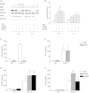
AhR and Arnt differentially regulate CXCL8 and CCL5 responses as well as p65 phosphorylation in Poly I:C-exposed BEAS-2B cells. Cells were transfected with siRNA against p65 (siP65), AhR (siAHR), Arnt (siARNT) or non-targeting control siRNA (siNT), and exposed to 10 μg/ml Poly I:C. Intracellular protein levels of total and phospho-p65 (Ser536) as well as AhR and Arnt were detected by Western blotting after 2 and 4 h exposure, as described under “Materials and methods”. The figure displays representative blots (A), as well as relative changes in phospho-p65 compared to total p65 quantified by densitometric analysis of the Western blots (B). CXCL8 (C and E) and CCL5 (D and F) protein levels in the medium were measured by ELISA after 18 h exposure, as described under “Materials and methods”. The results are expressed as mean ± SEM (n ≥ 3). *Significantly different from unexposed controls; †Significantly different from cells transfected with non-targeting siRNA.
Figure 6
Figure 6
Interaction between p65 and the CXCL8 and CCL5 promoters in Poly I:C exposed BEAS-2B cells. Cells were exposed to 10 μg/ml Poly I:C for 3 h. Interaction between p65 and the NF-κB responsive regions of the CXCL8 and CCL5 promoters were assessed by ChIP assay as described under Materials and methods”. Results are expressed as fold increase compared to p65 binding to the CXCL8 promoter after adjustment for the input control (GAPDH). The results are expressed as mean ± SEM ( n = 6). The insert figure shows the same data set on a log-scale. *Significantly different from unexposed controls.
Figure 7
Figure 7
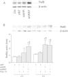
Depletion of AhR and Arnt increases RelB levels in Poly I:C-exposed BEAS-2B cells. Cells were transfected with siRNA against AhR (siAHR), Arnt (siARNT) or non-targeting control siRNA (siNT), and exposed to 10 μg/ml Poly I:C for 2 and 4 h. Intracellular protein levels of RelB and β-actin were detected by Western blotting as described under “Materials and methods”. The figure displays representative blots of RelB levels in unexposed cells (A) and Poly I:C-exposed cells (B). The graph depicts relative changes in RelB (B) compared to β-actin quantified by densitometric analysis of the Western blots. The results are expressed as mean ± SEM (n = 3). *Significantly different from unexposed controls; †Significantly different from cells transfected with non-targeting siRNA.
Figure 8
Figure 8
RelB suppresses CXCL8, but not CCL5, in BEAS-2B cells. Cells were transfected with siRNA against RelB (siRELB) or non-targeting control siRNA (siNT). Transfected cells were exposed to 20 μM 1-NP, 1-AP, or vehicle (DMSO) alone (A-D) or 10 μg/ml Poly I:C or vehicle (water) alone (E and F). CXCL8 (A) and CCL5 (B) gene expression were measured after 6 h by real-time PCR. CXCL8 (C and E) and CCL5 (D and F) protein levels in the medium were measured by ELISA after 18 h exposure as described under “Materials and methods”. Efficiency of transfection was assessed by Western blotting (G). The results are expressed as mean ± SEM (A/B: n = 1 (triplicate determinations); C-F: n ≥ 3; G: representative blot, n = 3) *Significantly different from unexposed controls; †Significantly different from cells transfected with non-targeting siRNA.
Figure 9
Figure 9
AhR-activation suppresses Poly I:C-induced CXCL8 and CCL5 responses, but stimulate CXCL8 responses alone in BEAS-2B cells. Cells were incubated with 1 μM of the AhR agonist β-naphtoflavone (BNF) for 30 min prior to exposure to 10 μg/ml Poly I:C for 18 h (A and B), or exposed to high concentrations of ANF or BNF alone for 18 h (C and D). CXCL8 (A andC) and CCL5 (B and D) levels in the medium were measured by ELISA as described under “Materials and methods”. The results are expressed as mean ± SEM (n = 3). *Significant increase induced by Poly I:C; †Significant reduction induced by BNF.
Figure 10
Figure 10
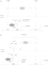
The constitutive activity of unligated AhR and Arnt differentially regulate CXCL8 and CCL5 in BEAS-2B cells. The model summarizes the possible pathways discussed for AhR- and Arnt-mediated regulation of CXCL8 and CCL5 in BEAS-2B cells exposed to 1-NP and 1-AP (A) or Poly I:C (B). “x/X” and “y/Y” represents hitherto unidentified signaling pathways/response elements involved in regulation of CXCL8 and CCL5, respectively. The positioning of the response elements in the CXCL8 or CCL5 promoters is not meant to be representative. Moreover, “y/Y” induced by 1-AP (A) and Poly I:C (B) are not necessarily identical and AhR may also affect other pathways involved in Poly I:C-induced CXCL8 in addition to NF-κB (B). The suggested position and association of Arnt and RelB is partly based on findings reported by Wright and Duckett [49]. The role of AhR and Arnt in chemokine regulation may differ in other cell types.

Similar articles

Cited by

References

    1. Rowlands JC, Gustafsson JA. Aryl hydrocarbon receptor-mediated signal transduction. Crit Rev Toxicol. 1997;27:109–134. doi: 10.3109/10408449709021615. - DOI - PubMed
    1. Ukena C, Mahfoud F, Kindermann M, Kindermann I, Bals R, Voors AA, van Veldhuisen DJ, Bohm M. The cardiopulmonary continuum systemic inflammation as ‘common soil’ of heart and lung disease. Int J Cardiol. 2010;145:172–176. doi: 10.1016/j.ijcard.2010.04.082. - DOI - PubMed
    1. Scrivo R, Vasile M, Bartosiewicz I, Valesini G. Inflammation as “common soil” of the multifactorial diseases. Autoimmun Rev. 2011;10:369–374. doi: 10.1016/j.autrev.2010.12.006. - DOI - PubMed
    1. Gomez-Mejiba SE, Zhai Z, Akram H, Pye QN, Hensley K, Kurien BT, Scofield RH, Ramirez DC. Inhalation of environmental stressors & chronic inflammation: autoimmunity and neurodegeneration. Mutat Res. 2009;674:62–72. doi: 10.1016/j.mrgentox.2008.09.016. - DOI - PMC - PubMed
    1. Bhatnagar A. Could dirty air cause diabetes? Circulation. 2009;119:492–494. doi: 10.1161/CIRCULATIONAHA.108.831404. - DOI - PubMed

Publication types

MeSH terms

LinkOut - more resources