. 2014 Nov;166(3):1312-28.
doi: 10.1104/pp.114.242487. Epub 2014 Sep 8.

Heterodimeric capping protein from Arabidopsis is a membrane-associated, actin-binding protein

Affiliations

Heterodimeric capping protein from Arabidopsis is a membrane-associated, actin-binding protein

Jose C Jimenez-Lopez et al. Plant Physiol. 2014 Nov.

Abstract

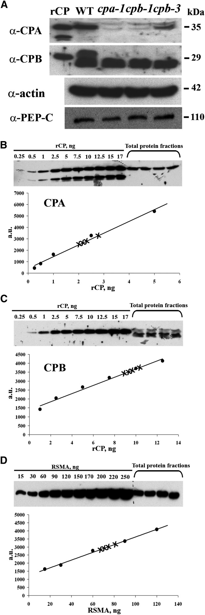
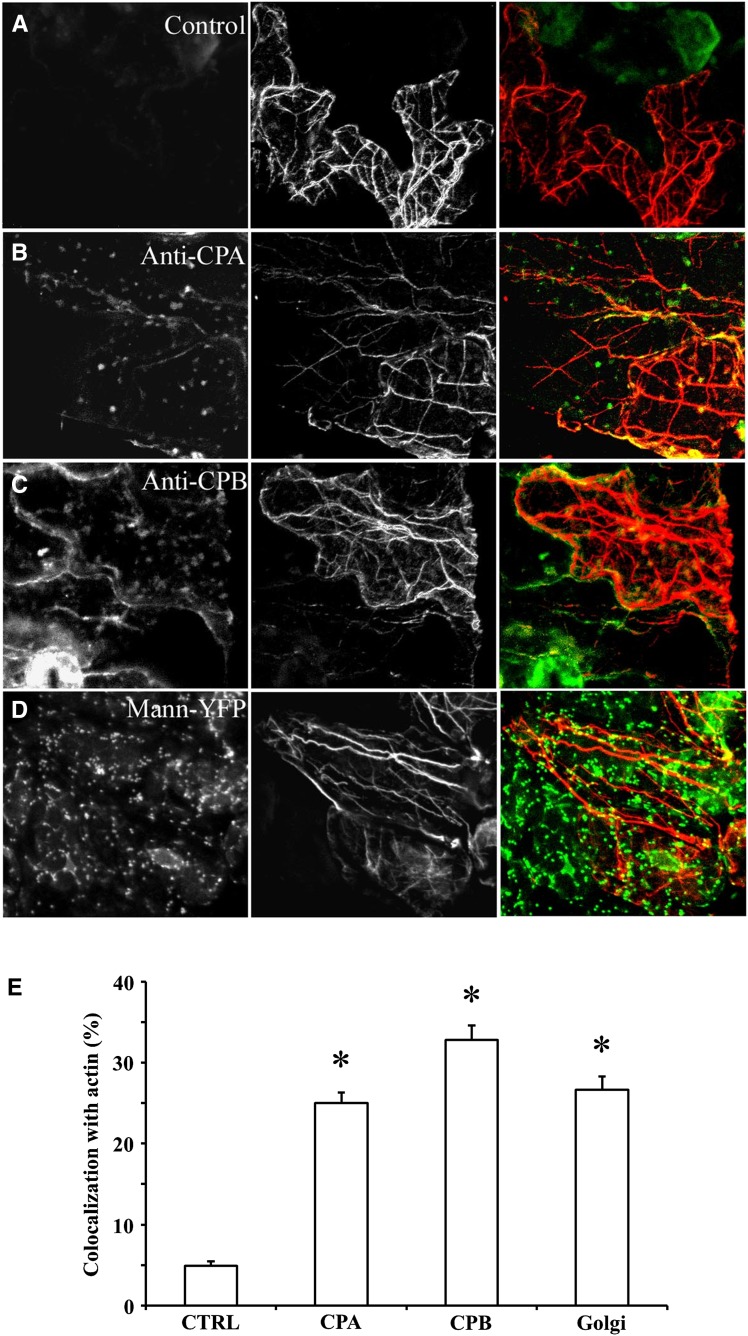
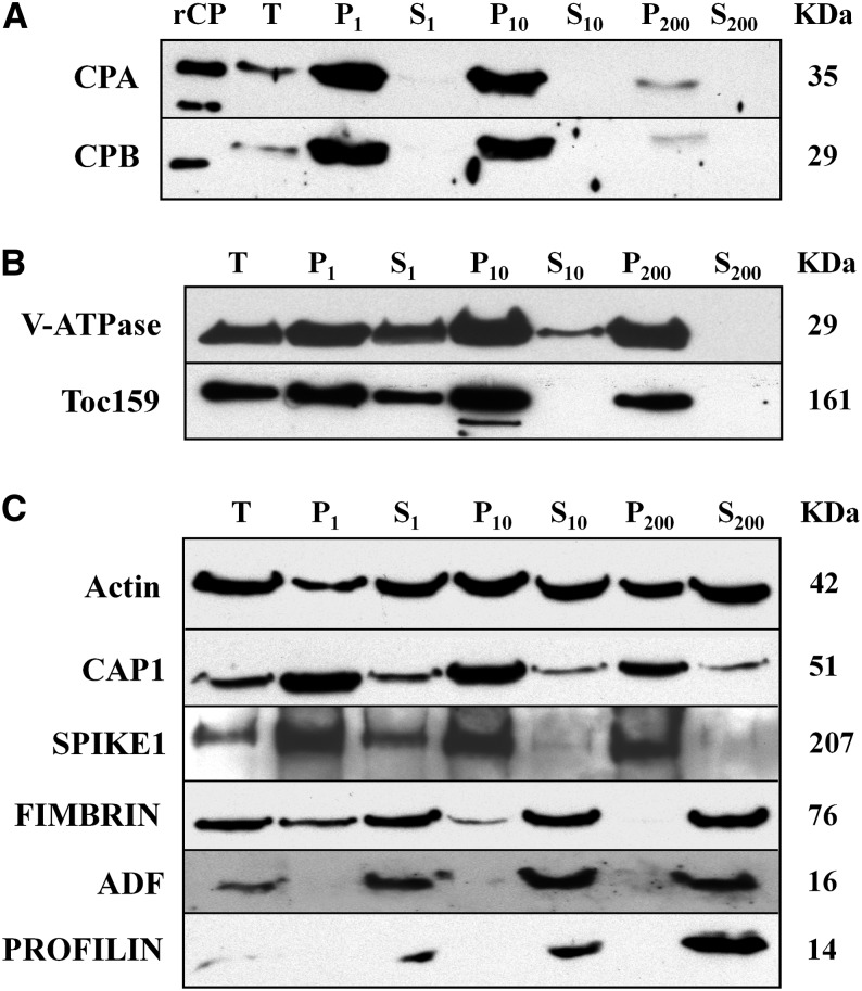
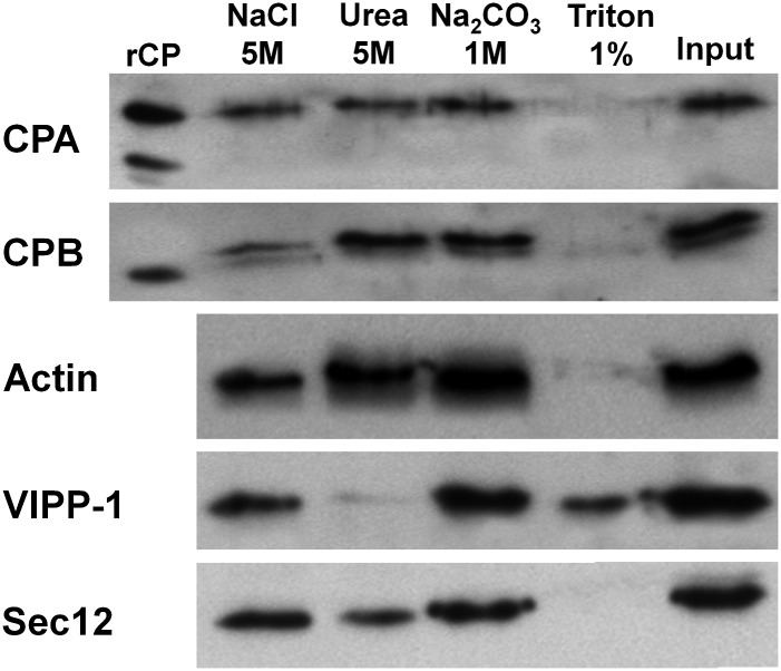
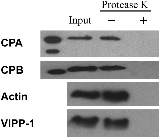
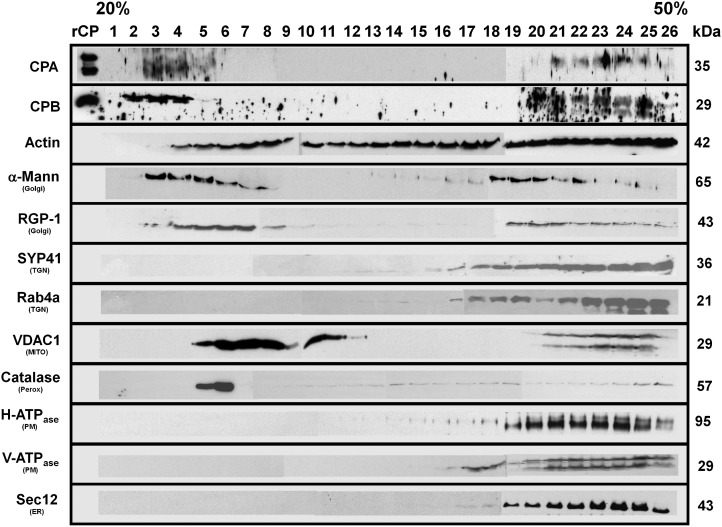
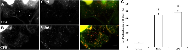
The actin cytoskeleton is a major regulator of cell morphogenesis and responses to biotic and abiotic stimuli. The organization and activities of the cytoskeleton are choreographed by hundreds of accessory proteins. Many actin-binding proteins are thought to be stimulus-response regulators that bind to signaling phospholipids and change their activity upon lipid binding. Whether these proteins associate with and/or are regulated by signaling lipids in plant cells remains poorly understood. Heterodimeric capping protein (CP) is a conserved and ubiquitous regulator of actin dynamics. It binds to the barbed end of filaments with high affinity and modulates filament assembly and disassembly reactions in vitro. Direct interaction of CP with phospholipids, including phosphatidic acid, results in uncapping of filament ends in vitro. Live-cell imaging and reverse-genetic analyses of cp mutants in Arabidopsis (Arabidopsis thaliana) recently provided compelling support for a model in which CP activity is negatively regulated by phosphatidic acid in vivo. Here, we used complementary biochemical, subcellular fractionation, and immunofluorescence microscopy approaches to elucidate CP-membrane association. We found that CP is moderately abundant in Arabidopsis tissues and present in a microsomal membrane fraction. Sucrose density gradient separation and immunoblotting with known compartment markers were used to demonstrate that CP is enriched on membrane-bound organelles such as the endoplasmic reticulum and Golgi. This association could facilitate cross talk between the actin cytoskeleton and a wide spectrum of essential cellular functions such as organelle motility and signal transduction.


Figures

Figure 1.
Figure 1.
CP is a moderately abundant protein in total cellular extracts. A, On protein immunoblots, CPA and CPB antisera recognized polypeptides from purified rCP (10-ng load), as well as polypeptides of appropriate size from total cellular extracts of wild-type Arabidopsis Col-0 seedlings (50-µg load). Total cellular extracts (50 µg each) prepared from three homozygous T-DNA insertion mutant lines (cpa-1, cpb-1, and cpb-3; Li et al., 2012) had reduced levels of CPA and CPB polypeptides. Probing of identical membranes with anti-actin antibodies revealed that total actin levels were not reduced in the cp homozygous mutant lines compared with the wild type. The same blot was reprobed with anti-phosphoenolpyruvate carboxylase antibody, which recognized a band of 110 kD, and verifies the equal loading of samples. B and C, CPA and CPB protein levels were estimated by semiquantitative immunoblotting using total protein extracts from wild-type Arabidopsis seedlings and a standard curve comprising varying amounts of rCP. The Coomassie-stained gel images at the top show results from the blotting of a standard curve and four biological replicates of seedling extracts (75 µg each) with anti-CPA (B) and anti-CPB (C). The intensity of each band from the standard curve as a function of protein amount is plotted. The data were fit with a linear function and the correlation coefficients for these representative examples were 0.99 and 0.98 for CPA and CPB, respectively. In this example, CPA represents 0.0015% ± 0.0001% of total cellular protein, whereas CPB represents 0.0013% ± 0.0002%. D, Total actin levels were estimated by immunoblotting of seedling extracts prepared from the wild type and a standard curve comprising different amounts of purified rabbit skeletal muscle actin. The gel image shows the result from blotting of a standard curve and four biological replicates of seedling extracts (25 µg each). The intensity of each band from the standard curve as a function of protein amount is plotted. The data were fit with a linear function and the correlation coefficient for this representative example was 0.99. In this experiment, actin represents 0.37% ± 0.02% of total cellular protein. a.u., Arbitrary units; RSMA, rabbit skeletal muscle actin; WT, wild type.
Figure 2.
Figure 2.
CP is present on cytoplasmic puncta that display only modest colocalization with actin filaments or cables in epidermal pavement cells. Seedlings of wild-type Arabidopsis plants (20 DAG) were fixed and prepared by the freeze-shattering method prior to incubation with affinity-purified CPA or CPB polyclonal antisera, as well as with a mouse monoclonal IgM against actin. Epidermal pavement cells were examined by confocal laser scanning microscopy and images shown are z-series projections. A, The left image shows a control with secondary antibody only (i.e. no CP primary antibody). The middle image shows actin labeling and the right image is a color overlay of the control (green) and actin (red) images. B, A representative epidermal pavement cell that is double labeled for CPA (left) and actin (middle). The right image is a color overlay of CPA (green) and actin (red). CPA is present on cytoplasmic puncta or foci of varying size and intensity. A small subset of these colocalize (right, yellow) with actin filaments or cables. C, A representative epidermal cell that is double labeled for CPB (left) and actin (middle). The right image is a color overlay of CPB (green) and actin (red). Similar to CPA, CPB is present on puncta that sometimes colocalize (yellow) with actin cables. D, Colocalization of Golgi and actin filaments. Arabidopsis seedlings expressing the Golgi marker mannosidase-YFP were prepared and immunolabeled as above with the actin monoclonal antibody. The left image shows mannosidase-YFP fluorescence and the middle image is actin. The right image is a color overlay of mannosidase-YFP (green) and actin (red), showing a substantial overlap (yellow) of Golgi on the actin cables (yellow). E, Quantitative analysis of CPA, CPB, and mannosidase-YFP association with actin filaments and cables. See “Materials and Methods” for details. The mean values (± sem) from analysis of more than 25 ROIs per treatment are plotted. Compared with controls, in which the CP primary antibody was excluded, the extent of colocalization between CPA, CPB, or mannosidase-YFP with actin was significant (*P < 0.01). CTRL, Control; Mann, mannosidase.
Figure 3.
Figure 3.
CP is present in membrane fractions after differential centrifugation of cellular extracts. Analysis of CP and several other ABPs during differential centrifugation of extracts prepared from 20 DAG Arabidopsis Col-0 seedlings. The individual lanes represent the pellet (P) and supernatant (S) fractions obtained after total cellular extracts (T) were subjected to differential centrifugation at 1,000g, 10,000g, and 200,000g, respectively. Lanes were loaded with equal amounts of protein (75 µg), separated by SDS-PAGE, and immunoblotted with antibodies against CP, V-ATPase, AtToc159, and various ABPs. The molecular weight in kilodaltons for each polypeptide is given at right. A, CPA and CPB were most abundant in the pellet fractions and were virtually undetectable in the soluble fractions. rCP loaded in the first lane verifies the size of the native protein in extracts. B, Antibodies against the tonoplast marker V-ATPase and the chloroplast outer envelope protein Toc159, were used as positive controls for differential centrifugation of membrane-associated proteins. C, Actin and several cytoskeletal-associated proteins also partitioned with membranes or organellar fractions. Antibodies were used to detect the following: actin; CAP1; the ROP-GEF, SPK1; an actin filament cross linking protein, FIMBRIN; and, two actin monomer-binding proteins, ADF and PROFILIN. Actin partitioned almost equally between soluble and pellet fractions, whereas CAP1 and SPK1 were mainly in pellet fractions. By contrast, FIMBRIN, ADF, and PROFILIN were predominantly soluble proteins.
Figure 4.
Figure 4.
CP behaves like an integral membrane-associated protein. The supernatant S1 fraction was centrifuged at 200,000g to give a P200 microsomal membrane fraction, which was resuspended and divided into five equal fractions in buffer containing either 5 m NaCl, 5 m urea, 1 m Na2CO3, pH 10.9, or 1% (v/v) Triton X-100 and incubated on a shaker for 30 min at 4°C. The resulting suspension was recentrifuged for 60 min at 200,000g, providing pellet and soluble fractions. Shown here are the pellet fractions that were blotted and probed with CPA and CPB antibodies, as well as with actin, VIPP-1, and Sec12 antibodies as controls for peripheral and integral membrane-associated proteins, respectively. Similar experiments were performed four independent times.
Figure 5.
Figure 5.
CP localizes on the cytoplasmic side of the membrane. The P200 fraction containing CP was incubated with and without PK. Immunoblots of the resulting samples were performed with antibodies against CPA and CPB, anti-actin, and anti-VIPP1, The P200 fraction prior to addition of protease was used as a loading control. rCP was loaded in the first lane as a molecular weight marker for CP.
Figure 6.
Figure 6.
CP is coenriched with several membrane-bound compartments in the microsomal fraction. Microsomal (P200) membrane fractions were separated on an isopycnic 20% to 50% (w/v) linear Suc gradient. Equal volumes of protein fractions collected from the gradient were separated on SDS-PAGE gels, blotted, and probed with antibodies against the following: CPA and CPB; actin; cis-Golgi, α-1,2-mannosidase; trans-Golgi, RGP1; plasma membrane, H+-ATPase; ER, Sec12; tonoplast, V-ATPase; mitochondrial outer membrane porin 1, VDAC1; trans-Golgi network, AtSYP41 and RabA4; and peroxisome, catalase. Protein names and sizes are indicated on the left and right, respectively. The entire gradient, fractions 1 to 26, required several gels and membranes for probing with each antibody. Separation between the individual blots or membranes comprising the full gradient is not shown on the figure, for clarity of presentation. Mann, Mannosidase; MITO, mitochondria; Perox, peroxisome; PM, plasma membrane; TGN, trans-Golgi network.
Figure 7.
Figure 7.
CP colocalizes with a cis-Golgi marker. A and B, Colocalization of CP with Golgi. Arabidopsis seedlings expressing the Golgi marker, mannosidase-YFP, were prepared and immunolabeled with CP polyclonal antibodies. The left image shows a representative image from an epidermal pavement cell labeled with CPA (A) and CPB (B), respectively. Middle images correspond to mannosidase-YFP fluorescence from the same cells. The right images show merged images depicting colocalization. C, Quantitative analysis of colocalization between CPA and CPB with mannosidase-YFP. See “Materials and Methods” for details. The mean values (± sem) from analysis of >41 ROIs within at least seven epidermal pavement cells per treatment are plotted. As a control, the primary anti-CPB antibody was left out and samples were processed in identical fashion. The extent of colocalization between both CP subunits and mannosidase-YFP was significantly different from the negative control (*P < 0.01). CTRL, Control. Bar = 10 μm.

Similar articles

Cited by

References

    1. Akin O, Mullins RD. (2008) Capping protein increases the rate of actin-based motility by promoting filament nucleation by the Arp2/3 complex. Cell 133: 841–851 - PMC - PubMed
    1. Amatruda JF, Cooper JA. (1992) Purification, characterization, and immunofluorescence localization of Saccharomyces cerevisiae capping protein. J Cell Biol 117: 1067–1076 - PMC - PubMed
    1. Amatruda JF, Gattermeir DJ, Karpova TS, Cooper JA. (1992) Effects of null mutations and overexpression of capping protein on morphogenesis, actin distribution and polarized secretion in yeast. J Cell Biol 119: 1151–1162 - PMC - PubMed
    1. Avisar D, Prokhnevsky AI, Makarova KS, Koonin EV, Dolja VV. (2008) Myosin XI-K is required for rapid trafficking of Golgi stacks, peroxisomes, and mitochondria in leaf cells of Nicotiana benthamiana. Plant Physiol 146: 1098–1108 - PMC - PubMed
    1. Baillie GS, Huston E, Scotland G, Hodgkin M, Gall I, Peden AH, MacKenzie C, Houslay ES, Currie R, Pettitt TR, et al. (2002) TAPAS-1, a novel microdomain within the unique N-terminal region of the PDE4A1 cAMP-specific phosphodiesterase that allows rapid, Ca2+-triggered membrane association with selectivity for interaction with phosphatidic acid. J Biol Chem 277: 28298–28309 - PubMed

Publication types

MeSH terms

Substances

Associated data

LinkOut - more resources