. 2014 Dec;64(6):1290-8.
doi: 10.1161/HYPERTENSIONAHA.114.03971. Epub 2014 Sep 8.

Elimination of vitamin D receptor in vascular endothelial cells alters vascular function

Affiliations

Elimination of vitamin D receptor in vascular endothelial cells alters vascular function

Wei Ni et al. Hypertension. 2014 Dec.

Abstract

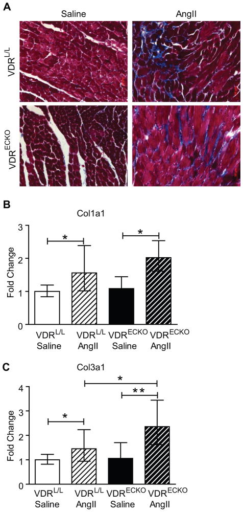
Vitamin D deficiency has been associated with cardiovascular dysfunction. We evaluated the role of the vitamin D receptor (VDR) in vascular endothelial function, a marker of cardiovascular health, at baseline and in the presence of angiotensin II, using an endothelial-specific knockout of the murine VDR gene. In the absence of endothelial VDR, acetylcholine-induced aortic relaxation was significantly impaired (maximal relaxation, endothelial-specific VDR knockout=58% versus control=73%; P<0.05). This was accompanied by a reduction in endothelial NO synthase expression and phospho-vasodilator-stimulated phosphoprotein levels in aortae from the endothelial-specific VDR knockout versus control mice. Although blood pressure levels at baseline were comparable at 12 and 24 weeks of age, the endothelial VDR knockout mice demonstrated increased sensitivity to the hypertensive effects of angiotensin II compared with control mice (after 1-week infusion: knockout=155±15 mm Hg versus control=133±7 mm Hg; P<0.01; after 2-week infusion: knockout=164±9 mm Hg versus control=152±13 mm Hg; P<0.05). By the end of 2 weeks, angiotensin II infusion-induced, hypertrophy-sensitive myocardial gene expression was higher in endothelial-specific VDR knockout mice (fold change compared with saline-infused control mice, type-A natriuretic peptide: knockout mice=3.12 versus control=1.7; P<0.05; type-B natriuretic peptide: knockout mice=4.72 versus control=2.68; P<0.05). These results suggest that endothelial VDR plays an important role in endothelial cell function and blood pressure control and imply a potential role for VDR agonists in the management of cardiovascular disease associated with endothelial dysfunction.

Keywords: gene targeting; hypertension; receptors, calcitriol; vitamin D.


Figures

Figure 1
Figure 1
Validation of expression of VDR in endothelial cells and generation of an endothelial cell-specific VDR knockout mouse. A. VDR expression in HUVECs. Parallel Western blots were incubated with antibody alone or antibody with a 5X competing peptide (CP). B. VDR protein was visualized by immunofluorescence (red, arrows) with anti-VDR in HUVECs. Nuclei (blue) are stained with DAPI. Cell borders (green) were defined with anti-CD31. C. Schematic representation of the generation of endothelial cell specific VDR gene knockout mouse. D. Mouse with homozygous LoxP-bordered VDR allele was crossed with a Tie-2 Cre transgenic mouse to generate VDRECKO. Each mouse with Cre transgene showed a minor band corresponding to VDR gene deletion. VDR genotyping was performed by PCR using primers indicated with arrows (Figure 1C). E. VDR mRNA expression in endothelial cells and non-endothelial cells isolated from hearts of control and VDRECKO mice. ***p<0.001 (n=4). F. VDR protein expression in endothelial cells isolated from hearts of control and VDRECKO mice. L= loxp; VDR= vitamin D receptor; VDRL/L= VDR floxed mouse, served as control; VDRECKO= endothelial cell specific VDR gene knockout mouse.
Figure 2
Figure 2
Impaired blood vessel relaxation in aorta from VDRECKO mice. A. Representative tracings of ACh-induced, concentration-dependent aortic relaxation. Nitric oxide donor sodium nitroprusside (SNP) relaxed aorta to baseline. B. Aortae from VDRECKO mice exhibit impaired acetylcholine-induced relaxation. Nitric oxide synthase inhibitor LNNA completely abolished ACh-induced aortic relaxation in aorta from control and VDRECKO mice. VDRL/L + LNNA (n=3); VDRECKO + LNNA (n=3); VDRL/L (n=5); VDRECKO (n=5). C. Comparable contractile response to PE in aorta from control and VDRECKO mice. *p<0.05 (n=5 for each group). VDRL/L= VDR floxed mouse, served as control; VDRECKO= endothelial cell specific VDR gene knockout mouse.
Figure 3
Figure 3
Reduced eNOS expression in aorta from VDRECKO mice. A. eNOS mRNA expression in aorta from control and VDRECKO mice (n=4). B. eNOS protein expression in aorta from control and VDRECKO mice (n=6). VDRL/L= VDR floxed mouse, served as control; VDRECKO= endothelial cell specific VDR gene knockout mouse. C. Representative Western blot (left) and quantification (right) of basal p-VASP level detected by Western blot in aorta from control and VDRECKO mice (n=4). VDRL/L= VDR floxed mouse, served as control; VDRECKO = endothelial cell specific VDR gene knockout mouse.
Figure 4
Figure 4
Tail cuff measurements of SBP (A), DBP (B) and MAP (C) in control and VDRECKO mice before, 1 week or 2 weeks after saline or AngII infusion. ANOVA was used for statistical analysis. *p<0.05; **p<0.01; ***p<0.001. VDRL/L + saline (n=6); VDRL/L + Ang II (n=6); VDRECKO + saline (n=6); VDRECKO +Ang II (n=5). VDRL/L= VDR floxed mouse, served as control; VDRECKO= endothelial cell specific VDR gene knockout mouse.
Figure 5
Figure 5
Similar changes in blood vessel morphology in aortae with AngII infusion. A. H&E staining of aortae from control and VDRECKO mice following treatment with saline or AngII infusion. B. Masson’s Trichrome staining of aortae from control and VDRECKO mice following treatment with saline or AngII infusion. VDRL/L + saline (n=4); VDRL/L + Ang II (n=4); VDRECKO + saline (n=5); VDRECKO +Ang II (n=5). VDRL/L= VDR floxed mouse, served as control; VDRECKO= endothelial cell specific VDR gene knockout mouse.
Figure 6
Figure 6
AngII infusion-induced cardiac hypertrophy in control and VDRECKO mice. A. H&E staining of hearts. B. Left ventricle weight over body weight ratio VDRL/L + saline (n=6); VDRL/L + Ang II (n=6); VDRECKO + saline (n=6); VDRECKO +Ang II (n=5). C. WGA staining of hearts. D. Quantification of cardiac myocyte size VDRL/L + saline (n=7); all other groups n=6. E. Expression of hypertrophic marker gene encoding ANP. VDRECKO +Ang II (n=6); all other groups =7. F. BNP gene expression. VDRL/L + saline (n=8); VDRL/L + Ang II (n=8); VDRECKO + saline (n=7); VDRECKO +Ang II (n=7). *p<0.05; **p<0.01; ***p<0.001. VDRL/L= VDR floxed mouse, served as control; VDRECKO= endothelial cell specific VDR gene knockout mouse.
Figure 7
Figure 7
AngII infusion-induced cardiac fibrosis in control and VDRECKO mice. A. Masson’s trichrome staining of hearts. B. Col 1a1 and Col 3a1 expression in hearts from control and VDRECKO mice after receiving saline or AngII-infusion for two weeks. VDRL/L + saline (n=6); VDRL/L + Ang II (n=6); VDRECKO + saline (n=6); VDRECKO +Ang II (n=5). *p<0.05; **p<0.01. VDRL/L= VDR floxed mouse, served as control; VDRECKO= endothelial cell specific VDR gene knockout mouse.

Comment in

Similar articles

Cited by

References

    1. Holick MF. Vitamin d deficiency. N Engl J Med. 2007;357:266–281. - PubMed
    1. Wootton AM. Improving the measurement of 25-hydroxyvitamin d. Clin Biochem Rev. 2005;26:33–36. - PMC - PubMed
    1. Haussler MR, Haussler CA, Bartik L, Whitfield GK, Hsieh J-C, Slater S, Jurutka PW. Vitamin d receptor: Molecular signaling and actions of nutritional ligands in disease prevention. Nutrition Reviews. 2008;66:S98–S112. - PubMed
    1. Forman JP, Giovannucci E, Holmes MD, Bischoff-Ferrari HA, Tworoger SS, Willett WC, Curhan GC. Plasma 25-hydroxyvitamin d levels and risk of incident hypertension. Hypertension. 2007;49:1063–1069. - PubMed
    1. Forman JP, Curhan GC, Taylor EN. Plasma 25-hydroxyvitamin d levels and risk of incident hypertension among young women. Hypertension. 2008;52:828–832. - PMC - PubMed

Publication types

MeSH terms