Review
. 2014 Dec;93(12):1222-31.
doi: 10.1177/0022034514549809. Epub 2014 Sep 8.

Tissue-engineering-based strategies for regenerative endodontics

Affiliations
Review

Tissue-engineering-based strategies for regenerative endodontics

M T P Albuquerque et al. J Dent Res. 2014 Dec.

Abstract

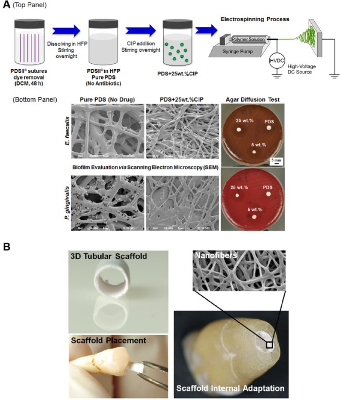
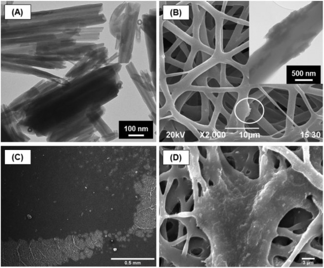
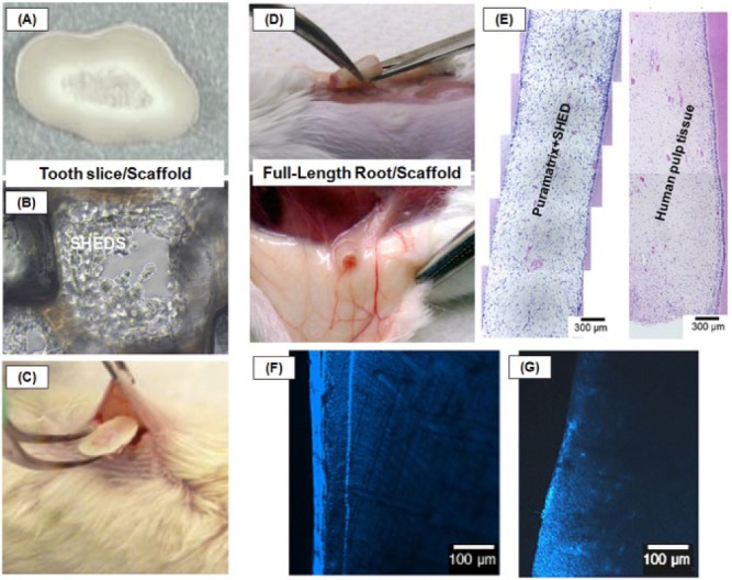
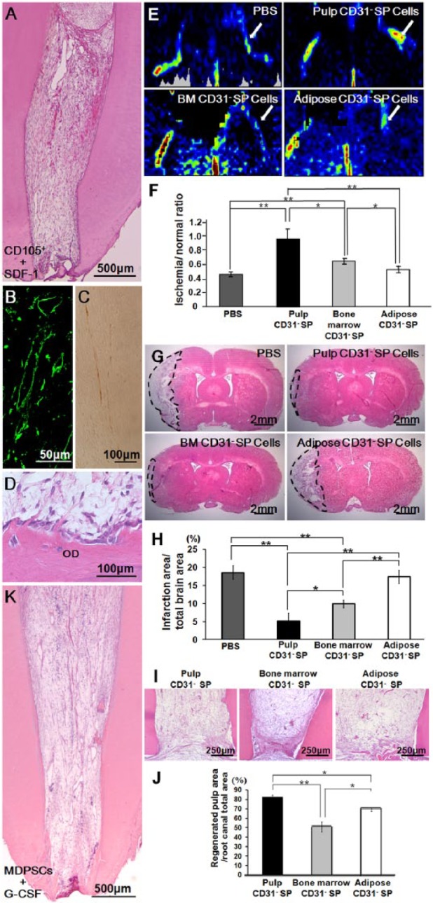
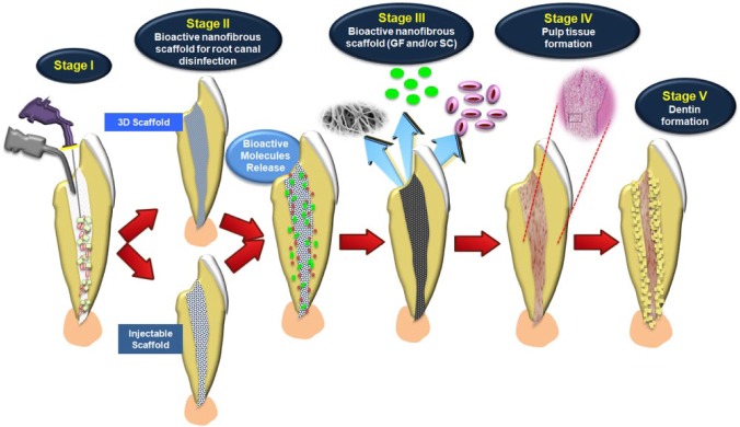
Stemming from in vitro and in vivo pre-clinical and human models, tissue-engineering-based strategies continue to demonstrate great potential for the regeneration of the pulp-dentin complex, particularly in necrotic, immature permanent teeth. Nanofibrous scaffolds, which closely resemble the native extracellular matrix, have been successfully synthesized by various techniques, including but not limited to electrospinning. A common goal in scaffold synthesis has been the notion of promoting cell guidance through the careful design and use of a collection of biochemical and physical cues capable of governing and stimulating specific events at the cellular and tissue levels. The latest advances in processing technologies allow for the fabrication of scaffolds where selected bioactive molecules can be delivered locally, thus increasing the possibilities for clinical success. Though electrospun scaffolds have not yet been tested in vivo in either human or animal pulpless models in immature permanent teeth, recent studies have highlighted their regenerative potential both from an in vitro and in vivo (i.e., subcutaneous model) standpoint. Possible applications for these bioactive scaffolds continue to evolve, with significant prospects related to the regeneration of both dentin and pulp tissue and, more recently, to root canal disinfection. Nonetheless, no single implantable scaffold can consistently guide the coordinated growth and development of the multiple tissue types involved in the functional regeneration of the pulp-dentin complex. The purpose of this review is to provide a comprehensive perspective on the latest discoveries related to the use of scaffolds and/or stem cells in regenerative endodontics. The authors focused this review on bioactive nanofibrous scaffolds, injectable scaffolds and stem cells, and pre-clinical findings using stem-cell-based strategies. These topics are discussed in detail in an attempt to provide future direction and to shed light on their potential translation to clinical settings.

Keywords: dental pulp; dentin; nanofibers; regeneration; stem cells; tissue scaffolds.


Conflict of interest statement

The authors declare no potential conflicts of interest with respect to the authorship and/or publication of this article.

Figures

Figure 1.
Figure 1.
Summarized schematic of nanofibrous antibiotic-containing scaffolds (e.g., ciprofloxacin [CIP]) processed via electrospinning and antimicrobial effects. (A) (top panel) FDA-approved polydioxanone suture filaments were used (PDS II®, Ethicon, Somerville, NJ, USA). First, the violet color of filament sutures is removed by immersion in dichloromethane. Then, the cleared PDS filaments are dissolved in 1,1,1,3,3,3-hexafluoro-2-propanol (HFP, Sigma-Aldrich, St. Louis, MO, USA) at optimized concentration under stirring conditions. CIP-containing PDS solution is prepared by the addition of CIP at a known concentration, being mixed together under vigorous stirring. (bottom panel) Representative scanning electron microscopy (SEM) micrographs showing the antimicrobial effects of antibiotic-containing PDS-based electrospun scaffolds on bacterial growth. Representative macrophotographs of the agar diffusion test show growth inhibition of E. faecalis and P. gingivalis) (adapted with permission from Bottino et al., 2013). (B) Potential clinical application of a three-dimensional (3D) tubular scaffold produced via electrospinning. Electrospun scaffolds can be fabricated in a cylindrical shape simulating the tubular and parallel format of immature root canals, making it easy to place and adapt into the root canal. Inset shows the nanofibrous structure of the 3D scaffold.
Figure 2.
Figure 2.
Polymer nanocomposite electrospun scaffolds synthesized with aluminosilicate clay nanotubes. (A) Representative transmission electron microscopy (TEM) micrograph of aluminosilicate clay Halloysite nanotubes (HNTs). (B) Representative scanning electron microscopy (SEM) micrograph of electrospun HNT-incorporated nanofibrous scaffolds. (inset) Representative TEM micrograph of HNTs protruding from the fiber structure. (C-D) Representative SEM micrographs showing the interaction between human-derived dental pulp fibroblast cells and PDS-HNT fibrous scaffolds (adapted with permission from Bottino et al., 2014b).
Figure 3.
Figure 3.
Summarized schematic of the (A-C) tooth slice and (D-G) full-length root/scaffold models. (A) Tooth slice provided from the cervical third of a human third molar with a highly porous PLLA scaffold placed within the pulp chamber. (B) SHED proliferation into the tooth slice/scaffold. (C) Insertion of a tooth slice and scaffold containing SHED into the subcutaneous space of the dorsum of an immunodeficient mouse. (D) Subcutaneous transplant of a human full-length root injected with hydrogel-based nanofibrous scaffolds containing SHEDs. (E) Photomicrographs of the engineered pulp-like tissue and human pulp tissue (control) in the root canal. (F) Layer of dentin formation after pulp tissue induction in PuraMatrix+SHEDs. (G) Dentin slice with no SHEDs (adapted with permission from Sakai et al., 2011; Casagrande et al., 2011; Rosa et al., 2013).
Figure 4.
Figure 4.
Clinical evidence of dentin-pulp complex regeneration. (A-D) Complete regeneration of pulp tissue after autologous transplantation of CD105+ cells with SDF-1 in the pulpectomized root canal in dogs. (B) Immunostaining with BS-1 lectin. (C) Immunostaining with PGP 9.5. (D) Odontoblastic cell lining to newly formed osteodentin/tubular dentin (OD), along with the dentinal wall. (E, F) Neovascularization in the ischemic hindlimb model 14 days after transplantation of pulp, bone marrow, and adipose-derived CD31- side-population (SP) cells. (E) Laser Doppler imaging. (F) Quantification of blood flow in mouse ischemic hindlimbs (n = 4 in each group). (G, H) Infarct area on day 21 after injection of PBS, pulp, bone marrow, and adipose CD31- SP cells. (H) Reduction of the infarct volume 21 days after injection (*p < .05, **p < .01). (I, J) Ectopic pulp regeneration 28 days after transplantation of pulp, bone marrow, and adipose-derived CD31- SP cells into porcine tooth root. (J) Ratio of regenerated pulp area to root canal area. Data are expressed as means ± SD of 5 determinations. *p < .05, **p < .01. (K) Complete regeneration of pulp tissue after autologous transplantation of mobilized dental pulp stem cells (MDPSCs) with G-CSF in the pulpectomized root canal in dogs (adapted with permission from Iohara et al., 2011; Ishizaka et al., 2013; Iohara et al., 2013).
Figure 5.
Figure 5.
Tissue-engineering-based strategies for regenerative endodontics in immature teeth. Strategies have included the incorporation of (i) therapeutic agents, such as antimicrobial drugs to be released and promote root canal disinfection, as well as (ii) bioactive molecules that can trigger stem cell differentiation to aid in regeneration of the pulp-dentin complex. Stage I: Disinfection of the root canal using irrigant solutions. Stage II: Bioactive nanofibrous scaffold with antibiotics as intracanal medication. Stage III: Nanofibrous scaffold with growth factors and/or stem cells. Stage IV: Pulp tissue formation. Stage V: Dentin formation. (Histology of pulp-like tissue formation, adapted with permission from Rosa et al., 2013.)

References

    1. Akkouch A, Zhang Z, Rouabhia M. (2013). Engineering bone tissue using human dental pulp stem cells and an osteogenic collagen-hydroxyapatite-poly (L-lactide-co-ϵ-caprolactone) scaffold. J Biomater Appl 28:922-936. - PubMed
    1. Banchs F, Trope M. (2004). Revascularization of immature permanent teeth with apical periodontitis: new treatment protocol? J Endod 30:196-200. - PubMed
    1. Baylan N, Bhat S, Ditto M, Lawrence JG, Lecka-Czernik B, Yildirim-Ayan E. (2013). Polycaprolactone nanofiber interspersed collagen type-I scaffold for bone regeneration: a unique injectable osteogenic scaffold. Biomed Mater 8:045011. - PubMed
    1. Becerra P, Ricucci D, Loghin S, Gibbs JL, Lin LM. (2014). Histologic study of a human immature permanent premolar with chronic apical abscess after revascularization/revitalization. J Endod 40:133-139. - PubMed
    1. Bose R, Nummikoski P, Hargreaves K. (2009). A retrospective evaluation of radiographic outcomes in immature teeth with necrotic root canal systems treated with regenerative endodontic procedures. J Endod 35:1343-1349. - PubMed

Publication types

LinkOut - more resources