. 2014 Oct 7;111(40):E4244-53.
doi: 10.1073/pnas.1409986111. Epub 2014 Sep 8.

Involvement of the host DNA-repair enzyme TDP2 in formation of the covalently closed circular DNA persistence reservoir of hepatitis B viruses

Affiliations

Involvement of the host DNA-repair enzyme TDP2 in formation of the covalently closed circular DNA persistence reservoir of hepatitis B viruses

Christian Königer et al. Proc Natl Acad Sci U S A. .

Abstract

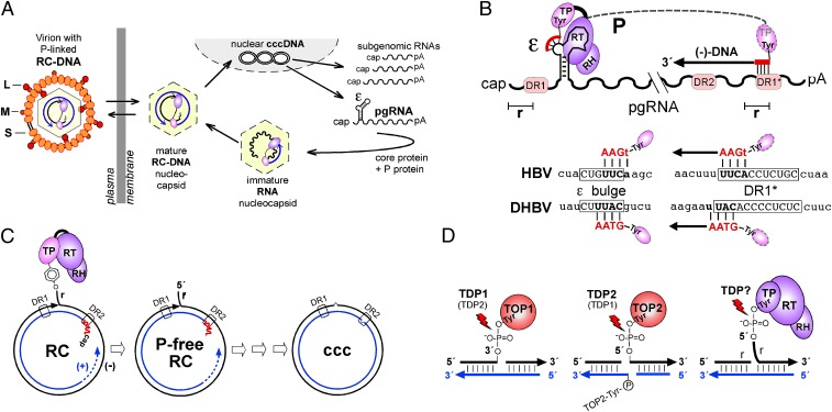
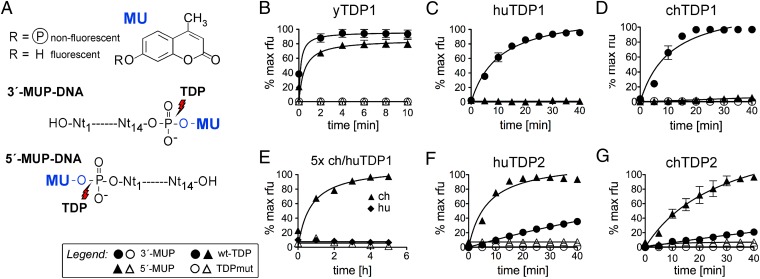
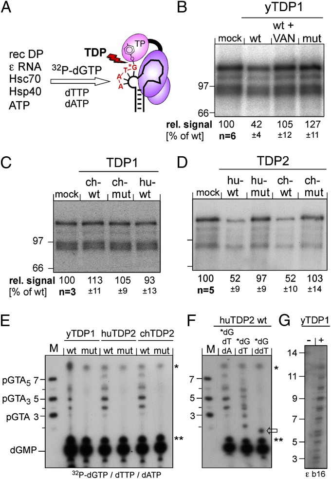
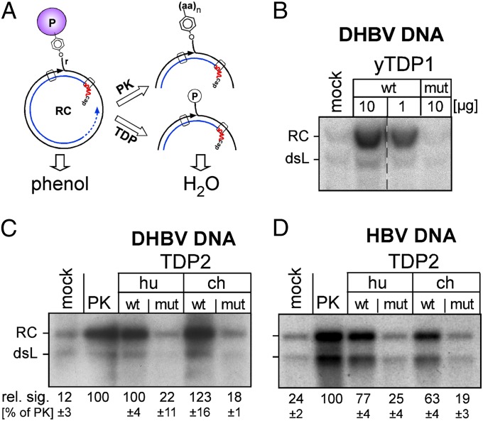
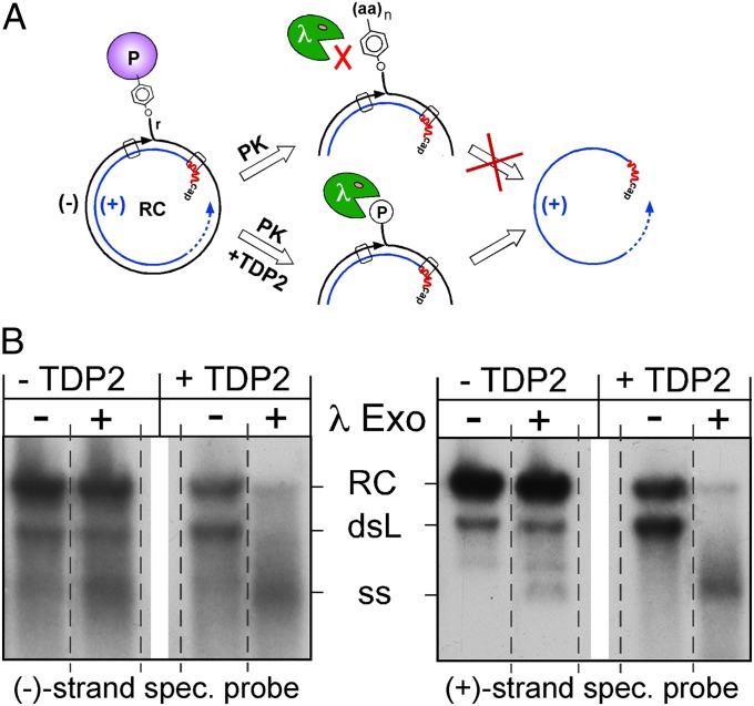
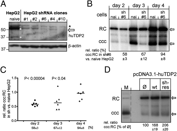
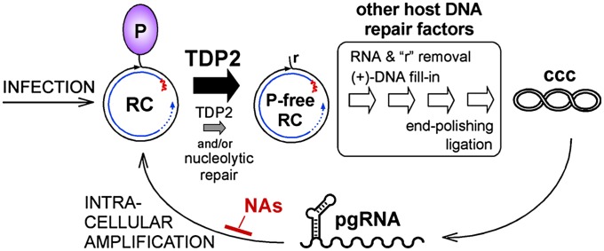
Hepatitis B virus (HBV), the causative agent of chronic hepatitis B and prototypic hepadnavirus, is a small DNA virus that replicates by protein-primed reverse transcription. The product is a 3-kb relaxed circular DNA (RC-DNA) in which one strand is linked to the viral polymerase (P protein) through a tyrosyl-DNA phosphodiester bond. Upon infection, the incoming RC-DNA is converted into covalently closed circular (ccc) DNA, which serves as a viral persistence reservoir that is refractory to current anti-HBV treatments. The mechanism of cccDNA formation is unknown, but the release of P protein is one mandatory step. Structural similarities between RC-DNA and cellular topoisomerase-DNA adducts and their known repair by tyrosyl-DNA-phosphodiesterase (TDP) 1 or TDP2 suggested that HBV may usurp these enzymes for its own purpose. Here we demonstrate that human and chicken TDP2, but only the yeast ortholog of TDP1, can specifically cleave the Tyr-DNA bond in virus-adapted model substrates and release P protein from authentic HBV and duck HBV (DHBV) RC-DNA in vitro, without prior proteolysis of the large P proteins. Consistent with TPD2's having a physiological role in cccDNA formation, RNAi-mediated TDP2 depletion in human cells significantly slowed the conversion of RC-DNA to cccDNA. Ectopic TDP2 expression in the same cells restored faster conversion kinetics. These data strongly suggest that TDP2 is a first, although likely not the only, host DNA-repair factor involved in HBV cccDNA biogenesis. In addition to establishing a functional link between hepadnaviruses and DNA repair, our results open new prospects for directly targeting HBV persistence.

Keywords: TDP substrate specificity; hepatitis B virus persistence; virus–DNA repair interface.


Conflict of interest statement

The authors declare no conflict of interest.

Figures

Fig. 1.
Fig. 1.
Study-relevant hepadnavirus features. (A) Replication cycle. Incoming P-protein–linked RC-DNA is converted into nuclear cccDNA from which viral RNAs are transcribed, exported, and translated. Via ε, pgRNA is copackaged with P into new nucleocapsids and reverse transcribed into RC-DNA. Mature nucleocapsids can be secreted as virions or reenter the intracellular cycle. (B) Establishment of the 5′-phosphotyrosyl linkage. (Upper) P protein uses a Tyr residue in its TP domain to initiate synthesis of an ε bulge-templated DNA oligo primer; its extension after translocation to 3′ DR1* yields minus-strand (−) DNA bearing a terminal redundancy (r). (Lower) HBV and DHBV primer and DR1* acceptor sequences are depicted. Subsequent RC-DNA formation is not depicted. DR, direct repeat; RH, RNase H domain of P protein; RT, reverse transcriptase domain of P protein. (C) P protein release is one of several obligatory steps in the conversion of RC-DNA to cccDNA. Before final ligation, all structural peculiarities of RC-DNA [i.e., linked P protein and r on minus-strand DNA, and RNA primer (red line) and the gap in plus-strand DNA] must be removed. (D) Similarity of abortive TOP1 and TOP2 cleavage complexes to P-protein–linked RC-DNA. TOP1 is bound to DNA via 3′- and TOP2 via 5′-phosphotyrosyl bonds. Major repair pathways include TDP1 for TOP1 and TDP2 for TOP2 adducts, but TDP2 may substitute for TDP1 and possibly vice versa. Alternative nucleolytic repair pathways are not indicated.
Fig. 2.
Fig. 2.
General structures of TDP1 and TDP2. (A) TDP1 contains two conserved HKN motifs; their sequences are shown for yTDP1, huTDP1, and chTDP1. (B) TDP2 contains conserved LQEV and GDXN motifs; their sequences for huTDP2 and chTDP2 are indicated. Numbers refer to amino acid positions. Catalytically important residues, for the chicken enzymes inferred by sequence homology, mutated to Ala are listed on the right. In preliminary model substrate assays, all four TDP2 mutants exerted no activity, hence only those shown in bold face were used in further experiments.
Fig. 3.
Fig. 3.
Differential activities of TDP1 and TDP2 from different species on 5′- and 3′-phosphotyrosyl model substrates. (A) Assay principle. MU as a Tyr mimic was coupled to a 5′- or 3′-phosphorylated 14-mer oligonucleotide representing the P-protein–linked 5′ end of DHBV minus-strand DNA (MUP-DNA). Specific cleavage of the nonfluorescent MUP-DNA substrate releases fluorescent MU. (BG) MU release activity of the indicated TDP proteins. Fluorescence development over time was recorded in a microplate fluorescence reader. Because of the bivalent metal ion dependence of TDP2, but not TDP1 (Fig. S2), TDP2 reactions were performed in the presence of 5 mM Mg2+. In all panels except E, 100 nM protein (filled symbols for wild-type, open symbols for the respective mutants) was reacted with 0.5 µM substrate (indicated by circles for the 3′ substrate and by triangles for the 5′ substrate). In E, protein concentrations were increased to 500 nM, and the observation time was extended to 5.5 h. Substrate conversions are expressed as the percent of the maximal fluorescence signal (in relative fluorescence units, rfu) obtained when the wild-type enzyme reaction with the optimal substrate reached a plateau. Error bars represent SD (n = 3). Formal progress curve fitting, serving only illustrative, not analytical purposes, was done by nonlinear regression using GraphPad Prism 5.
Fig. 4.
Fig. 4.
Both vertebrate TDP2s but only yTDP1 can cleave the P protein–DNA primer complex. (A) Assay principle. Recombinant DHBV P protein was activated in vitro to bind ε RNA, then incubated with [α32P]-dGTP plus dTTP plus dATP to initiate synthesis of the P-protein–linked primer. If P protein is a substrate for TDP, radiolabeling of P protein should be reduced by release of the labeled primer. (BD) Impact on the radiolabeled P protein. Equal aliquots of the priming reaction were incubated with the indicated wild-type (wt) or mutant (mut) proteins or in buffer only (mock); in B, one reaction was performed in the presence of vanadate (VAN). Reactions were separated by SDS/PAGE, and labeled P protein (plus smaller degradation products) was visualized by autoradiography. The top bands were quantified by phosphorimaging. Mean values ± SD are indicated at the bottom of each panel. (EG) Detection of released oligonucleotides. After incubation with the indicated TDP proteins, labeled nucleic acids were separated by electrophoresis in 20% polyacrylamide-7 M urea gels. M, 5′-32P–labeled marker DNA oligos of the indicated sequences and lengths. Bands marked with asterisks and double asterisks occurred in all reactions and are nonspecific. dGMP was identified by apyrase treatment of [α32P]-dGTP. In E, priming was performed with all three nucleotides required for the authentic primer. In F, [α32P]-dGTP was supplemented with only dTTP or ddTTP. In G, a variant ε RNA (ε b16) with a 16-nt bulge (5′ C in the bulge replaced by 5′ U2GU8) was used as priming template.
Fig. 5.
Fig. 5.
Vertebrate TDP2 and yTDP1 release P protein from authentic RC-DNA. (A) Assay principle. P-protein–linked viral DNA partitions into phenol unless the bulk of the protein is digested by PK or is released by TDP. Only the water-soluble DNA is detected by Southern blotting. (BD) The amount of water-soluble viral DNA is increased upon incubation with wild-type but not mutant TDP proteins. Cell-derived DHBV or HBV nucleocapsids were permeabilized (SI Materials and Methods and Fig. S3) and incubated with the indicated TDP proteins, with PK, or with buffer only (mock). After phenol extraction, DNAs were detected by Southern blotting using 32P-labeled DHBV or HBV DNA probes. The low signals in the mock reactions reflect a small fraction of RC-DNA that naturally lacks intact P protein. Numbers below panels C and D indicate mean RC-DNA signal intensities ± SD (n = 3) relative to intensities after PK treatment, which were set to 100%.
Fig. 6.
Fig. 6.
TDP2-mediated release of P protein from RC-DNA is caused by specific phosphotyrosyl bond cleavage. (A) Assay principle. Viral DNA with 5′-phosphorylated minus-strand ends (TDP2 pathway) but not 5′-aminoacylated minus-strand ends (PK pathway) is an efficient substrate for 5′→3′ λ exonuclease; plus-strand 5′ ends are protected by the RNA primer. Specific phosphotyrosyl bond cleavage should result in the accumulation of free plus-strands. (B) TDP2-treated viral DNA is sensitive to λ exonuclease. Viral DNA from cell-derived DHBV nucleocapsids was treated with PK only or with PK and subsequently with huTDP2. Thereafter, DNAs were incubated with λ exonuclease, and the products were detected by Southern blotting, using either a minus-strand–specific (Left) or plus-strand–specific (Right) riboprobe. A marked increase in single-stranded plus-strand DNA was seen only upon TDP2 treatment. All lanes within each panel are derived from the same exposure of the same blot, but the order of lanes has been rearranged (indicated by the dotted separation lines) to facilitate a direct comparison of the impact of the nuclease on TDP2-treated versus not TDP2-treated DNA. See text for further details.
Fig. 7.
Fig. 7.
Stable TDP2 knockdown in HepG2 cells significantly delays the conversion of RC-DNA to cccDNA. (A) Reduced huTDP2 levels in stable TDP2-knockdown HepG2 cell clones. TDP2 levels (bands marked with white asterisks) in naive HepG2 cells versus several HepG2 clones selected after transfection with a vector encoding shRNA si_TTRAP_4 were monitored by immunoblotting using rabbit anti-huTDP2 antiserum #6152 (SI Materials and Methods and Fig. S4). The black asterisk indicates a nonspecific signal. (B) TDP2 depletion slows the conversion of RC-DNA to cccDNA. Nuclear viral DNAs from naive or TDP2-depleted HepG2 cells transfected with the DHBVenv vector harvested at the indicated days posttransfection were analyzed by Southern blotting. RC-DNA and cccDNA signals were quantified by phosphorimaging. For each time point, the ratio of cccDNA to RC-DNA in the knockdown cells was compared with that in the naive cells (set at 100%); the resulting relative ratios ± SD indicated at the bottom are derived from C. (C) Statistical significance. Individual relative cccDNA:RC-DNA ratios in TDP2 knockdown vs. naive HepG2 cells on days 2, 3, and 4 posttransfection from six independent experiments are shown as scatter plots. The horizontal bar represents the median. P values were derived by a paired two-tailed t test. (D) Ectopic TDP2 expression restores faster conversion of RC-DNA to cccDNA. TDP2-knockdown cells were cotransfected with the DHBVenv vector plus pcDNA3.1 expression vectors for wild-type huTDP2 (wt), a variant with mutated shRNA target site (sh-res), or empty vector (Ø). RC-DNA and cccDNA were analyzed on day 3 posttransfection as in B. Relative cccDNA:RC-DNA ratios in cells transfected with theTDP2 expression vector and in cells transfected with empty vector (set at 100%), ± SD, are indicated at the bottom (n = 3).
Fig. 8.
Fig. 8.
Summary model and clinical implications. At normal levels, TDP2 efficiently releases P protein from RC-DNA (thick black arrow), and P-protein–free RC-DNA is processed further by other factors of the host DNA-repair machinery to yield cccDNA. P-protein release is not necessarily the first step in the pathway. In TDP2-depleted cells (smaller gray arrow), P-protein release becomes rate limiting, feeding less P-protein–free RC-DNA per time into the processing pathway and slowing the accumulation of cccDNA. Eventual formation of cccDNA levels similar to those in normal cells could be caused by residual TDP2 or, alternatively, by nucleolytic pathways. Clinically used NAs inhibit reverse transcription of cccDNA-derived pgRNA but not cccDNA formation and pgRNA production. Inhibition of TDP2 or other cccDNA-relevant host factors should provide a strategy for targeting the HBV persistence reservoir directly. A critical unresolved issue is whether the reported longevity of cccDNA holds on the individual-molecule level or reflects turnover and replenishment via intracellular amplification or infection.

References

    1. Ott JJ, Stevens GA, Groeger J, Wiersma ST. Global epidemiology of hepatitis B virus infection: New estimates of age-specific HBsAg seroprevalence and endemicity. Vaccine. 2012;30(12):2212–2219. - PubMed
    1. Ganem D, Prince AM. Hepatitis B virus infection—natural history and clinical consequences. N Engl J Med. 2004;350(11):1118–1129. - PubMed
    1. Zoulim F, Locarnini S. Hepatitis B virus resistance to nucleos(t)ide analogues. Gastroenterology. 2009;137(5):1593–1608 e1591-1592. - PubMed
    1. Scaglione SJ, Lok AS. Effectiveness of hepatitis B treatment in clinical practice. Gastroenterology. 2012;142(6):1360–1368 e1361. - PubMed
    1. Beck J, Nassal M. Hepatitis B virus replication. World J Gastroenterol. 2007;13(1):48–64. - PMC - PubMed

Publication types

MeSH terms