. 2014 Sep 23;111(38):13834-9.
doi: 10.1073/pnas.1412742111. Epub 2014 Sep 8.

Molecular basis for the recognition of methylated adenines in RNA by the eukaryotic YTH domain

Affiliations

Molecular basis for the recognition of methylated adenines in RNA by the eukaryotic YTH domain

Shukun Luo et al. Proc Natl Acad Sci U S A. .

Abstract

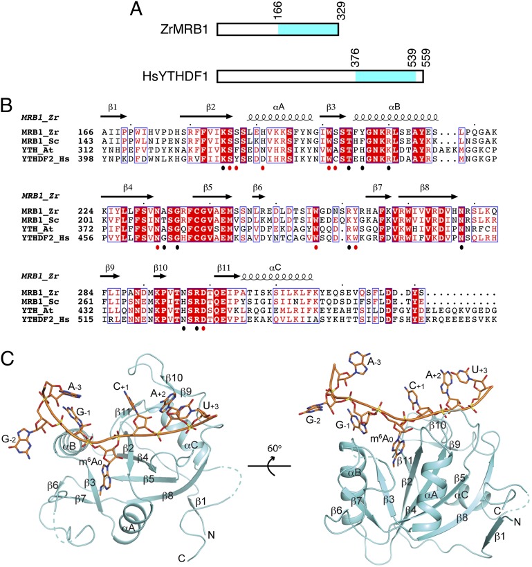
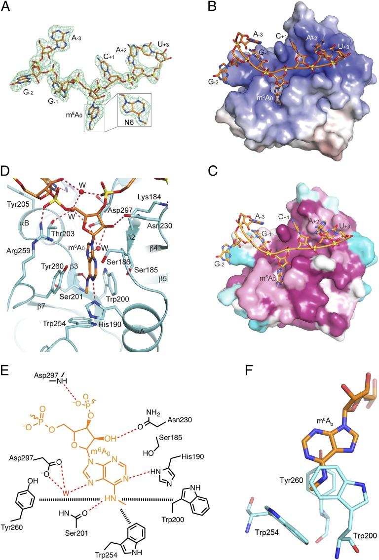
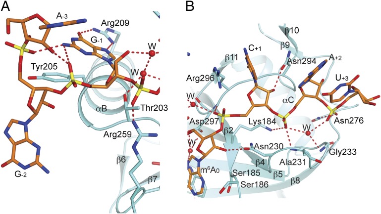
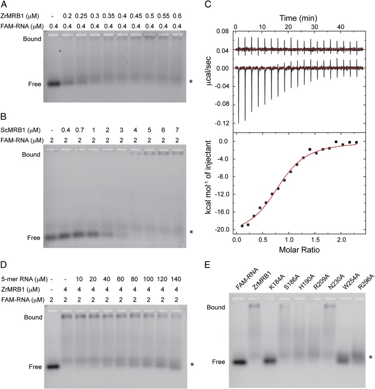
Methylation of the N6 position of selected internal adenines (m(6)A) in mRNAs and noncoding RNAs is widespread in eukaryotes, and the YTH domain in a collection of proteins recognizes this modification. We report the crystal structure of the splicing factor YT521-B homology (YTH) domain of Zygosaccharomyces rouxii MRB1 in complex with a heptaribonucleotide with an m(6)A residue in the center. The m(6)A modification is recognized by an aromatic cage, being sandwiched between a Trp and Tyr residue and with the methyl group pointed toward another Trp residue. Mutations of YTH domain residues in the RNA binding site can abolish the formation of the complex, confirming the structural observations. These residues are conserved in the human YTH proteins that also bind m(6)A RNA, suggesting a conserved mode of recognition. Overall, our structural and biochemical studies have defined the molecular basis for how the YTH domain functions as a reader of methylated adenines.


Conflict of interest statement

The authors declare no conflict of interest.

Figures

Fig. 1.
Fig. 1.
Crystal structure of the YTH domain of Z. rouxii MRB1 in complex with an m6A RNA. (A) Domain organization of ZrMRB1 and human YTHDF1. The YTH domain is shown in cyan. (B) Sequence alignment of the YTH domains of Z. rouxii (Zr) and S. cerevisiae (Sc) MRB1, A. thaliana (At) YTH protein, and human (Hs) YTHDF2. The secondary structure elements in the structure of the ZrMRB1 YTH domain are indicated. Residues that contact the m6A residue are indicated with the red dots below the alignment, and those that contact the rest of the RNA are indicated with the black dots. Produced with ESPript (25). (C) Two views of the structure of the YTH domain of ZrMRB1 (in cyan) in complex with a 7-mer m6A RNA (in orange). The secondary structure elements in the YTH domain are labeled. The structure figures were produced with PyMOL (www.pymol.org).
Fig. 2.
Fig. 2.
Recognition of the m6A RNA by the YTH domain. (A) Omit FoFc electron density for the RNA at 2.7 Å resolution, contoured at 2.5σ. (B) Electrostatic surface of the ZrMRB1 YTH domain in the region of m6A RNA binding. The RNA is located in a positively charged surface patch (blue) in the protein. (C) Sequence conservation of residues in the RNA binding site, generated based on an alignment of 50 sequences by the program ConSurf (26). Purple, conserved residues; cyan, variable residues. (D) Detailed interactions between the m6A nucleotide and the YTH domain. Water molecules are shown as red spheres. Hydrogen-bonding interactions are indicated by dashed lines in red. (E) Schematic drawing of the interactions between the m6A nucleotide and the YTH domain. The interactions between the m6A modification and the aromatic cage are indicated by the dashed lines in black. (F) Trp200 and Tyr260 flank the methylated N6 rather than the adenine base.
Fig. 3.
Fig. 3.
Interactions between the other nucleotides of the m6A RNA and the YTH domain. (A) Detailed interactions between the 5′-end of the RNA and the YTH domain. (B) Detailed interactions between the 3′-end of the RNA and the YTH domain.
Fig. 4.
Fig. 4.
Characterization of the interactions between the YTH domain and the m6A RNA. (A) A titration experiment assessing the affinity between the ZrMRB1 YTH domain and the 7-mer m6A RNA with a 5′ FAM fluorophore. The asterisk indicates a minor contaminating species in the RNA sample that does not interact with the YTH domain. (B) A titration experiment for the ScMRB1 YTH domain. (C) ITC data for the interaction between ZrMRB1 YTH domain and the labeled 7-mer RNA. Inset shows the heat release from the titration into buffer alone. (D) A competition binding assay testing the affinity of an unlabeled 5-mer m6A RNA for the ZrMRB1 YTH domain. (E) Effects of YTH domain mutations in the RNA interface on the formation of the complex. The FAM-labeled RNA is at 2 μM concentration, and the protein samples are at 4 μM concentration.

References

    1. Schwartz S, et al. High-resolution mapping reveals a conserved, widespread, dynamic mRNA methylation program in yeast meiosis. Cell. 2013;155(6):1409–1421. - PMC - PubMed
    1. Dominissini D, et al. Topology of the human and mouse m6A RNA methylomes revealed by m6A-seq. Nature. 2012;485(7397):201–206. - PubMed
    1. Meyer KD, et al. Comprehensive analysis of mRNA methylation reveals enrichment in 3′ UTRs and near stop codons. Cell. 2012;149(7):1635–1646. - PMC - PubMed
    1. Jia G, Fu Y, He C. Reversible RNA adenosine methylation in biological regulation. Trends Genet. 2013;29(2):108–115. - PMC - PubMed
    1. Niu Y, et al. N6-methyl-adenosine (m6A) in RNA: An old modification with a novel epigenetic function. Genomics Proteomics Bioinformatics. 2013;11(1):8–17. - PMC - PubMed

Publication types

MeSH terms

Associated data