. 2014 Sep 23;111(38):13876-81.
doi: 10.1073/pnas.1410602111. Epub 2014 Sep 8.

Altered mechanobiology of Schlemm's canal endothelial cells in glaucoma

Affiliations

Altered mechanobiology of Schlemm's canal endothelial cells in glaucoma

Darryl R Overby et al. Proc Natl Acad Sci U S A. .

Abstract

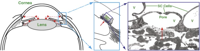
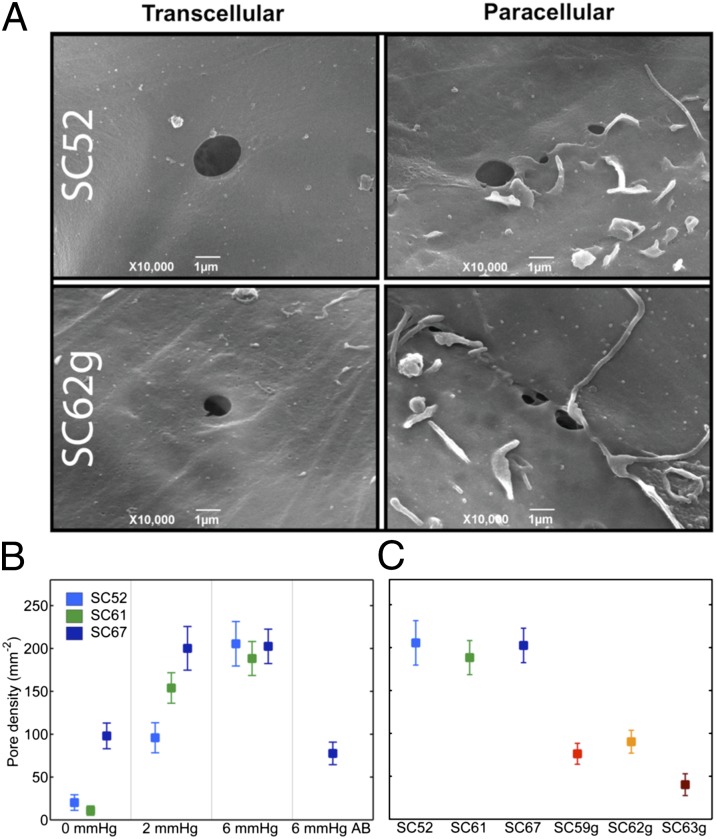
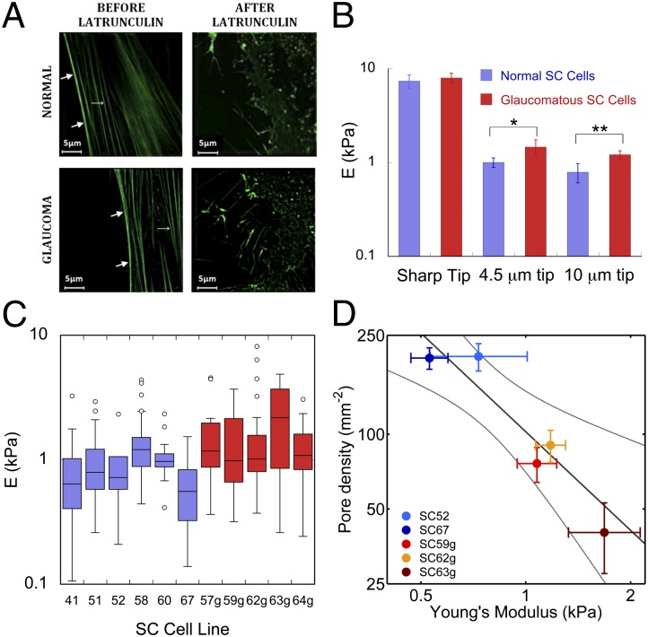
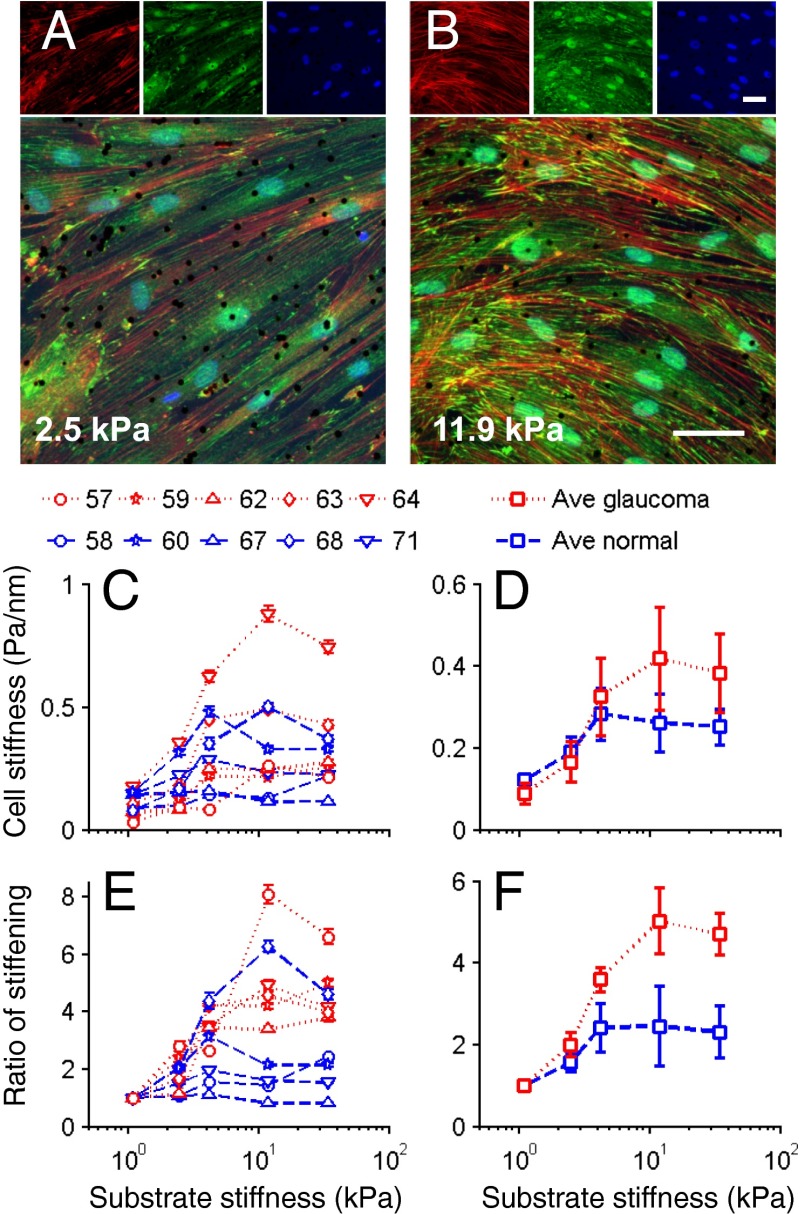
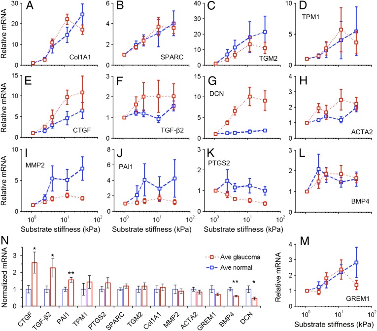
Increased flow resistance is responsible for the elevated intraocular pressure characteristic of glaucoma, but the cause of this resistance increase is not known. We tested the hypothesis that altered biomechanical behavior of Schlemm's canal (SC) cells contributes to this dysfunction. We used atomic force microscopy, optical magnetic twisting cytometry, and a unique cell perfusion apparatus to examine cultured endothelial cells isolated from the inner wall of SC of healthy and glaucomatous human eyes. Here we establish the existence of a reduced tendency for pore formation in the glaucomatous SC cell--likely accounting for increased outflow resistance--that positively correlates with elevated subcortical cell stiffness, along with an enhanced sensitivity to the mechanical microenvironment including altered expression of several key genes, particularly connective tissue growth factor. Rather than being seen as a simple mechanical barrier to filtration, the endothelium of SC is seen instead as a dynamic material whose response to mechanical strain leads to pore formation and thereby modulates the resistance to aqueous humor outflow. In the glaucomatous eye, this process becomes impaired. Together, these observations support the idea of SC cell stiffness--and its biomechanical effects on pore formation--as a therapeutic target in glaucoma.

Keywords: cell mechanics; cytoskeleton; modulus; primary open-angle glaucoma.


Conflict of interest statement

The authors declare no conflict of interest.

Figures

Fig. 1.
Fig. 1.
Aqueous humor flow pathway. (Left) Schematic of anterior segment of eye showing the direction of aqueous humor flow in red. (Center) Enlargement of the iris-cornea angle (boxed region in left panel) to show the conventional outflow pathway. (Right) Transmission electron micrograph of endothelial cells forming the inner wall of SC; aqueous humor crosses the endothelium through pores to enter the lumen of SC. V, giant vacuoles. C is reproduced with permission from ref. .
Fig. 2.
Fig. 2.
Pore density in perfused SC monolayers. (A) Representative image of transcellular and paracellular pores in normal (SC52) and glaucomatous SC (SC62g) cells. (B) Pore density increases in monolayers formed from three nonglaucomatous SC cell strains with transcellular (basal-to-apical) pressure drop; in one SC cell strain (SC67) perfused in the apical-to-basal direction (AB), pore densities are similar to unperfused controls at 0 mmHg. (C) Pore density is reduced in glaucomatous compared with normal SC cells following perfusion at 6 mmHg in the basal-to-apical direction. Bars are SEM.
Fig. 3.
Fig. 3.
Young’s modulus for normal and glaucomatous SC cells as measured by AFM. (A) Structured illumination microscopy images of normal and glaucomatous SC cells labeled with actin filament marker (42) rAV-LifeAct-TagGFP2 before and after application of latrunculin-A. Thick arrows, cortex; thin arrows, stress fibers. (B) Median and SEs of the modulus of six normal (blue) and five glaucomatous (red) nonconfluent SC cell strains as measured with three different AFM tips. Modulus is determined from force-deformation curves using a modified Hertzian analysis (10); *P = 0.117, **P = 0.017. (C) Box and whisker plot (43) of individual AFM measurements of cell modulus using a 10-µm tip for each of the six normal and five glaucomatous SC cell strains examined. (D) There is a significant correlation (dark line) between pore density and the modulus of the subcortical cytoskeleton, as measured by AFM using a 10-µm spherical tip. Bars represent SEM on pore density and modulus. Light curves in D represent 95% confident intervals on the slope of the GLM linear regression.
Fig. 4.
Fig. 4.
Influence of substrate stiffness on the biomechanical properties of SC cells. As the substrate stiffness increases, the stiffness of SC cells increases by different amounts in a donor- and disease-dependent manner. (A and B) Fluorescent micrographs of normal SC cells labeled for f-actin (red), vinculin (green), and DNA (blue) at two levels of substrate stiffness; black dots are 4.5-µm magnetic beads used for OMTC. (Scale bars: 50 µm.) (C and D) Cell stiffness index (g) of normal (blue) and glaucomatous (red) SC cells as measured by OMTC and expressed for individual cell strains (numbers above figure indicate cell strain) (C) or averages over all cell strains (D). (E and F) Stiffness index normalized by the value at the lowest substrate stiffness, expressed for individual cell strains (E) or averages over all cell strains (F). Median ± SEM with n > 600 beads for C and E; mean ± SEM with n = 5 cell strains each for D and F. Note that because the embedding depth of the beads in the cells is not known, an index of cell stiffness, g, is presented rather than an absolute value (44).
Fig. 5.
Fig. 5.
Increases in substrate stiffness modulated SC cell gene expression. (AM) The increases in substrate stiffness expression levels in normal or glaucomatous cell strains relative to that on the softest gel of that cell strain. Increased substrate stiffness led to increased expression in all genes except PTGS2 that showed constant or decreased expression. (N) The expression levels of 13 genes averaged across substrate stiffness and across donors were compared between normal and glaucomatous cell strains, normalized to the averaged expression level in the normal cells on the softest gel. Statistically significant differences between normal and glaucomatous cells indicated by *P < 0.05 and **P < 0.01. Mean ± SEM with n = 5 for AM; mean ± SEM with n = 25 for N.

Similar articles

Cited by

References

    1. Ethier CR. The inner wall of Schlemm’s canal. Exp Eye Res. 2002;74(2):161–172. - PubMed
    1. Johnson M. ‘What controls aqueous humour outflow resistance?’. Exp Eye Res. 2006;82(4):545–557. - PMC - PubMed
    1. Allingham RR, et al. The relationship between pore density and outflow facility in human eyes. Invest Ophthalmol Vis Sci. 1992;33(5):1661–1669. - PubMed
    1. Johnson M, et al. The pore density in the inner wall endothelium of Schlemm’s canal of glaucomatous eyes. Invest Ophthalmol Vis Sci. 2002;43(9):2950–2955. - PubMed
    1. Pedrigi RM, Simon D, Reed A, Stamer WD, Overby DR. A model of giant vacuole dynamics in human Schlemm’s canal endothelial cells. Exp Eye Res. 2011;92(1):57–66. - PMC - PubMed

Publication types

LinkOut - more resources