Macrophage arginase-1 controls bacterial growth and pathology in hypoxic tuberculosis granulomas
- PMID: 25201986
- PMCID: PMC4183271
- DOI: 10.1073/pnas.1408839111
Macrophage arginase-1 controls bacterial growth and pathology in hypoxic tuberculosis granulomas
Abstract
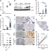
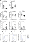
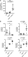
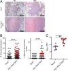
Lung granulomas develop upon Mycobacterium tuberculosis (Mtb) infection as a hallmark of human tuberculosis (TB). They are structured aggregates consisting mainly of Mtb-infected and -uninfected macrophages and Mtb-specific T cells. The production of NO by granuloma macrophages expressing nitric oxide synthase-2 (NOS2) via l-arginine and oxygen is a key protective mechanism against mycobacteria. Despite this protection, TB granulomas are often hypoxic, and bacterial killing via NOS2 in these conditions is likely suboptimal. Arginase-1 (Arg1) also metabolizes l-arginine but does not require oxygen as a substrate and has been shown to regulate NOS2 via substrate competition. However, in other infectious diseases in which granulomas occur, such as leishmaniasis and schistosomiasis, Arg1 plays additional roles such as T-cell regulation and tissue repair that are independent of NOS2 suppression. To address whether Arg1 could perform similar functions in hypoxic regions of TB granulomas, we used a TB murine granuloma model in which NOS2 is absent. Abrogation of Arg1 expression in macrophages in this setting resulted in exacerbated lung granuloma pathology and bacterial burden. Arg1 expression in hypoxic granuloma regions correlated with decreased T-cell proliferation, suggesting that Arg1 regulation of T-cell immunity is involved in disease control. Our data argue that Arg1 plays a central role in the control of TB when NOS2 is rendered ineffective by hypoxia.
Conflict of interest statement
The authors declare no conflict of interest.
Figures


Similar articles
-
Arginase-1 expression in granulomas of tuberculosis patients.FEMS Immunol Med Microbiol. 2012 Nov;66(2):265-8. doi: 10.1111/j.1574-695X.2012.01012.x. Epub 2012 Aug 21. FEMS Immunol Med Microbiol. 2012. PMID: 22827286
-
Microenvironments in tuberculous granulomas are delineated by distinct populations of macrophage subsets and expression of nitric oxide synthase and arginase isoforms.J Immunol. 2013 Jul 15;191(2):773-84. doi: 10.4049/jimmunol.1300113. Epub 2013 Jun 7. J Immunol. 2013. PMID: 23749634 Free PMC article.
-
Helminth-induced arginase-1 exacerbates lung inflammation and disease severity in tuberculosis.J Clin Invest. 2015 Dec;125(12):4699-713. doi: 10.1172/JCI77378. Epub 2015 Nov 16. J Clin Invest. 2015. PMID: 26571397 Free PMC article.
-
Immunometabolism within the tuberculosis granuloma: amino acids, hypoxia, and cellular respiration.Semin Immunopathol. 2016 Mar;38(2):139-52. doi: 10.1007/s00281-015-0534-0. Epub 2015 Oct 21. Semin Immunopathol. 2016. PMID: 26490974 Free PMC article. Review.
-
Perspectives on host adaptation in response to Mycobacterium tuberculosis: modulation of inflammation.Semin Immunol. 2014 Dec;26(6):533-42. doi: 10.1016/j.smim.2014.10.002. Epub 2014 Oct 25. Semin Immunol. 2014. PMID: 25453228 Review.
Cited by
-
Interleukin-10 Family and Tuberculosis: An Old Story Renewed.Int J Biol Sci. 2016 Apr 27;12(6):710-7. doi: 10.7150/ijbs.13881. eCollection 2016. Int J Biol Sci. 2016. PMID: 27194948 Free PMC article. Review.
-
Biphasic Dynamics of Macrophage Immunometabolism during Mycobacterium tuberculosis Infection.mBio. 2019 Mar 26;10(2):e02550-18. doi: 10.1128/mBio.02550-18. mBio. 2019. PMID: 30914513 Free PMC article. Review.
-
Immunometabolism: new insights and lessons from antigen-directed cellular immune responses.Semin Immunopathol. 2020 Jun;42(3):279-313. doi: 10.1007/s00281-020-00798-w. Epub 2020 Jun 9. Semin Immunopathol. 2020. PMID: 32519148 Free PMC article. Review.
-
Metabolic Regulation of Immune Responses to Mycobacterium tuberculosis: A Spotlight on L-Arginine and L-Tryptophan Metabolism.Front Immunol. 2021 Feb 9;11:628432. doi: 10.3389/fimmu.2020.628432. eCollection 2020. Front Immunol. 2021. PMID: 33633745 Free PMC article. Review.
-
Macrophage defense mechanisms against intracellular bacteria.Immunol Rev. 2015 Mar;264(1):182-203. doi: 10.1111/imr.12266. Immunol Rev. 2015. PMID: 25703560 Free PMC article. Review.
References
-
- Ramakrishnan L. Revisiting the role of the granuloma in tuberculosis. Nat Rev Immunol. 2012;12(5):352–366. - PubMed
-
- Reece ST, Kaufmann SH. Floating between the poles of pathology and protection: Can we pin down the granuloma in tuberculosis? Curr Opin Microbiol. 2012;15(1):63–70. - PubMed
-
- Spector WG. The granulomatous inflammatory exudate. Int Rev Exp Pathol. 1969;8:1–55. - PubMed
-
- Paige C, Bishai WR. Penitentiary or penthouse condo: The tuberculous granuloma from the microbe’s point of view. Cell Microbiol. 2010;12(3):301–309. - PubMed
Publication types
MeSH terms
Substances
Grants and funding
LinkOut - more resources
Full Text Sources
Other Literature Sources
Molecular Biology Databases
Research Materials
Miscellaneous

