. 2014 Oct 24;289(43):29506-18.
doi: 10.1074/jbc.M114.577684. Epub 2014 Sep 8.

The LQLP calcineurin docking site is a major determinant of the calcium-dependent activation of human TRESK background K+ channel

Affiliations

The LQLP calcineurin docking site is a major determinant of the calcium-dependent activation of human TRESK background K+ channel

Gábor Czirják et al. J Biol Chem. .

Abstract

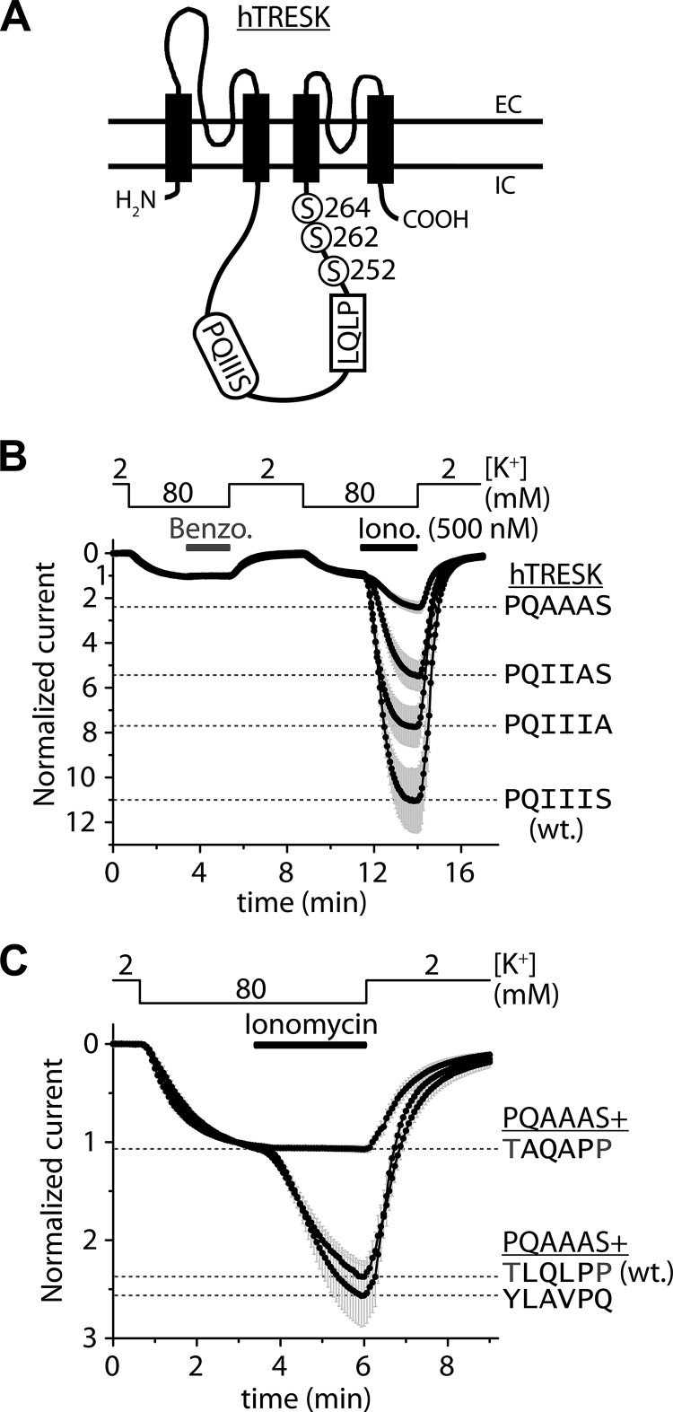
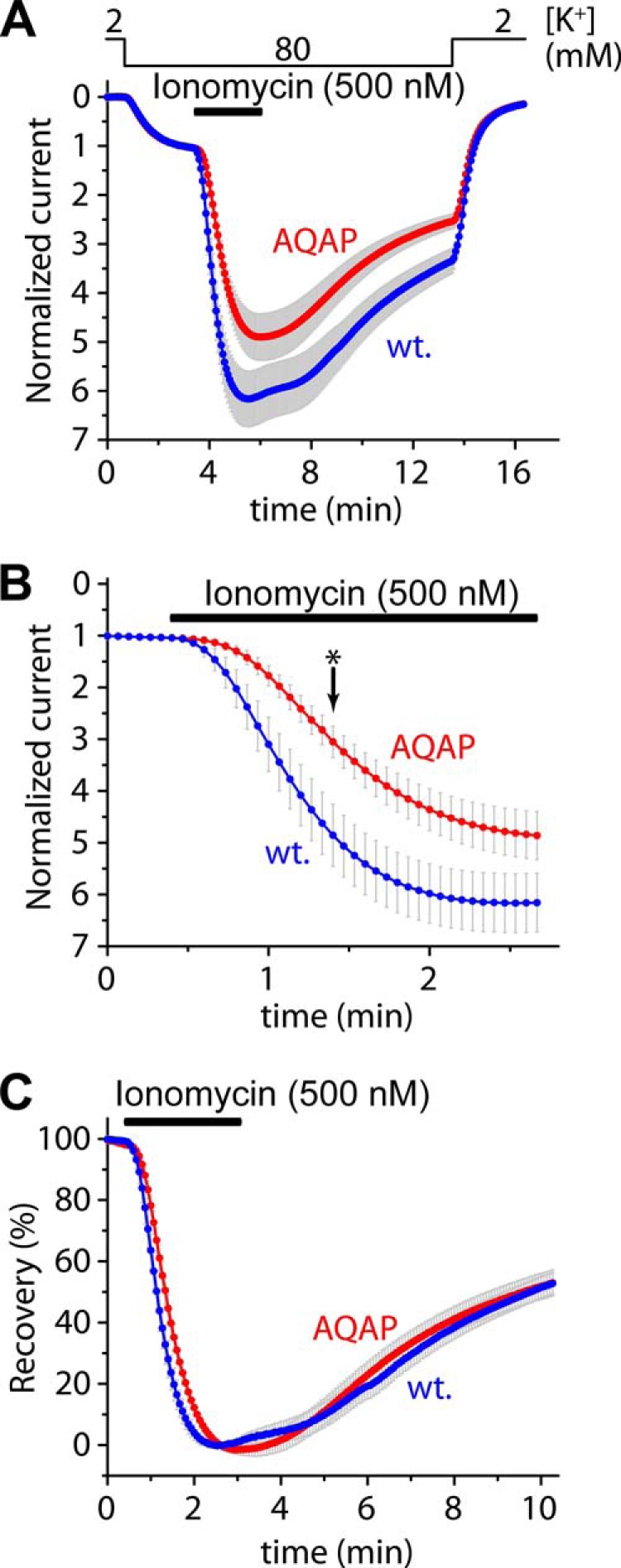
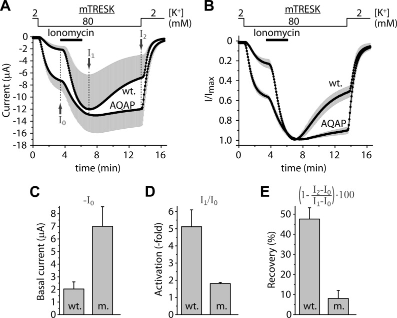
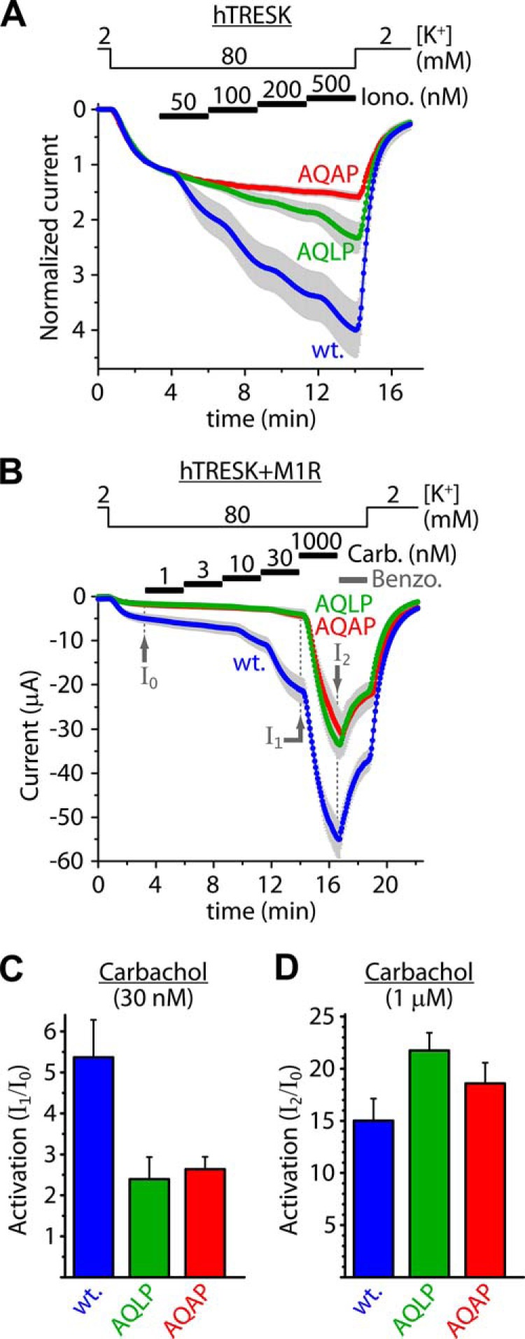
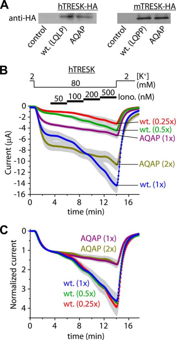
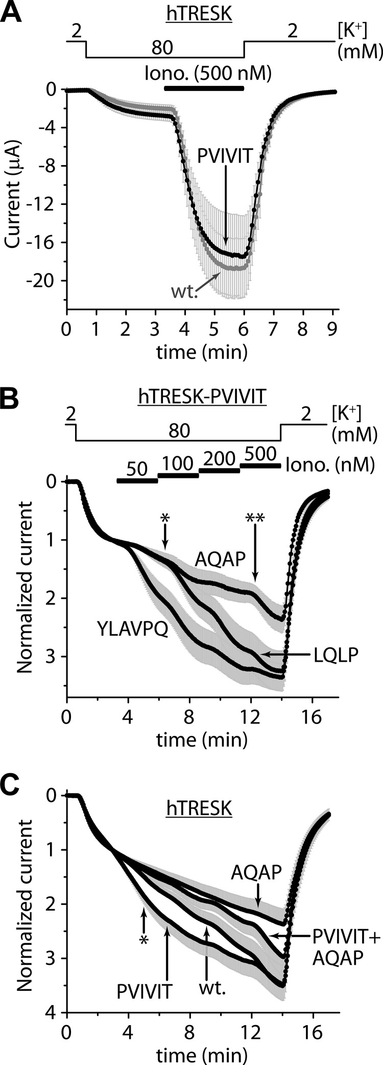
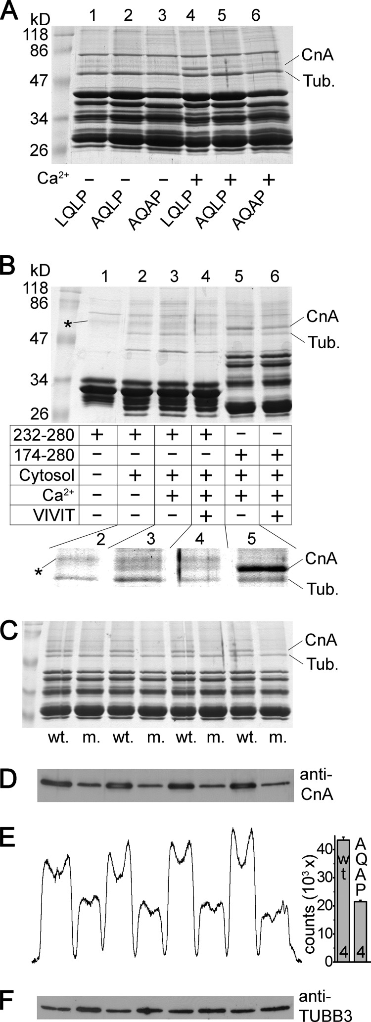
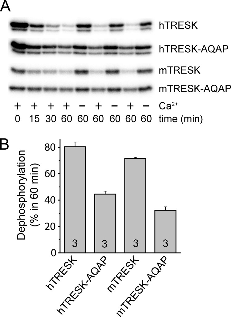
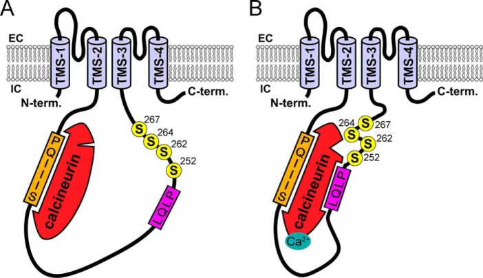
Calcium-dependent activation of human TRESK (TWIK-related spinal cord K(+) channel, K2P18.1) depends on direct targeting of calcineurin to the PQIIIS motif. In the present study we demonstrate that TRESK also contains another functionally relevant docking site for the phosphatase, the LQLP amino acid sequence. Combined mutations of the PQIIIS and LQLP motifs were required to eliminate the calcium-dependent regulation of the channel. In contrast to the alanine substitutions of PQIIIS, the mutation of LQLP to AQAP alone did not significantly change the amplitude of TRESK activation evoked by the substantial elevation of cytoplasmic calcium concentration. However, the AQAP mutation slowed down the response to high calcium. In addition, modest elevation of [Ca(2+)], which effectively regulated the wild type channel, failed to activate TRESK-AQAP. This indicates that the AQAP mutation diminished the sensitivity of TRESK to calcium. Even if PQIIIS was replaced by the PVIVIT sequence of high calcineurin binding affinity, the effect of the AQAP mutation was clearly detected in this TRESK-PVIVIT context. Substitution of the LQLP region with the corresponding fragment of NFAT transcription factor, perfectly matching the previously described LXVP calcineurin-binding consensus sequence, increased the calcium-sensitivity of TRESK-PVIVIT. Thus the enhancement of the affinity of TRESK for calcineurin by the incorporation of PVIVIT could not compensate for or prevent the effects of LQLP sequence modifications, suggesting that the two calcineurin-binding regions play distinct roles in the regulation. Our results indicate that the LQLP site is a fundamental determinant of the calcium-sensitivity of human TRESK.

Keywords: Calcineurin; Calcium; Ion Channel; K2P; KCNK18; PXIXIT; Potassium Channel; Protein Phosphatase 2B (PP2B); Two-pore Domain.


Figures

FIGURE 1.
FIGURE 1.
Combined mutations of the PQIIIS and LQLP sites are required for the complete elimination of the calcium-dependent activation of human TRESK. A, transmembrane topology of human TRESK subunit and the PQIIIS and LQLP calcineurin docking motifs are illustrated. The regulatory serine residues of the channel are shown in the vicinity of the LQLP site. (The drawing is not to scale.) B, Xenopus oocytes expressing wild type (PQIIIS, wt.) or mutant (PQIIIA, PQIIAS and PQAAAS) TRESK channels were stimulated with the calcium ionophore ionomycin (0.5 μm, Iono.) after verifying the insensitivity of the basal K+ currents to benzocaine (1 mm, Benzo.). The extracellular [K+] was changed from 2 to 80 mm and back as indicated above the graph. The currents were measured at −100 mV by two-electrode voltage clamp, and normalized to their resting value in 80 mm [K+]. Note that the mutations of the PQIIIS site gradually deteriorated but did not completely eliminate the activation; the PQAAAS mutant was activated more than 2-fold by ionomycin. The gray error bars represent S.E. C, normalized activation in response to ionomycin (0.5 μm) is shown for three different TRESK constructs, all containing the PQAAAS mutation. The LQLP motif was not modified in TRESK-PQAAAS (see TLQLPP, wt. curve); it was disintegrated by AQAP mutation in TRESK-PQAAAS-TAQAPP (TAQAPP curve); or it was substituted with the corresponding sequence of NFATc1 (NFAT2) in TRESK-PQAAAS-YLAVPQ (YLAVPQ curve). Maximum activation levels at the end of the stimulation with ionomycin are indicated by dashed lines for the different mutants, as labeled on the right side of the panel. Note that the combined mutations of the PQIIIS and LQLP sites eliminated the calcium-dependent regulation (TAQAPP curve), but YLAVPQ functionally substituted for the TLQLPP sequence of TRESK (YLAVPQ curve).
FIGURE 2.
FIGURE 2.
The AQAP mutation slows down the activation of human TRESK evoked by high concentration of ionomycin, but the inhibitory kinase reaction is not affected by this mutation. A, cells expressing wild type or AQAP mutant TRESK channels were stimulated with high (0.5 μm) concentration of ionomycin (as indicated by the horizontal black bar). Recovery from the activation was evaluated by a long washout period in 80 mm extracellular [K+], as indicated above the graph. The activations of the wild type and AQAP mutant channels were not significantly different at the end of the stimulation with ionomycin. B, activation kinetics of the currents (same as in panel A) during the stimulation with ionomycin are plotted on a shorter time scale. The AQAP mutation slowed down the activation process; the partial activation of the AQAP mutant is significantly smaller than that of the wild type channel at the time point indicated with an asterisk. C, percent recovery values were calculated from the recordings represented in panel A. The recovery from activation was identical in the cases of the wild type and AQAP mutant TRESK channels.
FIGURE 3.
FIGURE 3.
In mouse TRESK, the AQAP mutation also interferes with the calcium-dependent activation. A, average currents of two groups of oocytes expressing wild type or AQAP mutant mouse TRESK are plotted. The cells were stimulated with ionomycin (Iono., 0.5 μm, as indicated by the horizontal black bar) in 80 mm extracellular [K+] (as shown above the graph). Basal K+ currents were estimated as I0, maximum activations in response to ionomycin as I1, and the recovery at the end of the measurement as I2. The small nonspecific leak currents measured in 2 mm [K+] were subtracted from the values of I0, I1 and I2 in further calculations but not in this graph. (Only plus or minus error bars (gray, S.E.) are shown.) B, same currents as in panel A were normalized to their peak values. The major part of the variation of data in panel A came from the different channel expression of the oocytes, and the response to ionomycin was rather uniform within the groups. C, average basal K+ currents of the wild type (wt.) and AQAP mutant (m.) channels (I0 in panel A) are illustrated in this column diagram. Basal currents of the AQAP mutant were significantly larger than those of the wild type channel. D, normalized activations (I1/I0) of the wild type, and AQAP mutant channels were calculated from the same data as represented in panel A. The apparent activation of the wild type channel exceeded that of the AQAP mutant. E, percent recovery from activation of the K+ currents at the end of the measurement (I2 in panel A) was calculated as indicated by the expression above the columns. The AQAP mutation diminished the return of the K+ current to the resting state after the stimulation with ionomycin.
FIGURE 4.
FIGURE 4.
Alanine substitutions in the LQLP site reduce the sensitivity of human TRESK to calcium. A, cells expressing wild type (wt., LQLP) or mutant (AQLP and AQAP) TRESK channels were stimulated with stepwise increasing (50, 100, 200, and 500 nm) concentrations of ionomycin as indicated by the horizontal black bars. Note that the AQAP mutant was unresponsive to the modest elevation of cytoplasmic [Ca2+], in sharp contrast to the wild type channel. B, wild type (wt.), AQLP and AQAP mutant TRESK channels were coexpressed with M1 muscarinic receptor. Modest calcium signals were evoked by the application of progressively increasing (1, 3, 10, and 30 nm) concentrations of carbachol as indicated by the horizontal black bars. Subsequently, robust calcium signal was induced by 1 μm concentration of the agonist. Finally the sensitivity to benzocaine (1 mm) was tested as indicated by the horizontal gray bar. Note that the AQLP and AQAP mutants have not been activated by the low concentrations of carbachol, but they were responsive to high calcium. (The green AQLP and red AQAP curves overlap.) C, cumulative activations of the wild type (wt.), AQLP and AQAP mutant channels by low (1–30 nm) concentrations of carbachol were calculated as I1/I0. The wild type channel was more strongly activated than the AQLP and AQAP mutants. D, activations of the wild type, AQLP and AQAP mutant channels in response to the robust calcium signal (1 μm carbachol) were estimated as I2/I0. By high calcium, the AQLP and AQAP mutants were activated similarly to (or more than) the wild type channel.
FIGURE 5.
FIGURE 5.
The effect of the AQAP mutation is independent of the expression levels. A, expression levels of TRESK channels containing the wild type (LQLP, wt.) sequence or the AQAP mutation in their intracellular loops, and the influenza hemagglutinin epitope (HA) at their C termini, were compared by anti-HA immunoblots. Expression levels of the LQLP (wt.) and AQAP constructs were similar both in the case of the human (hTRESK) and the mouse (mTRESK) channels. Control membrane preparations were prepared from an identical number of non-injected oocytes as used for the groups expressing hTRESK-HA and hTRESK-HA-AQAP (n = 17, 17) or mTRESK-HA and mTRESK-HA-AQAP (n = 40, 40). Identical amounts of injected cRNAs of the respective constructs were verified on denaturing agarose gels and identical protein content of the membrane preparations were checked by SDS-PAGE (not shown). B, expression levels of hTRESK (wt.) and hTRESK-AQAP (AQAP) were controlled by adjusting the amounts of cRNAs injected into the oocytes (as indicated on the right side). The five groups of cells (n = 22 for wt.(1x) and n = 11 or 12 for the other four groups), characterized by basal current amplitudes varying in line with the injected cRNA quantity, were stimulated by stepwise increasing concentrations of ionomycin (as in Fig. 4A). C, normalized currents from the same five groups of oocytes as in panel B. Note that the calcium sensitivity of the wild type and AQAP channels is independent of the expression levels. The wild type (blue, green, and red) and the AQAP (purple and olive) curves, respectively, overlap.
FIGURE 6.
FIGURE 6.
The loss- and gain-of-function mutations of the LQLP site are effective in the TRESK-PVIVIT context; the PVIVIT mutation is more effective in the wild type (LQLP) than in the AQAP background. A, average currents of oocytes expressing wild type TRESK (wt., gray curve) or TRESK-PVIVIT (PVIVIT, black curve) are compared. The PQIIIS site was replaced with the PVIVIT sequence of high calcineurin-affinity in the TRESK-PVIVIT mutant. The activation of the currents was evoked by ionomycin (0.5 μm, as indicated by the horizontal black bar). B, normalized responses of three different mutants of TRESK to stepwise increasing (50, 100, 200, and 500 nm) concentrations of ionomycin are illustrated. The PQIIIS motif was replaced with PVIVIT in all three mutants. The LQLP motif was not modified in TRESK-PVIVIT (see the LQLP curve); it was disabled by AQAP mutation in TRESK-PVIVIT-AQAP (AQAP curve); or the TLQLPP region was replaced by the corresponding fragment of NFAT in TRESK-PVIVIT-YLAVPQ (YLAVPQ curve). The partial activation of TRESK-PVIVIT-YLAVPQ (YLAVPQ) was significantly higher than that of TRESK-PVIVIT (LQLP) at the time point indicated with an asterisk, whereas the partial activation of TRESK-PVIVIT-AQAP (AQAP) was significantly reduced, compared with TRESK-PVIVIT (LQLP) at the time point indicated with a double asterisk. C, normalized responses of TRESK (wt.), TRESK-PVIVIT (PVIVIT), TRESK-AQAP (AQAP), and TRESK-PVIVIT-AQAP (PVIVIT+AQAP) to the same stimulation as in panel B are plotted. The partial activation of TRESK-PVIVIT (PVIVIT) was significantly higher than that of the wild type (wt.) channel at the time point indicated with an asterisk. In contrast, the sensitivity of TRESK-AQAP to low calcium was not enhanced by the PVIVIT mutation (compare PVIVIT+AQAP to AQAP curve). (Only plus or minus error bars are shown. The currents in panel C were measured from another oocyte preparation than those in panel B.)
FIGURE 7.
FIGURE 7.
The LQLP motif of TRESK is a calcineurin binding site. A, GST pulldown assays were performed from mouse brain cytosol with the cytoplasmic loop of human TRESK (wild type fragment 174–280, LQLP, lanes 1 and 4), and the AQLP (AQLP, lanes 2 and 5) and AQAP (AQAP, lanes 3 and 6) mutant versions of this bait protein. The binding of calcineurin was tested in the presence (1 mm, lanes 4–6) or in the absence of calcium (2 mm EGTA, lanes 1–3). The two known interacting proteins were marked on the right side of the Coomassie Blue-stained gel as tubulin (Tub.) and calcineurin A subunit (CnA). Note the abundant binding of calcineurin to the wild type bait protein in the presence of calcium (lane 4) and the drastic reduction of the interaction by the AQLP and AQAP mutations (lanes 5 and 6) or by the chelation of Ca2+ (lane 1). (The bait protein preparations contained several incompletely translated fragments in addition to the full-length product below 47 kDa, and a prominent contaminating bacterial protein band below 86 kDa.) B, GST pulldown reactions were performed with fragment 232–280 retaining only the LQLP motif (lanes 1–4), or with fragment 174–280 containing both intact (PQIIIS and LQLP) binding sites (lanes 5 and 6). The addition of mouse brain cytosol to the reaction (lane 1 is an only bait control), the presence of calcium (1 mm) or EGTA (2 mm) and the administration of VIVIT peptide (75 μm) were controlled as indicated in the table. The interacting partners, tubulin (Tub.) and calcineurin A (CnA), are labeled on the right side, and a faint contaminating band from the purification of fragment 232–280 is indicated with asterisks. The relevant region of the Coomassie Blue-stained gel was magnified and the contrast was adjusted for better visibility of the bands in the inset below the table. Note that the binding of calcineurin to fragment 232–280 was detected in the presence (lane 3), but not in the absence of calcium (lane 2). VIVIT peptide attenuated the interaction of calcineurin with fragment 174–280 (lane 5 versus 6), but did not enhance the binding of calcineurin to fragment 232–280 (lane 3 versus 4). C, four pairs of independent pulldown reactions were performed with GST-hTRESK(174–280) (wt.) and GST-hTRESK(174–280)-AQAP mutant (m.) as in lanes 4 and 6 of panel A. The samples were analyzed on SDS-PAGE gel followed by Coomassie Blue staining. D, immunoblot of the same samples as in panel C with anti-calcineurin A (anti-CnA) antibody. E, densitometry curve of the immunoblot in panel D, and column diagram of the densitometry counts. Significantly more calcineurin interacted with the wild type (LQLP) construct than with the AQAP mutant (p < 10−5). F, same membrane as in panel D was stripped and re-probed with anti-tubulin β3 (anti-TUBB3) antibody. The loaded amounts of the AQAP pulldown reactions were not less than those of the wild type.
FIGURE 8.
FIGURE 8.
The in vitro calcium-dependent dephosphorylation of the wild type TRESK sequence is more rapid than that of the AQAP mutant. A, different substrate proteins, GST-hTRESK(174–280) (hTRESK), GST-hTRESK(174–280)-AQAP (hTRESK-AQAP), GST-mTRESK(164–292) (mTRESK), GST-mTRESK(164–292)-AQAP (mTRESK-AQAP) immobilized on glutathione resin, were in vitro phosphorylated in the presence of [32P-γ]ATP by constitutively active recombinant MARK2 kinase (Trx-His6-MARK2-T208E). Subsequently, they were dephosphorylated with diluted mouse brain cytosol in the presence or absence of calcium for different time periods (as indicated below the autoradiograms of SDS-PAGE gels). Note that the wild type TRESK sequences were more rapidly dephosphorylated than the AQAP mutants (see the 0, 15, 30, 60 min reactions in the left 4 columns of bands). For statistical analysis, three independent pairs of dephosphorylation reactions were performed in the presence or absence of calcium for 60 min with each substrate proteins (see the right 6 columns of bands). B, calcium-dependent dephosphorylation (illustrated in the column graph in percent) was calculated from the radioactive counts of these bands in panel A. The wild type versions of the substrate proteins were dephosphorylated more efficiently than the AQAP mutants (p < 0.002 for both hTRESK and mTRESK).
FIGURE 9.
FIGURE 9.
Interplay of the PQIIIS and LQLP calcineurin-docking motifs in the regulation of human TRESK. A, calcineurin binds to the PQIIIS motif under resting conditions, depending on the concentration of the phosphatase in the cell, but independently of the cytoplasmic [Ca2+]. B, LQLP-binding site of calcineurin becomes available in response to the calcium signal. The LQLP motif binds to calcineurin and brings the adjacent substrate residues in the proximity of the active site of the enzyme.

Similar articles

Cited by

References

    1. Enyedi P., Czirják G. (2010) Molecular background of leak K+ currents: two-pore domain potassium channels. Physiol. Rev. 90, 559–605 - PubMed
    1. Noël J., Sandoz G., Lesage F. (2011) Molecular regulations governing TREK and TRAAK channel functions. Channels 5, 402–409 - PMC - PubMed
    1. Mathie A., Al-Moubarak E., Veale E. L. (2010) Gating of two pore domain potassium channels. J. Physiol. 588, 3149–3156 - PMC - PubMed
    1. Plant L. D. (2012) A Role for K2P Channels in the Operation of Somatosensory Nociceptors. Front. Mol. Neurosci. 5, 21. - PMC - PubMed
    1. Lotshaw D. P. (2007) Biophysical, pharmacological, and functional characteristics of cloned and native mammalian two-pore domain K+ channels. Cell Biochem. Biophys. 47, 209–256 - PubMed

Publication types

MeSH terms

LinkOut - more resources