. 2014 Oct 24;289(43):29827-35.
doi: 10.1074/jbc.M114.597161. Epub 2014 Sep 8.

Osmotic stress-induced phosphorylation of H2AX by polo-like kinase 3 affects cell cycle progression in human corneal epithelial cells

Affiliations

Osmotic stress-induced phosphorylation of H2AX by polo-like kinase 3 affects cell cycle progression in human corneal epithelial cells

Ling Wang et al. J Biol Chem. .

Abstract

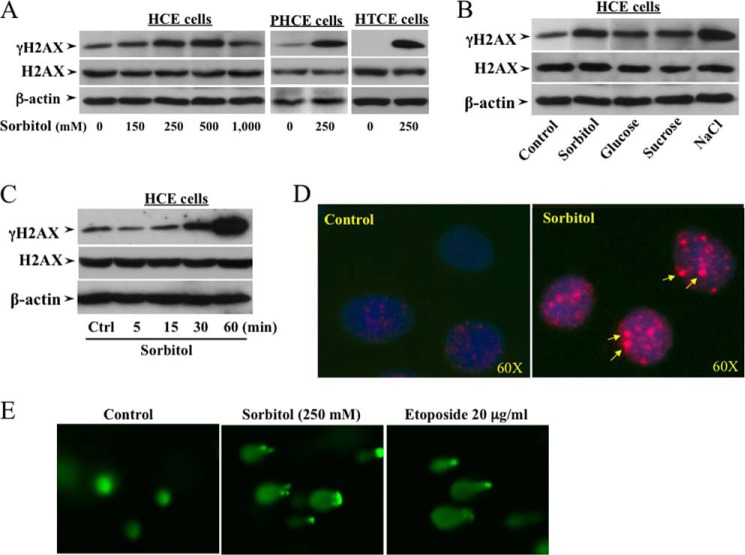
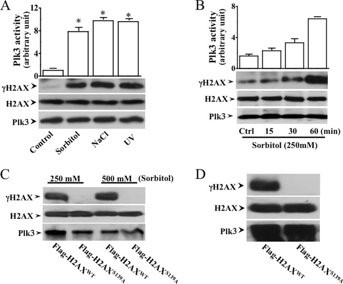
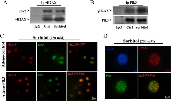
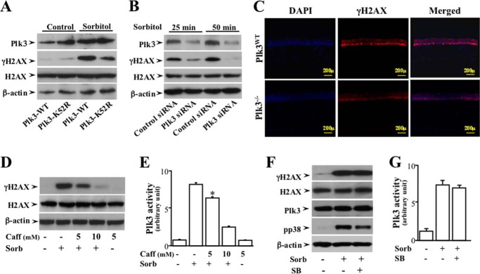
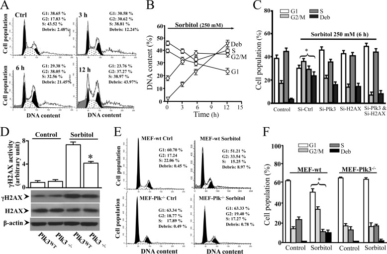
Increased concentrations of extracellular solutes affect cell function and fate by stimulating cellular responses, such as evoking MAPK cascades, altering cell cycle progression, and causing apoptosis. Our study results here demonstrate that hyperosmotic stress induced H2AX phosphorylation (γH2AX) by an unrevealed kinase cascade involving polo-like kinase 3 (Plk3) in human corneal epithelial (HCE) cells. We found that hyperosmotic stress induced DNA-double strand breaks and increased γH2AX in HCE cells. Phosphorylation of H2AX at serine 139 was catalyzed by hyperosmotic stress-induced activation of Plk3. Plk3 directly interacted with H2AX and was colocalized with γH2AX in the nuclei of hyperosmotic stress-induced cells. Suppression of Plk3 activity by overexpression of a kinase-silencing mutant or by knocking down Plk3 mRNA effectively reduced γH2AX in hyperosmotic stress-induced cells. This was consistent with results that show γH2AX was markedly suppressed in the Plk3(-/-) knock-out mouse corneal epithelial layer in response to hyperosmotic stimulation. The effect of hyperosmotic stress-activated Plk3 and increased γH2AX in cell cycle progression showed an accumulation of G2/M phase, altered population in G1 and S phases, and increased apoptosis. Our results for the first time reveal that hyperosmotic stress-activated Plk3 elicited γH2AX. This Plk3-mediated activation of γH2AX subsequently regulates the cell cycle progression and cell fate.

Keywords: Cell Cycle; Cornea; Epithelial Cell; H2A Histone Family, Member X (H2AFX); Phosphorylation; Signaling.


Figures

FIGURE 1.
FIGURE 1.
Effects of altered osmotic stresses on DNA DSBs and H2AX phosphorylation. A, increases in γH2AX following increased sorbitol concentrations in various human corneal epithelial cells. B, effects of high concentrations of extracellular solutes: sorbitol (300 mm), glucose (300 mm), sucrose (300 mm), and NaCl (300 mm) on increases in γH2AX in HCE cells. C, high concentration sorbitol-induced γH2AX following a time course in HCE cells. D, localization of hyperosmotic stress-induced γH2AX in nuclei of HCE cells. E, hyperosmotic stress-induced DNA DSBs in HCE cells. DNA DSBs were detected with Comet assays in sorbitol (250 mm)-treated cells, and the anti-tumor drug etoposide (20 μg/ml)-treated cells served as positive controls.
FIGURE 2.
FIGURE 2.
Hyperosmotic stress-activated Plk3 phosphorylated H2AX. A, phosphorylation of H2AX by activated Plk3 from HCE cells stimulated with hyperosmotic stress and UV irradiation in HCE cells. B, phosphorylation of H2AX fusion protein by hyperosmotic stress-activated Plk3 following a time course in HCE cells. C, hyperosmotic stress-activated Plk3 failed to phosphorylate the H2AX mutant (H2AXS139A) in which Ser-139 was substituted by an alanine residue. The Plk3 level was detected by Western blot and served as the loading control in HCE cells. D, effect of constitutively activated Plk3 upon phosphorylation of wild type H2AX and H2AXS139A mutant fusion proteins in HCE cells. The asterisk represents a significant difference between untreated control and hyperosmotic stress-induced HCE cells (p < 0.05, n = 3).
FIGURE 3.
FIGURE 3.
Determining interaction between Plk3 and H2AX in hyperosmotic stress-induced HCE cells. A, detection of Plk3 and H2AX interaction by immunoprecipitation using an anti-phospho-H2AX (anti-γH2AX) antibody. Plk3 was detected by Western analysis. B, detection of Plk3 and H2AX interaction by immunoprecipitation using an anti-Plk3 antibody. γH2AX was detected by Western analysis using an anti-γH2AX antibody. C, immunocolocalization of Plk3 and γH2AX in adeno-GFP-Plk3-transfected and hyperosmotic stress-induced cells. D, high amplification view (60×) to show colocalization of Plk3 and γH2AX in the nucleus of hyperosmotic stress-induced HCE cells. Immunocolocalization experiments were done by immunostaining of hyperosmotic stress-induced human corneal epithelial cells using antibodies specific to γH2AX. Photo images were taken using a Nikon fluorescent microscope at 20× and 60×, respectively. Data were analyzed with a Nikon software program.
FIGURE 4.
FIGURE 4.
Effects of altered Plk3 activity on hyperosmotic stress-induced γH2AX in HCE cells. A, effect of overexpressing wild type Plk3 and kinase-silencing Plk3k52R mutant on hyperosmotic stress-induced γH2AX in HCE cells. B, effect of knocking down Plk3 mRNAs on hyperosmotic stress-induced Plk3 γH2AX in HCE cells. C, suppression of hyperosmotic stress-induced γH2AX in the epithelial layer of the Plk3-deficient (Plk3−/−) mouse corneas. Immunostaining experiments were performed to detect hyperosmotic stress-induced activation of γH2AX using a γH2AX specific antibody, and cell nuclei were stained by DAPI in corneas of wild type and Plk3−/− knock-out mice. Photo images were taken using a Nikon fluorescent microscope at 10×. D, effect of inhibiting ATM/ATR with Caff (caffeine) on hyperosmotic stress-induced H2AX phosphorylation. E, effect of inhibiting ATM/ATR with Caff on hyperosmotic stress-induced Plk3 activity. F, effects of inhibiting p38 in the absence or presence of 500 μm SB (SB202190, an inhibitor of p38) on hyperosmotic stress-induced ATF-2 phosphorylation. G, effect of hyperosmotic stress on Plk3 activity. Plk3 activity was determined by immunocomplex kinase assay, and the H2AX fusion protein was used as the substrate. The asterisk symbol indicates a significant difference (p < 0.05, n = 3).
FIGURE 5.
FIGURE 5.
Effect of altered Plk3 activity on cell cycle distribution in hyperosmotic stress-induced HCE cells and MEFs. A, effect of hyperosmotic stimulation on cell cycle progression in HCE cells. B, time course of hyperosmotic stress-induced alteration in cell cycle progression in HCE cells. C, effects of knocking down Plk3 mRNA and/or H2AX mRNA on alterations of cell cycle progress in hyperosmotic stress-induced HCE cells. D, effect of hyperosmotic stress on γH2AX levels in MEFs obtained from wild type (Plk3wt) and Plk3−/− knock-out mice. E, effect of hyperosmotic stimulation on cell cycle progression in Plk3wt and Plk3−/− MEFs. F, comparisons of cell cycle distributions between wild type and Plk3−/− knock-out MEFs in the absence or presence of hyperosmotic stimulation. Asterisk symbols represent a significant difference between hyperosmotic stress-induced control cells and Plk3 and/or H2AX activities suppressed cells (p < 0.05, n = 3).

Similar articles

Cited by

References

    1. Foulks G. N. (2007) The correlation between the tear film lipid layer and dry eye disease. Surv. Ophthalmol. 52, 369–374 - PubMed
    1. Wu S. G., Jeng F. R., Wei S. Y., Su C. Z., Chung T. C., Chang W. J., Chang H. W. (1998) Red blood cell osmotic fragility in chronically hemodialyzed patients. Nephron. 78, 28–32 - PubMed
    1. Mitono H., Endoh H., Okazaki K., Ichinose T., Masuki S., Takamata A., Nose H. (2005) Acute hypoosmolality attenuates the suppression of cutaneous vasodilation with increased exercise intensity. J. Appl. Physiol. 99, 902–908 - PubMed
    1. Ito T., Itoh T., Hayano T., Yamauchi K., Takamata A. (2005) Plasma hyperosmolality augments peripheral vascular response to baroreceptor unloading during heat stress. Am. J. Physiol. Regul. Integr. Comp. Physiol. 289, R432–R440 - PubMed
    1. Burg M. B., Ferraris J. D., Dmitrieva N. I. (2007) Cellular response to hyperosmotic stresses. Physiol. Rev. 87, 1441–1474 - PubMed

Publication types

LinkOut - more resources