. 2014 Oct 31;289(44):30318-30332.
doi: 10.1074/jbc.M114.586438. Epub 2014 Sep 8.

Cytoplasmic domain interactions of syndecan-1 and syndecan-4 with α6β4 integrin mediate human epidermal growth factor receptor (HER1 and HER2)-dependent motility and survival

Affiliations

Cytoplasmic domain interactions of syndecan-1 and syndecan-4 with α6β4 integrin mediate human epidermal growth factor receptor (HER1 and HER2)-dependent motility and survival

Haiyao Wang et al. J Biol Chem. .

Abstract

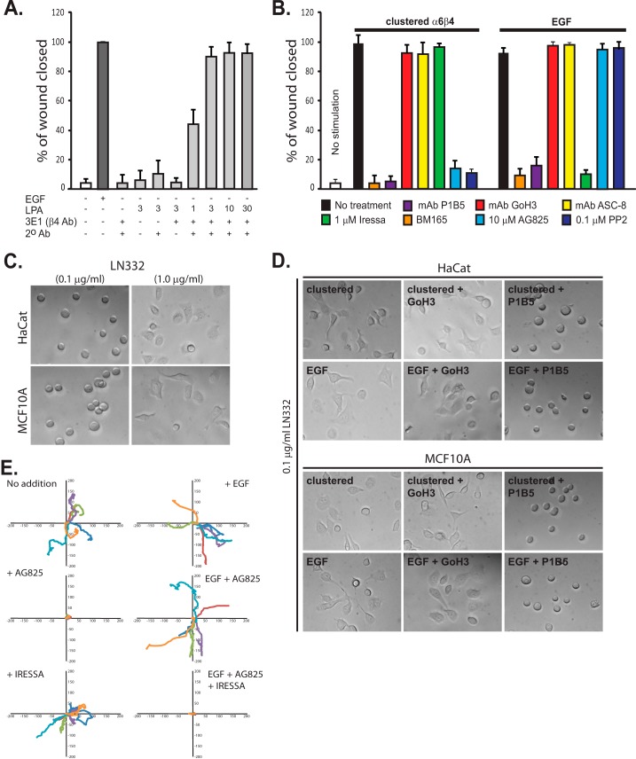
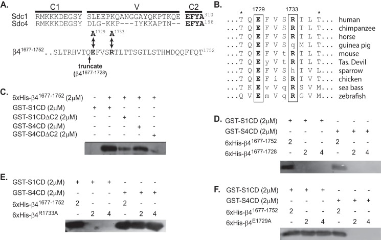
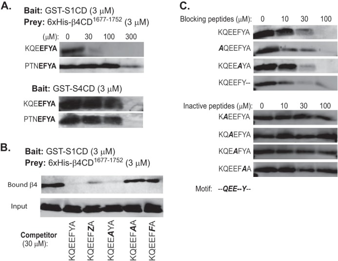
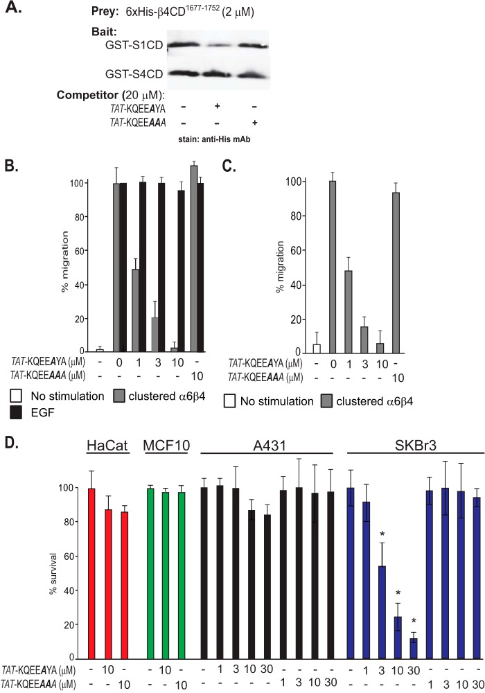
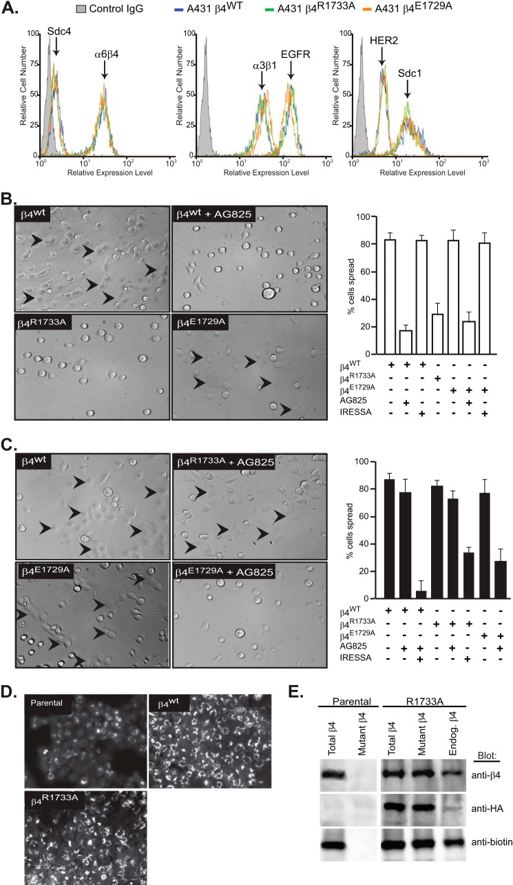
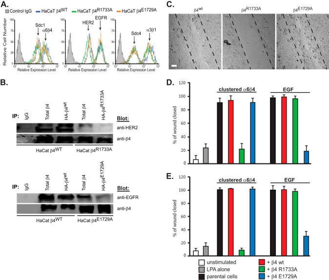
Epithelial cells are highly dependent during wound healing and tumorigenesis on the α6β4 integrin and its association with receptor tyrosine kinases. Previous work showed that phosphorylation of the β4 subunit upon matrix engagement depends on the matrix receptor syndecan (Sdc)-1 engaging the cytoplasmic domain of the β4 integrin and coupling of the integrin to human epidermal growth factor receptor-2 (HER2). In this study, HER2-dependent migration activated by matrix engagement is compared with migration stimulated by EGF. We find that whereas HER2-dependent migration depends on Sdc1, EGF-dependent migration depends on a complex consisting of human epidermal growth factor receptor-1 (HER1, commonly known as EGFR), α6β4, and Sdc4. The two syndecans recognize distinct sites at the extreme C terminus of the β4 integrin cytoplasmic domain. The binding motif in Sdc1 is QEEXYX, composed in part by its syndecan-specific variable (V) region and in part by the second conserved (C2) region that it shares with other syndecans. A cell-penetrating peptide containing this sequence competes for HER2-dependent epithelial migration and carcinoma survival, although it is without effect on the EGFR-stimulated mechanism. β4 mutants bearing mutations specific for Sdc1 and Sdc4 recognition act as dominant negative mutants to block cell spreading or cell migration that depends on HER2 or EGFR, respectively. The interaction of the α6β4 integrin with the syndecans appears critical for it to be utilized as a signaling platform; migration depends on α3β1 integrin binding to laminin 332 (LN332; also known as laminin 5), whereas antibodies that block α6β4 binding are without effect. These findings indicate that specific syndecan family members are likely to have key roles in α6β4 integrin activation by receptor tyrosine kinases.

Keywords: Carcinogenesis; Cell Invasion; Epidermal Growth Factor Receptor (EGFR); Integrin; Laminin; Syndecan; Wound Healing.


Figures

FIGURE 1.
FIGURE 1.
HER2 and EGFR are involved in distinct α6β4-dependent cell motility mechanisms. A, confluent HaCat keratinocytes are starved and then stimulated to migrate for 15–18 h in a scratch wound assay by the addition of 10 ng/ml EGF (EGF chemokinesis) or by clustering of the integrin using mAb3E1 plus a secondary anti-mouse IgG in the presence of 1–30 μm LPA to mimic matrix chemokinesis. Maximal haptotactic migration is observed in the presence of 3 μm LPA, which is used for all remaining experiments. B, HaCat keratinocytes are stimulated to undergo matrix chemokinesis (clustered α6β4) or EGF-stimulated chemokinesis in the presence or absence of mAb P1B5 to block binding by α3β1 integrin, mAb GoH3, or ASC-8 to block α6β4 integrin binding, BM165 to block the integrin-binding sites in LN332, 1 μm Iressa to block EGFR, and 10 μm tyrphostin AG825 to block HER2 or the general Src kinase inhibitor PP2. C, HaCat human keratinocytes or MCF10A mammary epithelial cells are plated on 0.1 or 1 μg/ml LN332. D, HaCat or MCF10A cells are treated with EGF, or α6β4 integrin is clustered to activate the EGFR- or HER2-dependent integrin activation mechanisms. GoH3 or P1B5 is used to inhibit α6β4 or α3β1 binding to the laminin. E, MCF-10A cells are plated on 1 μg/ml LN332 under serum-free conditions, and the motility of individual cells is tracked over 10 h in the presence or absence of EGF. Relative migration distances are shown on spatial plot maps. Cells are treated with 10 μm AG825 to block HER2, 1 μm Iressa to block EGFR, or a combination of both inhibitors.
FIGURE 2.
FIGURE 2.
Sdc1 is required for HER2-dependent haptotaxis, whereas Sdc4 is required for EGFR-dependent chemotaxis. A, potential roles for Sdc1 or Sdc4 in HaCat wound closure are tested by silencing expression of the endogenous human syndecans with siRNA, together with rescue from either wild-type mouse Sdc1 or Sdc4, or mouse mutants unable to engage the β4 cytoplasmic domain (mSdc1ΔC2 or mSdc4ΔC2). Migration data represent the mean of six wells ± S.D. B, lysates of HaCat cells expressing either mSdc4 or the mSdc4ΔC2 mutant were subjected to immunoprecipitation (IP) using mAb KY8.2 specific for mouse Sdc4, P1B5 specific for α3β1 integrin, or 3E1 specific for α6β4 integrin. Immunoprecipitates were analyzed on Western blots probing for wild-type Sdc4 or mutant mSdc4ΔC2 with KY8.2. C, HaCat cell lysates were subjected to immunoprecipitation using mAb B-A38 specific for human Sdc1 (hSdc1), F94-8G3 specific for human Sdc4, 3E1 specific for β4 integrin, and anti-neu (c-18) for HER2 or anti-EGFR (1005). Blots were probed for HER2 using anti-neu (c-18) or anti-EGFR (1005).
FIGURE 3.
FIGURE 3.
Sdc1 and Sdc4 engage distinct sites at the C terminus of the β4 integrin cytoplasmic domain. A, complete cytoplasmic domains of human Sdc1 (S1CD) and Sdc4 (S4CD) are shown, denoting the juxtamembrane region conserved across the syndecan family (C1), the distal conserved region (C2) composed of the amino acids EFYA, and the V region unique to each syndecan family member. Also shown are the C-terminal 32 amino acids of the β4 integrin subunit contained in a His6-tagged β4 integrin construct (β41677–1752) used in binding studies. Double-headed arrows denote Glu-1729 and Arg-1733 (in bold), which were mutated to alanine to disrupt syndecan-specific binding to this construct. An arrow also denotes the site at which the construct is truncated to remove the last 24 amino acids (His6-β4(1677–1728)) necessary for syndecan binding. B, conservation of amino acids 1727–1736 in the β4 integrin cytoplasmic domain across species is shown, with conserved Glu-1729 and Arg-1733 in bold. Asterisks denote threonines that disrupt binding of the β4 cytoplasmic domain to plectin when phosphorylated (66). C, glutathione beads preincubated with 2 μm GST-S1CD, GST-S4CD, GST-S1CDΔC2, or GST-S4CDΔC2 were tested for their ability to capture 2 μm His6-β4(1677–1752), detected on Western blots with anti-penta-His. D–F, glutathione beads preloaded with GST-S1CD or S4CD are tested for the capture of either 2 or 4 μm His6-β4(1677–1752) or His6-β4(1677–1728) (D), or constructs in which point mutations have been introduced at Arg-1733 (His6-β4R1733A) (E) or Glu-1729 (His6-β4E1729A) (F).
FIGURE 4.
FIGURE 4.
Distinct cytoplasmic motifs in Sdc1 and Sdc4 engage the β4 cytoplasmic domain. The glutathione bead pulldown assay is used to assess the capture of His6-β4(1677–1752) (prey) either by S1CD or S4CD (bait) in the presence of competing peptides. Captured β4 construct is visualized on Western blots by staining with anti-penta-His. A, capture by GST-S1CD or GST-S4CD in the presence of 0–300 μm KQEEFYA peptide sequence derived from the Sdc1 or PTNEFYA derived from Sdc4. The conserved C2 region (EFYA) is shown in bold. B, capture by GST-S1CD in the presence of Sdc1-specific peptide in which Phe-308 is mutated to Ala (A), or Tyr-309 is phosphorylated (Z), or is mutated to Ala or Phe (F). C, capture by GST-S1CD in the presence of Sdc1-specific peptide in which individual amino acids are mutated to Ala or Ala-310 is deleted.
FIGURE 5.
FIGURE 5.
Cell-penetrating peptide containing the Sdc1-specific β4-binding motif disrupts HER2-dependent haptotaxis and cell survival. TAT peptide is constructed containing the Sdc1-specific binding motif in which Phe-308 necessary for PDZ binding but not for β4 binding is mutated (KQEEAYA) (active peptide) or Phe-308 is mutated along with Tyr-309 necessary for β4 binding (KQEEAAA) (inactive control peptide). A, active or control TAT peptides are tested as competitors of His6-β4(1677–1752) capture by GST-S1CD or GST-S4CD. B, HaCat keratinocytes are stimulated with LPA and α6β4 integrin clustering antibodies to mimic HER2-dependent matrix chemokinesis or stimulated with EGF to undergo EGFR-dependent chemokinesis in the presence of 0–10 μm TAT peptides. C, MCF10A human breast epithelial cells are stimulated to undergo matrix chemokinesis in the presence of TAT peptides ranging from 0 to 10 μm. D, HaCat keratinocytes, MCF10A mammary epithelium, A431 cervical carcinoma, or HER2-positive SKBr3 breast carcinoma cells were grown for 1 week in the presence of TAT peptide ranging from 0 to 30 μm. Cell death was quantified by trypan blue exclusion. Data represent the mean of six wells ± S.D. (* indicates difference between control and active peptide >0.05.)
FIGURE 6.
FIGURE 6.
α6β4R1733A and α6β4E1729A integrin constructs act as dominant negative mutants to block the HER2- and EGFR-dependent mechanisms, respectively, during A431 cell spreading. A, A431 cells expressing HA-tagged β4WT, β4R1733A, or β4E1729A constructs were subjected to flow cytometry to quantify relative expression of cell surface α6β4 integrin (3E1), Sdc1 (B-A38), and Sdc4 (F4–8G3), α3β1 integrin (ASC-1), and HER2 (9C6) or EGFR (EGFR.1). B, A431 cells expressing the β4WT, β4R1733A, or β4E1729A constructs are plated on 1 μg/ml LN332 for 2 h to activate the HER2-dependent signaling mechanism (spread cells denoted by arrowheads) with or without addition of 10 μm AG825 or 1 μm Iressa to inhibit HER2 or EGFR, respectively. The percentage of spread cells in triplicate wells from two experiments is quantified. C, A431 cells expressing β4 receptor constructs are plated on 1 μg/ml LN332 for 2 h in the presence of 10 ng/ml EGF with or without AG825 or Iressa and quantified as in B. D, A431 parental cells, β4WT-expressing cells, or β4R1733A-expressing cells are fixed, permeabilized, and stained for β4 integrin with mAb 3E1. E, relative amounts of endogenous α6β4 and HA-tagged α6β4R1733A (mutant) integrin at the cell surface is determined by biotinylation of parental or α6β4R1733A-expressing (R1733A) cells, followed by immunoprecipitation with mAb 3E1 to obtain “total β4,” with anti-HA (mAb 12CAS) to obtain “mutant β4,” or 3E1 precipitation of integrin remaining in the supernatant after anti-HA precipitation to quantify native endogenous β4. The relative amounts of total α6β4, HA-tagged α6β4R1733A, and biotinylated (cell surface) α6β4 integrin in each pool are assessed by staining a single blot with anti-β4 (AB1922), anti-HA (C29F4), and anti-biotin (212.26.A2) by stripping and reprobing.
FIGURE 7.
FIGURE 7.
Dominant negative α6β4R1733A integrin disrupts HER2-dependent haptotaxis and α6β4E1729A disrupts EGFR-dependent chemotaxis of HaCat and MCF10A cells. A, HaCat cells expressing β4WT, β4R1733A, or β4E1729A mutants are subjected to flow cytometry to determine relative cell surface expression of α6β4 integrin, Sdc1 and Sdc4, α3β1 integrin, and HER2 or EGFR. B, either total or HA-tagged β4 integrin was immunoprecipitated (IP) with mAb 3E1 or anti-HA from lysates of HaCat cells expressing HA-β4WT, HA-β4R1733A, or HA-β4E1729A mutants. Immunoprecipitates divided into duplicate samples were probed for co-immunoprecipitation of integrin and either HER2 or EGFR. C, confluent HaCat cells expressing either β4WT, β4R1733A, or β4E1729A integrin subunits are stimulated to close a scratch wound during a 15–18-h treatment with 3 μm LPA, 10 μg/ml 3E1, and 30 μg/ml anti-mouse secondary antibody to stimulate HER2-dependent haptotaxis. Bar, 40 μm. D, quantification of scratch wound closure by either parental HaCat cells or cells transfected with β4 integrin constructs and stimulated to undergo haptotaxis or chemotaxis in response to 10 ng/ml EGF. The migration of parental cells in the absence of stimulation or stimulated with LPA alone is shown as a control. E, quantification of scratch wound closure by either parental MCF10A cells or cells transfected with β4 integrin constructs and stimulated as described in C. Data represent the mean of six wells ± S.D.

Similar articles

Cited by

References

    1. Aumailley M., Bruckner-Tuderman L., Carter W. G., Deutzmann R., Edgar D., Ekblom P., Engel J., Engvall E., Hohenester E., Jones J. C., Kleinman H. K., Marinkovich M. P., Martin G. R., Mayer U., Meneguzzi G., Miner J. H., Miyazaki K., Patarroyo M., Paulsson M., Quaranta V., Sanes J. R., Sasaki T., Sekiguchi K., Sorokin L. M., Talts J. F., Tryggvason K., Uitto J., Virtanen I., von der Mark K., Wewer U. M., Yamada Y., Yurchenco P. D. (2005) A simplified laminin nomenclature. Matrix Biol. 24, 326–332 - PubMed
    1. Nievers M. G., Schaapveld R. Q., Sonnenberg A. (1999) Biology and function of hemidesmosomes. Matrix Biol. 18, 5–17 - PubMed
    1. Hopkinson S. B., Jones J. C. (2000) The N terminus of the transmembrane protein BP180 interacts with the N-terminal domain of BP230, thereby mediating keratin cytoskeleton anchorage to the cell surface at the site of the hemidesmosome. Mol. Biol. Cell 11, 277–286 - PMC - PubMed
    1. Wilhelmsen K., Litjens S. H., Sonnenberg A. (2006) Multiple functions of the integrin α6β4 in epidermal homeostasis and tumorigenesis. Mol. Cell. Biol. 26, 2877–2886 - PMC - PubMed
    1. Rabinovitz I., Tsomo L., Mercurio A. M. (2004) Protein kinase C-α phosphorylation of specific serines in the connecting segment of the β4 integrin regulates the dynamics of type II hemidesmosomes. Mol. Cell. Biol. 24, 4351–4360 - PMC - PubMed

Publication types

MeSH terms