. 2014 Nov 15;34(22):4143-64.
doi: 10.1128/MCB.00480-14. Epub 2014 Sep 8.

MicroRNAs 206 and 21 cooperate to promote RAS-extracellular signal-regulated kinase signaling by suppressing the translation of RASA1 and SPRED1

Affiliations

MicroRNAs 206 and 21 cooperate to promote RAS-extracellular signal-regulated kinase signaling by suppressing the translation of RASA1 and SPRED1

Sriganesh B Sharma et al. Mol Cell Biol. .

Abstract

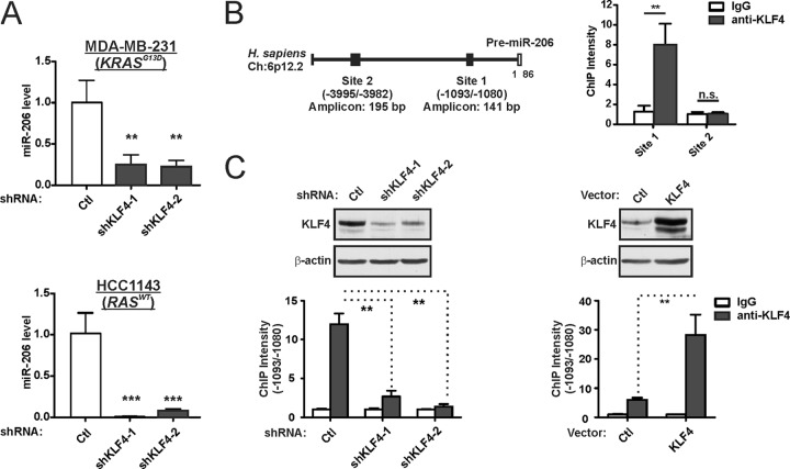
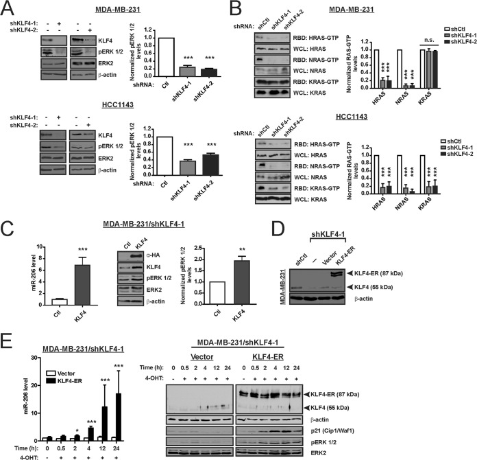
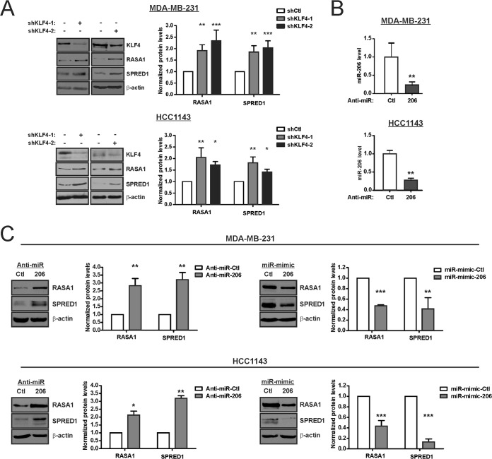
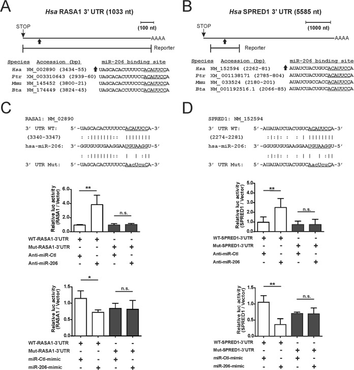
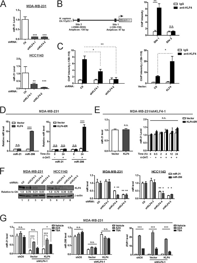
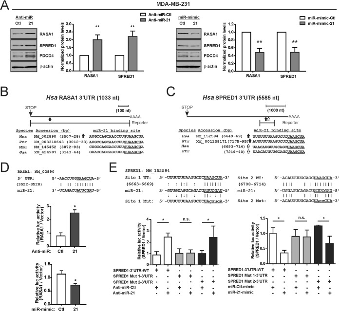
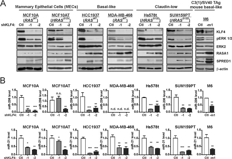
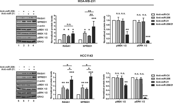
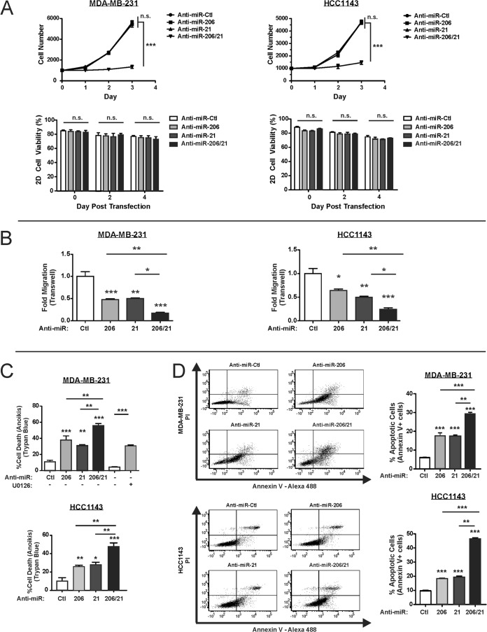
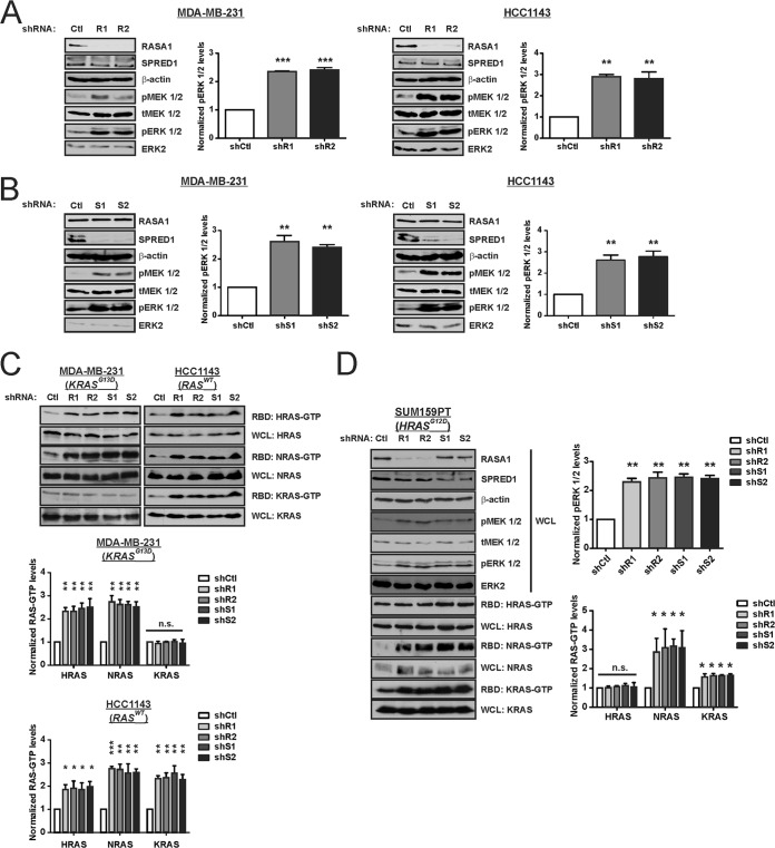
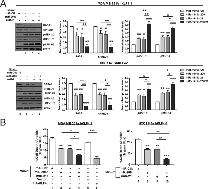
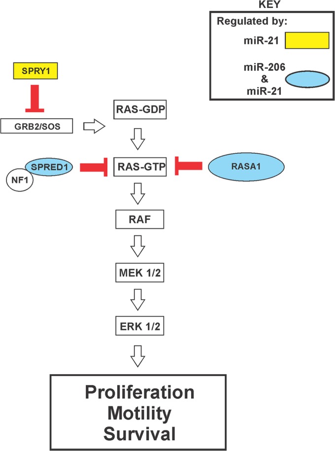
Despite the low prevalence of activating point mutation of RAS or RAF genes, the RAS-extracellular signal-regulated kinase (ERK) pathway is implicated in breast cancer pathogenesis. Indeed, in triple-negative breast cancer (TNBC), there is recurrent genetic alteration of pathway components. Using short hairpin RNA (shRNA) methods, we observed that the zinc finger transcription factor Krüppel-like factor 4 (KLF4) can promote RAS-ERK signaling in TNBC cells. Endogenous KLF4 bound to the promoter regions and promoted the expression of two microRNAs (miRs), miR-206 and miR-21 (i.e., miR-206/21). Antisense-mediated knockdown (anti-miR) revealed that miR-206/21 coordinately promote RAS-ERK signaling and the corresponding cell phenotypes by inhibiting translation of the pathway suppressors RASA1 and SPRED1. In TNBC cells, including cells with mutation of RAS, the suppression of either RASA1 or SPRED1 increased the levels of GTP-bound, wild-type RAS and activated ERK 1/2. Unlike the control cells, treatment of RASA1- or SPRED1-suppressed cells with anti-miR-206/21 had little or no impact on the level of activated ERK 1/2 or on cell proliferation and failed to suppress tumor initiation. These results identify RASA1 and SPRED1 mRNAs as latent RAS-ERK pathway suppressors that can be upregulated in tumor cells by anti-miR treatment. Consequently, KLF4-regulated miRs are important for the maintenance of RAS-ERK pathway activity in TNBC cells.


Figures

FIG 1
FIG 1
KLF4 is bound to the MIR206 promoter region and induces miR-206 expression. (A) TNBC cells were transduced with lentiviral vectors expressing KLF4 shRNA or a nontargeting control (Ctl). Endogenous miR-206 levels in stably selected cells were measured by stem-loop reverse transcriptase real-time quantitative PCR. miR-206 levels were measured relative to U6 snRNA. Mean values are shown; error bars represent standard errors (SE). **, P < 0.01; ***, P < 0.001. (B) Schematic of the MIR206 locus (left panel) indicating KLF4 consensus binding sites that were analyzed by ChIP analysis of MDA-MB-231 cells (right panel). n.s., not significant. (C) KLF4 protein levels (upper panels) and the KLF4-MIR206 ChIP intensity levels (lower panels) were analyzed in MDA-MB-231 cells expressing shKLF4, a KLF4 transgene, or controls. β-Actin served as a loading control for immunoblot analysis.
FIG 2
FIG 2
KLF4 rapidly induces miR-206 and RAS-ERK signaling. (A) The indicated proteins, including phospho-ERK 1/2 (pERK 1/2) and ERK2, were analyzed by immunoblot analysis of TNBC cells expressing shKLF4 or shCtl. Bar graph data indicate the average of three independent experiments. Error bars represent standard deviations (SD). (B) The levels of GTP-bound (active) HRAS, NRAS, and KRAS in cell extracts were analyzed by affinity precipitation using the RAS binding domain (RBD) of Raf-1. RAS proteins were analyzed by immunoblotting. The level of each RAS isoform in the whole-cell lysate (WCL) served as the loading control. (C) KLF4-deficient MDA-MB-231 cells were transduced with retroviral vector expressing hemagglutinin (HA) epitope-tagged KLF4 or empty vector (Ctl). miR-206 levels (left panel) and levels of the indicated proteins (right panel) were analyzed. (D) KLF4-deficient MDA-MB-231 cells were transduced with a 4-hydroxytamoxifen (4-OHT)-dependent transgene, coding for KLF4-ER, or empty vector. KLF4 and KLF4-ER levels were analyzed by immunoblotting. (E) miR-206 levels (left panel) and levels of the indicated proteins (right panel) were analyzed. CDKN1A is regulated by KLF4, and p21Cip1/Waf1 therefore served as an indicator of KLF4 activity. *, P < 0.05; **, P < 0.01; ***, P < 0.001.
FIG 3
FIG 3
KLF4 and miR-206 regulate the levels of two RAS-ERK pathway suppressors, RASA1 and SPRED1. (A) RASA1 and SPRED1 levels were determined by immunoblot analysis of KLF4-deficient cells and control cells. (B) TNBC cells were transfected with either anti-miR-206 or a nontargeting anti-miR (Ctl), and miR-206 levels were determined. (C) TNBC cells were transfected with the indicated anti-miR or miR mimic. RASA1 and SPRED1 levels were analyzed by immunoblotting. *, P < 0.05; **, P < 0.01; ***, P < 0.001.
FIG 4
FIG 4
miR-206 represses the translation of RASA1 and SPRED1 by directly targeting the 3′ UTRs. (A and B) Schematic of the RASA1 3′ UTR (A) and the SPRED1 3′ UTR (B) indicating potential miR-206 binding sites. The portion of each 3′ UTR that was cloned into the translational reporter is indicated relative to the stop codon and polyadenylation signal. The sequence of the miR-206 candidate binding site is indicated below each schematic for several mammals: Homo sapiens (Hsa), Pan troglodytes (Ptr), Mus musculus (Mmu), and Bos taurus (Bta). (The seed sequence complement is underlined.) (C and D) For analysis of protein translation, WT and mutant versions of the RASA1 3′ UTR (C) or SPRED1 3′ UTR (D) were inserted into the 3′ UTR of firefly luc (top panels, 3′ UTR WT and 3′ UTR Mut). MDA-MB-231 cells were cotransfected with reporters in combination with either anti-miR (middle panels) or the miR mimic (bottom panels). The normalized activity of the reporters relative to empty luc vector was analyzed at 24 h posttransfection. *, P < 0.05; **, P < 0.01; n.s., not significant.
FIG 5
FIG 5
Endogenous KLF4 is bound to the MIR21 promoter region and maintains miR-21 expression in TNBC cells. (A) miR-21 levels were analyzed in KLF4-deficient or control TNBC cells. (B) Schematic of the MIR21 locus (left panel) indicating a KLF4 consensus binding site that was analyzed in MDA-MB-231 cells by ChIP (right panel). (C) Similarly as shown in Fig. 1C, the KLF4-MIR21 ChIP intensity levels were analyzed in MDA-MB-231 cells expressing shKLF4, a KLF4 transgene, or controls. (D) TNBC cells were transduced with empty vector or with vector encoding KLF4 or KLF4-ER. Where indicated, cells were treated with 4-OHT, and the levels of the indicated miRs were analyzed. (E) Similarly to what is shown in Fig. 2C to E, miR-21 levels were analyzed in KLF4-deficient TNBC cells following rescue with exogenous KLF4 (left panel) or KLF4-ER (right panel). (F) TNBC cells were transduced with the indicated shRNA vector. Proteins were analyzed by immunoblotting (left panels), and miR levels were determined (right panels). (G) Cells were treated with 5-aza-2′-deoxycytidine (AZA; 96 h) or trichostatin A (TSA; 12 h). ESR1 mRNA levels were analyzed as a positive control for drug activity (right panel). *, P < 0.05; **, P < 0.01; ***, P < 0.001; n.s., not significant.
FIG 6
FIG 6
miR-21 directly represses the translation of RASA1 and SPRED1. (A) MDA-MB-231 cells were transfected with the indicated anti-miR or miR mimic, and RASA1 and SPRED1 levels were determined by immunoblotting. PDCD4 is encoded by an miR-21-targeted transcript and was analyzed in parallel. (B) To construct a WT translational reporter, a portion of the RASA1 3′ UTR containing an established miR-21 binding site was inserted into the 3′ UTR of firefly luc (64). The sequences of the miR-21 binding site are indicated for several vertebrates: Homo sapiens (Hsa), Pan troglodytes (Ptr), Mus musculus (Mmu), and Gallus gallus (Gga). (The seed sequence complement is underlined.) (C) Schematic of the SPRED1 3′ UTR indicating potential miR-21 binding sites. (D and E) For analysis of protein translation, WT or mutant versions of the indicated 3′ UTR were inserted into the 3′ UTR of firefly luc (top panels). MDA-MB-231 cells were cotransfected with reporters in combination with either anti-miR or the miR mimic. The normalized activity of the reporters relative to that of the empty luc vector was analyzed at 24 h posttransfection. *, P < 0.05; **, P < 0.01; n.s., not significant.
FIG 7
FIG 7
KLF4 promotes activated ERK 1/2 levels and miR-206 and miR-21 expression in a panel composed of human mammary epithelial cells and TNBC cell lines. (A) Cells were transduced with the indicated shRNA vector. Stably selected cells were analyzed for the indicated proteins by immunoblotting. (B) miR levels were analyzed in the indicated cells. For MDA-MB-468 cells, the miR-206 cycle threshold exceeded 40, and the expression level was therefore designated “not detected” (n.d.). *, P < 0.05; **, P < 0.01; ***, P < 0.001.
FIG 8
FIG 8
Endogenous miR-206 and miR-21 cooperate to promote RAS-ERK signaling. TNBC cells were transfected with the indicated anti-miR, and protein levels were analyzed by immunoblotting. pMEK 1/2, phospho-MEK 1/2; tMEK 1/2, total MEK 1/2. Transfections were performed using a 12.5 nM concentration of the indicated anti-miR, with 25 nM as the final concentration of all species combined, using anti-miR-Ctl where indicated (−). *, P < 0.05; **, P < 0.01; ***, P < 0.001; n.s., not significant.
FIG 9
FIG 9
Inhibition of miR-206 and miR-21 cooperatively suppresses ERK 1/2-dependent phenotypes in TNBC cells. (A) TNBC cells were transfected with the indicated anti-miR and plated 24 h posttransfection (i.e., day 0). Cell proliferation was measured using an ATP-based luminescence assay (n = 3; error bars, SE). (B) Migration of anti-miR-transfected TNBC cells was measured in a Boyden chamber transwell assay (n = 3; error bars, SE). (C) Anti-miR-transfected cells were analyzed in an anoikis assay. After 24 h in suspension, cell death was analyzed by trypan blue staining (n = 3; error bars, SE). In parallel, cells were treated with MEK inhibitor U0126. (D) Three experiments were performed independently of those in panel C, and anoikis was assessed by annexin V-propidium idodide (PI) staining. The percentage of annexin V+ cells, representing both early and late apoptotic cells, is depicted in the right panel for each treatment group (n = 3; error bars, SD). *, P < 0.05; **, P < 0.01; ***, P < 0.001; n.s., not significant.
FIG 10
FIG 10
RASA1 and SPRED1 are limiting factors for RAS-ERK signaling in TNBC cells. (A) TNBC cells were stably transduced with shRNA vector targeting RASA1 (R1 or R2) or with a nontargeting control (Ctl). The indicated proteins were analyzed by immunoblotting. (B) Cells were stably transduced with shRNA vector targeting SPRED1 (S1 or S2) or with a nontargeting Ctl. The indicated proteins were analyzed by immunoblotting. (C) RAS-GTP levels were analyzed by the RBD pulldown assay in RASA1 and SPRED1 knockdown cells and control cells. (D) SUM159PT TNBC cells were stably transduced with the indicated shRNA expression vector, and the indicated proteins and RAS-GTP levels were analyzed. *, P < 0.05; **, P < 0.01; ***, P < 0.001; n.s., not significant.
FIG 11
FIG 11
RASA1 and SPRED1 mediate the regulation of RAS-ERK pathway signaling by miR-206 and miR-21. (A) TNBC cells expressing the indicated shRNAs were treated with anti-miR-206 and anti-miR-21 in combination (anti-miR-206/21) or with anti-miR-Ctl. Whole-cell extracts were prepared, and the indicated proteins were analyzed by immunoblotting. Two distinct cell culture models were analyzed (MDA-MB-231 versus HCC1143) using independent shRNAs (R1, S1, R2, and S2). For pERK 1/2, both short (S) and long (L) exposures are indicated. (B) Absent any anti-miR transfection (Untransfected), baseline cell proliferation was analyzed for cells expressing the indicated shRNA and for the untransduced, parental cells (P). Following anti-miR transfection, cell proliferation was measured for control TNBC cells (shCtl) or cells deficient in RASA1 (shR1 and shR2) or SPRED1 (shS1 and shS2). Assays were performed following transfection of anti-miR-Ctl (AC) or anti-miR-206/21 (A+). (C) MDA-MB-231 cells expressing the indicated shRNAs were transfected with either anti-miR-206/21 or anti-miR-Ctl. A total of 2 × 10⁶ cells were orthotopically injected into the left fourth mammary gland of athymic mice. Biweekly tumor measurements were made using calipers. For each treatment group, the number of mice in which tumor initiation occurred by day 28 is indicated (Initiated/Total). *, P < 0.05; **, P < 0.01; ***, P < 0.001; n.s., not significant.
FIG 12
FIG 12
Exogenous miR-206 and miR-21 cooperate to promote RAS-ERK signaling and cell survival in KLF4-depleted cells. (A) KLF4-depleted TNBC cells were transfected with the indicated miR mimic, and protein levels were analyzed by immunoblotting. (B) Anoikis assays were performed following transfection of the indicated miR mimic into KLF4-depleted TNBC cells. For MDA-MB-231, cells transduced with KLF4 vector or empty vector were analyzed in parallel. *, P < 0.05; **, P < 0.01; ***, P < 0.001.
FIG 13
FIG 13
KLF4-dependent miRs cooperatively promote RAS-ERK pathway activity by cotargeting of pathway inhibitors. The schematic shows the organization of the RAS-ERK pathway. miR-206/21-cotargeted repressors of RAS-ERK signaling are shown in blue ovals. The GAP protein NF1 is indicated as a likely catalytic partner of SPRED1 (13).

References

    1. Weinberg RA. 2007. Growth factors, receptors, and cancer, p 119–158 In Weinberg RA. (ed), The biology of cancer. Garland Science, Taylor and Francis Group, LLC, New York, NY.
    1. Johnson GL, Lapadat R. 2002. Mitogen-activated protein kinase pathways mediated by ERK, JNK, and p38 protein kinases. Science 298:1911–1912. 10.1126/science.1072682. - DOI - PubMed
    1. Downward J. 2003. Targeting RAS signalling pathways in cancer therapy. Nat. Rev. Cancer 3:11–22. 10.1038/nrc969. - DOI - PubMed
    1. Kolch W. 2005. Coordinating ERK/MAPK signalling through scaffolds and inhibitors. Nat. Rev. Mol. Cell Biol. 6:827–837. 10.1038/nrm1743. - DOI - PubMed
    1. McCubrey JA, Steelman LS, Abrams SL, Lee JT, Chang F, Bertrand FE, Navolanic PM, Terrian DM, Franklin RA, D'Assoro AB, Salisbury JL, Mazzarino MC, Stivala F, Libra M. 2006. Roles of the RAF/MEK/ERK and PI3K/PTEN/AKT pathways in malignant transformation and drug resistance. Adv. Enzyme Regul. 46:249–279. 10.1016/j.advenzreg.2006.01.004. - DOI - PubMed

Publication types

MeSH terms