. 2014 Aug 25:6:220.
doi: 10.3389/fnagi.2014.00220. eCollection 2014.

n-3 polyunsaturated fatty acids supplementation enhances hippocampal functionality in aged mice

Affiliations

n-3 polyunsaturated fatty acids supplementation enhances hippocampal functionality in aged mice

Debora Cutuli et al. Front Aging Neurosci. .

Abstract

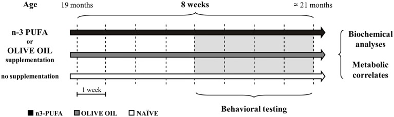
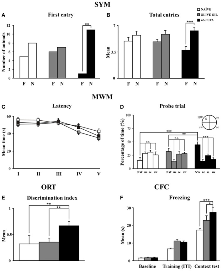
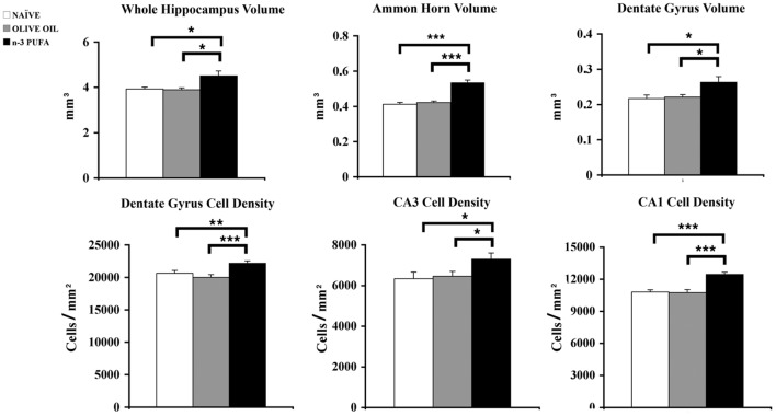
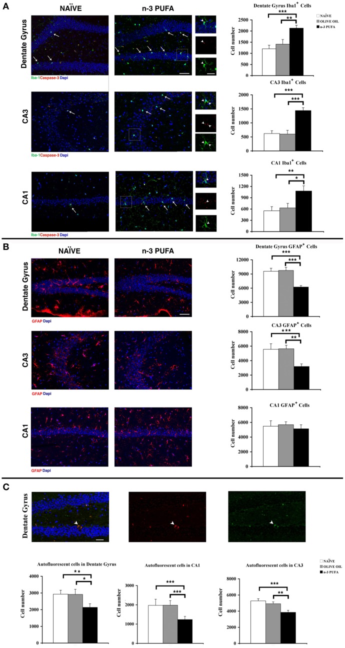
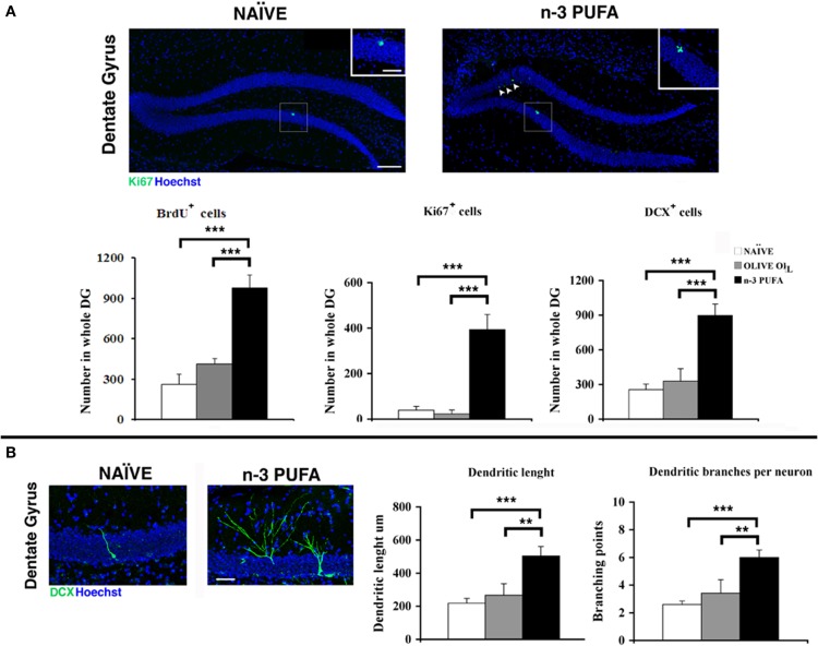
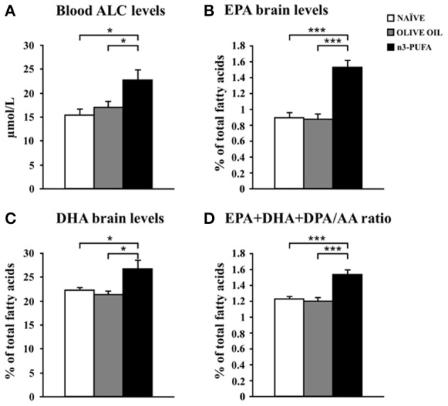
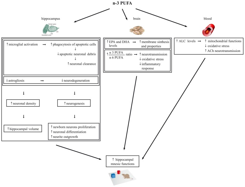
As major components of neuronal membranes, omega-3 polyunsaturated acids (n-3 PUFA) exhibit a wide range of regulatory functions, modulating from synaptic plasticity to neuroinflammation, from oxidative stress to neuroprotection. Recent human and animal studies indicated the n-3 PUFA neuroprotective properties in aging, with a clear negative correlation between n-3 PUFA levels and hippocampal deficits. The present multidimensional study was aimed at associating cognition, hippocampal neurogenesis, volume, neurodegeneration and metabolic correlates to verify n-3 PUFA neuroprotective effects in aging. To this aim 19 month-old mice were given n-3 PUFA mixture, or olive oil or no dietary supplement for 8 weeks during which hippocampal-dependent mnesic functions were tested. At the end of behavioral testing morphological and metabolic correlates were analyzed. n-3 PUFA supplemented aged mice exhibited better object recognition memory, spatial and localizatory memory, and aversive response retention, without modifications in anxiety levels in comparison to controls. These improved hippocampal cognitive functions occurred in the context of an enhanced cellular plasticity and a reduced neurodegeneration. In fact, n-3 PUFA supplementation increased hippocampal neurogenesis and dendritic arborization of newborn neurons, volume, neuronal density and microglial cell number, while it decreased apoptosis, astrocytosis and lipofuscin accumulation in the hippocampus. The increased levels of some metabolic correlates (blood Acetyl-L-Carnitine and brain n-3 PUFA concentrations) found in n-3 PUFA supplemented mice also pointed toward an effective neuroprotection. On the basis of the present results n-3 PUFA supplementation appears to be a useful tool in health promotion and cognitive decline prevention during aging.

Keywords: aging; cognitive decline; hippocampus; neuroprotection; omega-3 fatty acids.


Figures

Figure 1
Figure 1
Global timing of the experimental procedure. Experimental groups of aged mice (n-3 PUFA, OLIVE OIL, and NAÏVE), type of dietary supplementation (duration: 8 weeks), behavioral testing (EPM, Elevated Plus Maze; SYM, Spatial Y-Maze; MWM, Morris Water Maze; ORT, Object Recognition Maze; CFC, Contextual Fear Conditioning), morphological analyses (hippocampal volumes, cell density, caspase-3 levels, astroglia and microglial cells, lipofuscin concentrations, neurogenesis) and metabolic correlates (blood amino acids and carnitines levels; brain n-3 PUFA levels) are indicated.
Figure 2
Figure 2
n-3 PUFA effects on behavioral performances. (A,B) First entry (A) and total entries (B) in the familiar (F) and novel (N) arm displayed by the three experimental groups in Spatial Y-Maze (SYM). (C,D) Mean escape latency to reach the hidden platform during Place phase of Morris Water Maze (MWM) (C) and time spent in the rewarded quadrant during Probe trial (D). (E) Discrimination index in Object Recognition Test (ORT). (F) Mean freezing time exhibited during Baseline, Training (Inter-Trial Intervals: ITI) and Context Test of Contextual Fear Conditioning (CFC). Asterisks inside the graphs indicate the significance of comparisons between groups: *p < 0.05, **p ≤ 0.01, or ***p ≤ 0.001.
Figure 3
Figure 3
n-3 PUFA effects on hippocampal volumes and cell density. Volumes of whole hippocampus, Ammon Horn (CA1+CA3) and Dentate Gyrus resulted significantly enhanced in n-3 PUFA group in comparison to NAÏVE and OLIVE OIL groups. Also cell density was significantly increased in Dentate Gyrus, CA3 and CA1 of n-3 PUFA group in comparison to both control groups. *p < 0.05, **p < 0.01, or ***p < 0.001; Student's T test.
Figure 4
Figure 4
n-3 PUFA effects on hippocampal glial populations and lipofuscin deposits. (A) Representative fluorescence images of confocal optical sections of Dentate Gyrus, CA3 and CA1 in NAÏVE and n-3 PUFA groups, showing Iba-1+ (green, arrows) and caspase-3+ (red) cells. Scale bar 50 μm. In the central small panels, the higher magnifications (square boxes) show the neuronal caspase-3+ debris (red, arrowheads) phagocytated by the microglial cells (green, arrowheads). Scale bar 25 μm. The histograms on the right show the Iba1+ cell number of the Dentate Gyrus, CA3 and CA1 in n-3 PUFA, NAÏVE and OLIVE OIL groups. (B) Representative fluorescence images of confocal optical sections representing GFAP+ cells (red) in Dentate Gyrus, CA3, and CA1 of NAÏVE and n-3 PUFA groups, showing decreased astrocytosis in the n-3 PUFA group in Dentate gyrus and CA3 but not in CA1. Scale bar 50 μm. The histograms on the right show the GFAP+ cell number of the Dentate Gyrus, CA3, and CA1 in n-3 PUFA, NAÏVE, and OLIVE OIL groups. (C) Representative fluorescence images of confocal optical sections representing lipofuscin autofluorescent cells in the Dentate Gyrus of n-3 PUFA group (arrowhead). Scale bar 50 μm. The bottom histograms show quantitative analysis of the lipofuscin-loaded cells in Dentate Gyrus, CA3 and CA1. *p < 0.05, **p ≤ 0.01, or ***p ≤ 0.001; Student's T test.
Figure 5
Figure 5
n-3 PUFA effects on hippocampal neurogenesis. (A) Representative images showing the increase of the Ki67+ cells in the dentate gyrus of the mice treated with n-3 PUFA, when compared with NAÏVE group. At higher magnification (square boxes) the presence of a cluster of Ki67+ cells, which is a sign of intense proliferative activity can be noted for the n-3 PUFA treated mice (right panel), but not for the NAÏVE mice (left panel). Scale bars 50 μm and 25 μm. The histograms show the increase of BrdU±, Ki67+ and DCX+ cells in the n-3 PUFA mice, when compared with NAÏVE and OLIVE OIL mice. (B) Representative morphology of DCX cells, analyzed from NAÏVE and n-3 PUFA experimental groups, showing an increase of dendritic complexity in the n-3 PUFA neuronal progenitors. Scale bar 50 μm. Quantification of dendritic length and dendritic branching points measured in NAÏVE, OLIVE OIL, and n-3 PUFA experimental groups. **p ≤ 0.01, or ***p ≤ 0.001; Student's T test.
Figure 6
Figure 6
n-3 PUFA effects on metabolic correlates. (A) Average blood levels of acetylcarnitine (ALC) for the three experimental groups. (B) EPA brain levels. (C) DHA brain levels. (D) EPA+DHA+DPA/AA ratio. *p < 0.05, or ***p < 0.001.
Figure 7
Figure 7
Schematic representation of the cellular mechanisms underlying the beneficial effects of n-3 PUFA supplementation on hippocampal-dependent mnesic functions in aged mice.

Similar articles

Cited by

References

    1. Abdul H. M., Calabrese V., Calvani M., Butterfield D. A. (2006). Acetyl-L-carnitine-induced up-regulation of heat shock proteins protects cortical neurons against amyloid-beta peptide 1-42-mediated oxidative stress and neurotoxicity: implications for Alzheimer's disease. J. Neurosci. Res. 84, 398–408 10.1002/jnr.20877 - DOI - PubMed
    1. Ames B. N., Liu J. (2004). Delaying the mitochondrial decay of aging with acetylcarnitine. Ann. N.Y. Acad. Sci. 1033, 108–116 10.1196/annals.1320.010 - DOI - PubMed
    1. Arsenault D., Julien C., Tremblay C., Calon F. (2011). DHA improves cognition and prevents dysfunction of entorhinal cortex neurons in 3xTg-AD mice. PLoS ONE 6:e17397 10.1371/journal.pone.0017397 - DOI - PMC - PubMed
    1. Barhwal K., Hota S. K., Jain V., Prasad D., Singh S. B., Ilavazhagan G. (2009). Acetyl-l-carnitine (ALCAR) prevents hypobaric hypoxia-induced spatial memory impairment through extracellular related kinase-mediated nuclear factor erythroid 2-related factor 2 phosphorylation. Neuroscience 161, 501–514 10.1016/j.neuroscience.2009.02.086 - DOI - PubMed
    1. Bonomini M., Di Liberato L., Del Rosso G., Stingone A., Marinangeli G., Consoli A., et al. (2013). Effect of an L-Carnitine containing peritoneal dialysate on insulin sensitivity in patients treated with continuous ambulatory peritoneal dialysis: a 4-month prospective multicenter randomized trial. Am. J. Kidney Dis. 62, 929–938 10.1053/j.ajkd.2013.04.007 - DOI - PubMed