Review
. 2014 Aug 25:5:315.
doi: 10.3389/fphys.2014.00315. eCollection 2014.

Investigating the role of uncoupling of troponin I phosphorylation from changes in myofibrillar Ca(2+)-sensitivity in the pathogenesis of cardiomyopathy

Affiliations
Review

Investigating the role of uncoupling of troponin I phosphorylation from changes in myofibrillar Ca(2+)-sensitivity in the pathogenesis of cardiomyopathy

Andrew E Messer et al. Front Physiol. .

Abstract

Contraction in the mammalian heart is controlled by the intracellular Ca(2+) concentration as it is in all striated muscle, but the heart has an additional signaling system that comes into play to increase heart rate and cardiac output during exercise or stress. β-adrenergic stimulation of heart muscle cells leads to release of cyclic-AMP and the activation of protein kinase A which phosphorylates key proteins in the sarcolemma, sarcoplasmic reticulum and contractile apparatus. Troponin I (TnI) and Myosin Binding Protein C (MyBP-C) are the prime targets in the myofilaments. TnI phosphorylation lowers myofibrillar Ca(2+)-sensitivity and increases the speed of Ca(2+)-dissociation and relaxation (lusitropic effect). Recent studies have shown that this relationship between Ca(2+)-sensitivity and TnI phosphorylation may be unstable. In familial cardiomyopathies, both dilated and hypertrophic (DCM and HCM), a mutation in one of the proteins of the thin filament often results in the loss of the relationship (uncoupling) and blunting of the lusitropic response. For familial dilated cardiomyopathy in thin filament proteins it has been proposed that this uncoupling is causative of the phenotype. Uncoupling has also been found in human heart tissue from patients with hypertrophic obstructive cardiomyopathy as a secondary effect. Recently, it has been found that Ca(2+)-sensitizing drugs can promote uncoupling, whilst one Ca(2+)-desensitizing drug Epigallocatechin 3-Gallate (EGCG) can reverse uncoupling. We will discuss recent findings about the role of uncoupling in the development of cardiomyopathies and the molecular mechanism of the process.

Keywords: Ca sensitivity; cardiomyopathies; heart muscle; myofilament; phosphorylation; troponin I.


Figures
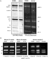
Figure 1
Figure 1
SDS-PAGE analysis of myofibrillar proteins and phosphoproteins. (A) Myofibril fraction of heart muscle separated by SDS-PAGE and stained successively with Pro-Q Diamond phosphoprotein stain and SYPRO Ruby total protein stain. Left, NTG and ACTC E99K mouse heart myofibrils (38 weeks male). Right, donor and ACTC E99K heart sample 2 myofibrils (Song et al., 2011). The TnI band is strongly stained together with MyBP-C, TnT and MLC-2. (B) Phosphate affinity SDS-PAGE analysis of TnI phospho-species. Left, comparison of TnI phosphorylation in 21-week-old female ACTC E99K and NTG mouse myofibrils. Right, comparison of TnI phosphorylation in myofibrils from ACTC E99K sample 2, donor heart and a typical interventricular septum sample from a patient with hypertrophic obstructive cardiomyopathy (HOCM) (Song et al., 2011). All samples show a high level of phosphorylation, except for the failing heart sample.
Figure 2
Figure 2
(A) Ca2+-regulation of thin filament motility by non-failing (donor) and failing heart troponin. Thin filament motility was measured by motility assay over a range of [Ca2+] in paired cells. The percentage of filaments motile is plotted as a function of [Ca2+] for a representative experiment. In blue lines and points, non-failing thin filaments, red lines and open points, failing thin filaments. TnI phosphorylation levels are shown in Figure 1B. The points ± s.e.m. are the mean of four determinations of percentage motile measured in one motility cell. The curves are fits of the data to the Hill equation (Messer et al., 2014). (B) Relationship between EC50 for Ca2+-activation of thin filaments and TnI bisphosphorylation. EC50 for thin filaments containing human heart Tn was plotted against the level of TnI bis-phosphorylation. EC50 was measured by IVMA and bis-phosphorylation of TnI (approximates to phosphorylation at Ser22 and 23) was measured by phosphate affinity SDS-PAGE. D, donor heart Tn, XI donor Tn with TnI exchanged, XI:D mixed donor and cTnI-exchanged donor, XI P PKA-treated cTnI-exchanged donor, D P PKA treated donor heart Tn. The gray band corresponds to the phosphorylation level range of human donor and wild-type mouse Tn, see Figure 1B (Memo et al., 2013).
Figure 3
Figure 3
Demonstration of uncoupling due to DCM and HCM mutations and secondary to HOCM. Thin filament motility was measured by motility assay over a range of [Ca2+] in paired cells. The percentage of filaments motile is plotted as a function of [Ca2+] for a representative experiment. Solid lines and points, phosphorylated thin filaments, dotted line and open points, unphosphorylated thin filaments (obtained by phosphatase treatment). The points ± s.e.m. are the mean of four determinations of percentage motile measured in one motility cell. The curves are fits of the data to the Hill equation. (A) Natively phosphorylated and unphosphorylated human donor heart Tn with DCM related α- tropomyosin D230N (100%) and rabbit skeletal muscle α-actin (Memo et al., 2013). (B) Natively phosphorylated and unphosphorylated human donor heart Tn with exchanged recombinant HCM related TnT K280N, human heart tropomyosin and rabbit skeletal muscle α-actin (Messer et al., 2014). (C) Thin filaments containing Tn from a HOCM heart (0.1 mol Pi/mol TnI) or protein kinase A-treated HOCM heart Tn (1.6 mol Pi/mol TnI) (Bayliss et al., 2012b). (D) EC50 of phosphorylated Tn relative to EC50 of unphosphorylated Tn is plotted for wild-type thin filaments and thin filaments containing DCM and HCM-causing mutations. Error bars show s.e.m. of up to 7 replicate comparative measurements (Memo et al., ; Messer et al., 2014).
Figure 4
Figure 4
Effect of Ca2+-sensitizers and desensitizers on coupling in thin filaments. Thin filament motility was measured by motility assay over a range of [Ca2+] in paired cells. The percentage of filaments motile is plotted as a function of [Ca2+] for representative experiments. Solid lines and points, phosphorylated thin filaments, dotted line and open points, unphosphorylated thin filaments (obtained by phosphatase treatment). The points ± s.e.m. are the mean of four determinations of percentage motile measured in one motility cell. The curves are fits of the data to the Hill equation. (A) The effect of the Ca2+-sensitizer EMD57033 (EMD). In dark blue, thin filaments in the absence of EMD and in light blue are thin filaments in the presence of 30 μM EMD (Messer et al., 2014). (B) The effect of the Ca2+-desensitizer EGCG. In dark blue, thin filaments in the absence of EGCG and in orange, thin filaments in the presence of 100 μM EGCG (Messer et al., 2014).
Figure 5
Figure 5
Modulation of the interaction of TnI Ser 22 and 23 with the N terminal lobe of TnC by phosphorylation, determined by molecular dynamics simulations. Average structure after 700 ns simulation for unphosphorylated Tn, left. The Tn was phosphorylated in silico after 550 ns of simulation and the average structure after a further 200 ns is shown, right. The N-terminal lobe of cTnC is shown as spheres with color coding along the peptide chain as indicated by the sequence below; Ser 69 and Thr 71 are colored according to their atoms (oxygen red, hydrogen white). The backbone of cTnI N-terminus is shown in blue; Ser 22 and 23 are shown in yellow in stick representation. Molecular dynamics suggests that with unphosphorylated Tn there is a close interaction of Ser 22 and 23 with cTnC Ser 69 and Thr 71 which is much less prominent when phosphorylated (Gould et al., 2014).
Figure 6
Figure 6
(A) The ACTC E361G mutation blunts the lusitropic, inotropic and chronotropic response to dobutamine in vivo: mice were examined using a pressure volume catheter. The dobutamine-induced acceleration of relaxation (peak rate of relaxation and time constant of relaxation) was significantly lower in ACTC E361G mice indicating a blunted lusitropic response. The inotropic response to dobutamine was also blunted in ACTC E361G mice as indicated by a blunted increase in maximum pressure and the peak rate of pressure increase. Furthermore, dobutamine-induced increase in heart rate (chronotropic effect) was also blunted. Taken together with the attenuated increase in cardiac output these data suggest a significantly diminished cardiac reserve in ACTC E361G mice in vivo (Wilkinson, 2014). (B) The ACTC E361G mutation predisposes TG mice to systolic heart failure under chronic stress: chronic stress was achieved using subcutaneously implanted Alzet osmotic mini pumps to deliver a 4-week infusion of angiotensin II (2 mg/kgBW/day, in saline). Sham controls received mini pumps carrying saline only. At the end of the 4-week infusion period the mice were examined using a pressure volume catheter. The chronic stress treatment evoked symptoms of systolic heart failure in ACTC E361G mice, characterized by decreased ejection fraction, cardiac output and maximum rates of contraction and ejection compared to NTG mice (Wilkinson, 2014).

Similar articles

Cited by

References

    1. Al-Hillawi E., Bhandari D. G., Trayer H. R., Trayer I. P. (1995). The effects of phosphorylation of cardiac troponin-I on its interactions with actin and troponin-C. Eur. J. Biochem. 228, 962–970 10.1111/j.1432-1033.1995.tb20347.x - DOI - PubMed
    1. Al-Hillawi E., Chilton D., Trayer I. P., Cummins P. (1998). Phosphorylation-specific antibodies for human cardiac troponin-I. Eur. J. Biochem. 256, 535–540 10.1046/j.1432-1327.1998.2560535.x - DOI - PubMed
    1. Alves M. L., Dias F. A. L., Gaffin R. D., Simon J. N., Montminy E. M., Wolska B. M. (2014). Desensitization of myofilaments to Ca2+ as a therapeutic target for hypertrophic cardiomyopathy with mutations in thin filament proteins. Circ. Cardiovasc. Gen. 7, 132–143 10.1161/CIRCGENETICS.113.000324 - DOI - PMC - PubMed
    1. Ardelt P., Dorka P., Jaquet K., Heilmeyer L. M. G., Kortke H., Korfer R., et al. (1998). Microanalysis and distribution of cardiac troponin I phosphospecies in heart areas. Biol. Chem. 379, 341–347 10.1515/bchm.1998.379.3.341 - DOI - PubMed
    1. Ayaz-Guner S., Zhang J., Li L., Walker J. W., Ge Y. (2009). In vivo phosphorylation site mapping in mouse cardiac troponin i by high resolution top-down electron capture dissociation mass spectrometry: Ser22/23 are the only sites basally phosphorylated. Biochemistry 48, 8161–8170 10.1021/bi900739f - DOI - PMC - PubMed