Review
. 2014:2014:765451.
doi: 10.1155/2014/765451. Epub 2014 Aug 19.

The utilization of imaging features in the management of intraductal papillary mucinous neoplasms

Affiliations
Review

The utilization of imaging features in the management of intraductal papillary mucinous neoplasms

Stefano Palmucci et al. Gastroenterol Res Pract. 2014.

Abstract

Intraductal papillary mucinous neoplasms (IPMNs) represent a group of cystic pancreatic neoplasms with large range of clinical behaviours, ranging from low-grade dysplasia or borderline lesions to invasive carcinomas. They can be grouped into lesions originating from the main pancreatic duct, main duct IPMNs (MD-IPMNs), and lesions which arise from secondary branches of parenchyma, denominated branch-duct IPMNs (BD-IPMNs). Management of these cystic lesions is essentially based on clinical and radiological features. The latter have been very well described in the last fifteen years, with many studies published in literature showing the main radiological features of IPMNs. Currently, the goal of imaging modalities is to identify "high-risk stigmata" or "worrisome feature" in the evaluation of pancreatic cysts. Marked dilatation of the main duct (>1 cm), large size (3-5 cm), and intramural nodules have been associated with increased risk of degeneration. BD-IPMNs could be observed as microcystic or macrocystic in appearance, with or without communication with main duct. Their imaging features are frequently overlapped with cystic neoplasms. The risk of progression for secondary IPMNs is lower, and subsequently an imaging based follow-up is very often proposed for these lesions.


Figures
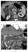
Figure 1
Figure 1
CT postcontrast examination in a patient who was suffering from jaundice and abdominal pain: axial images after contrast administration (a) and curved-MPR images (b). White arrows in (a) and (b) show a marked dilatation of the entire main pancreatic duct, from the head to the tail of the gland, associated with subtotal parenchymal atrophy. No dilatation of secondary branches was observed, and radiological diagnosis of MD-IPMN was formulated. The high degree of main pancreatic duct dilatation (>1 cm) was considered as high-risk stigmata and required surgical treatment. In addition, white arrows show mild wall enhancement. Final diagnosis of invasive cancer (adenocarcinoma) in IPMN was reported.
Figure 2
Figure 2
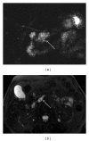
BD-IPMN in a 67-year-old female. MRCP acquisition clearly shows a cystic lesion centred on the body of pancreatic parenchyma (white arrows). The cyst shows a curved tubular shape. Due to the absence of high-risk-stigmata and worrisome features, lesions were safely managed.
Figure 3
Figure 3
MRCP images (a and b), obtained using 2D FSE sequence and 3D FRFSE technique, respectively. BD-IPMN of about 3 centimeters located in the uncinate process of pancreas, with a typical microcystic appearance. No other worrisome features were found by EUS; the patient was successfully enrolled in a follow-up program.
Figure 4
Figure 4
Axial T1-weighted spoiled gradient echo after gadolinium administration (a). 3D FRFSE MRCP sequence obtained using MIP reconstruction (b). Surgical specimen (c), from poster EPOS C-2228 presented in [15]. (a) shows a homogeneous cystic lesion centered in the head of pancreas. No intralesion solid components were observed. In (b), MIP reconstruction was useful to better appreciate the cystic morphology of the lesion due to main pancreatic duct enlargement. Again, high-risk stigmata (main duct caliber >1 centimeter) suggested surgical management. A pancreatoduodenectomy was performed and final diagnosis deposed for borderline IPMN.
Figure 5
Figure 5
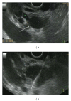
Linear EUS image of a MD-IPMN (a): a lobulated anechoic cystic lesion is clearly depicted (white arrow). (b) shows EUS-FNA of the same lesions. In this lesion (about 3 cm in size), the absence of mural nodules and positive or suspicious cytology allowed a conservative management.
Figure 6
Figure 6
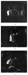
Coronal MRCP acquisitions in an asymptomatic 70-year-old female patient with an incidental radiological finding of multiple pancreatic cystic lesions; MRCP exams were performed in 2009 (a), in 2012 (b), and in 2013 (c). Multiple small cystic lesions in the pancreatic parenchyma are clearly depicted in the three MRCP acquisitions, some of them showing a typical connection to the main pancreatic duct. This typical radiological pattern suggests the diagnosis of multifocal BD-IPMNs. No main pancreatic duct dilatation is observed. Cystic lesions do not show intraluminal solid components or mural nodules. Over time the MRI monitoring initially showed a mild enlargement of the lesions (from a to b) and then a size-reduction (from b to c). As reported in literature, IPMNs in a multifocal setting could also be managed in a safe and reliable mode.

Similar articles

Cited by

References

    1. Acar M, Tatli S. Cystic tumors of the pancreas: a radiological perspective. Diagnostic and Interventional Radiology. 2011;17(2):143–149. - PubMed
    1. Suyama M, Ariyama H, Ogawa K. Clinical diagnosis of mucin producing pancreatic carcinoma. Tan to Sui. 1986;7:739–745.
    1. Sahani DV, Kadavigere R, Saokar A, Fernandez-del Castillo C, Brugge WR, Hahn PF. Cystic pancreatic lesions: a simple imaging-based classification system for guiding management. Radiographics. 2005;25(6):1471–1484. - PubMed
    1. Manfredi R, Mehrabi S, Motton M, et al. MR imaging and MR cholangiopancreatography of multifocal intraductal papillary mucinous neoplasms of the side branches: MR pattern and its evolution. Radiologia Medica. 2008;113(3):414–428. - PubMed
    1. Salvia R, Crippa S, Falconi M, et al. Branch-duct intraductal papillary mucinous neoplasms of the pancreas: to operate or not to operate? Gut. 2007;56(8):1086–1090. - PMC - PubMed

LinkOut - more resources