

Usefulness of 11C-methionine positron emission tomography for detecting intracranial ameloblastic carcinoma: A case report
- PMID: 25202358
- PMCID: PMC4156171
- DOI: 10.3892/ol.2014.2352
Usefulness of 11C-methionine positron emission tomography for detecting intracranial ameloblastic carcinoma: A case report
Abstract
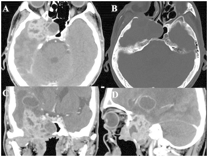
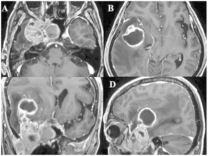
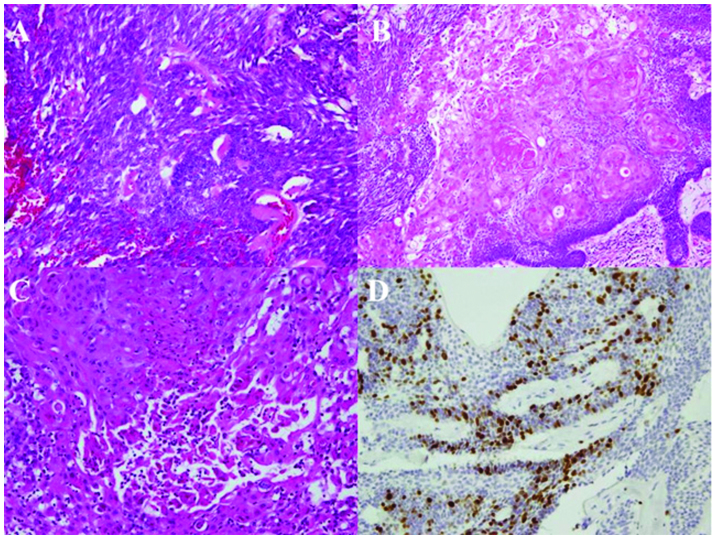
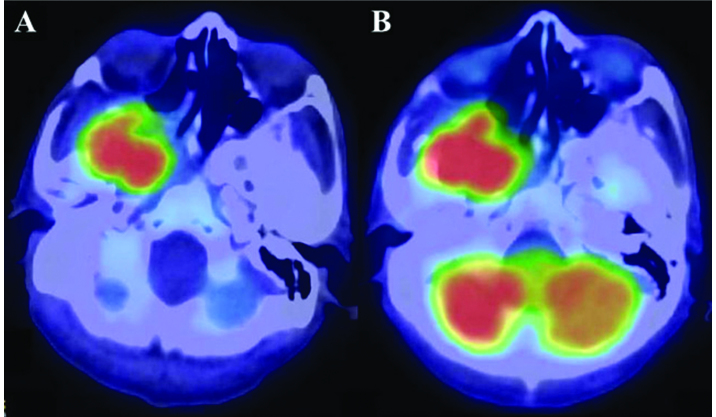
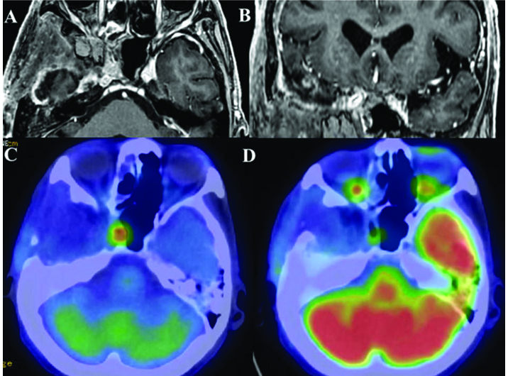
Ameloblastic carcinoma, secondary type, is an extremely rare odontogenic malignant tumor. The present study reports the case of a 58-year-old male with ameloblastic carcinoma that extended into the intracranial space close to the internal carotid artery. Surgical excision was performed, as headaches were being caused via compression by the mass. Small remnants of the tumor remained surrounding the internal carotid artery following surgical resection. Although the remnant tissue was not detected on magnetic resonance imaging or 18F-fluorodeoxyglucose (FDG)-positron emission tomography (PET), it was clearly visualized on 11C-methionine PET in the early post-operative follow-up period. No neurological deficits were exhibited during the follow-up period, and 11C-methionine PET was able to detect the remnant lesion distribution in the intracranial space. The current study presents a rare case of ameloblastic carcinoma that extended into the intracranial space. In addition, several diagnostic imaging tools were compared in order to determine the most suitable imaging modality. At present, the patient is continuing a therapeutic course of radiation and evident mass reduction has been observed. However, the therapeutic effects are currently under consideration. To the best of our knowledge, this is the first study on the effectiveness of using 11C-methionine PET for detecting ameloblastic carcinoma with intracranial extension.
Keywords: ameloblastic carcinoma; intracranial extension; methionine-labeled positron emission tomography.
Figures
Similar articles
-
More advantages in detecting bone and soft tissue metastases from prostate cancer using 18F-PSMA PET/CT.Hell J Nucl Med. 2019 Jan-Apr;22(1):6-9. doi: 10.1967/s002449910952. Epub 2019 Mar 7. Hell J Nucl Med. 2019. PMID: 30843003
-
Metabolic assessment of intracranial tuberculomas using 11C-methionine and 18F-FDG PET/CT.Nucl Med Commun. 2012 Apr;33(4):408-14. doi: 10.1097/MNM.0b013e32834f9b14. Nucl Med Commun. 2012. PMID: 22301451
-
Ameloblastic carcinoma: a case report with radiological features of computed tomography and magnetic resonance imaging and positron emission tomography.Oral Surg Oral Med Oral Pathol Oral Radiol Endod. 2011 Jul;112(1):e40-7. doi: 10.1016/j.tripleo.2011.01.023. Epub 2011 Mar 31. Oral Surg Oral Med Oral Pathol Oral Radiol Endod. 2011. PMID: 21458329
-
Ameloblastic carcinoma of the mandible: Report of a case and review.J Oral Maxillofac Pathol. 2014 Sep;18(Suppl 1):S96-S102. doi: 10.4103/0973-029X.141336. J Oral Maxillofac Pathol. 2014. PMID: 25364189 Free PMC article. Review.
-
Literature review of 86 cases of mandibular ameloblastic carcinoma.Natl J Maxillofac Surg. 2018 Jan-Jun;9(1):2-7. doi: 10.4103/njms.NJMS_33_16. Natl J Maxillofac Surg. 2018. PMID: 29937652 Free PMC article. Review.
Cited by
-
Ameloblastic carcinoma of the maxilla: A case report and an updated review of the literature.Oncol Lett. 2016 Dec;12(6):4339-4350. doi: 10.3892/ol.2016.5272. Epub 2016 Oct 17. Oncol Lett. 2016. PMID: 28105148 Free PMC article.
References
-
- Yoshioka Y, Toratani S, Ogawa I, Okamaoto T. Ameloblastic carcinoma, secondary type, of the mandible: a case report. J Oral Maxillofac Surg. 2013;71:e58–e62. - PubMed
-
- Barnes L, Eveson J, Reichat P, Sidransky D. Pathology and Genetics of Head and Neck Tumours. IARC Press; Lyon, France: 2005. World Health Organization Classification of Tumours; pp. 286–291.
-
- Karakida K, Aoki T, Sakamoto H, et al. Ameloblastic carcinoma, secondary type: a case report. Oral Surg Oral Med Oral Pathol Oral Radiol Endod. 2010;110:e33–e37. - PubMed
LinkOut - more resources
Full Text Sources
Other Literature Sources