

Focal fibrocartilaginous dysplasia in the thoracic vertebra: A case report
- PMID: 25202364
- PMCID: PMC4156278
- DOI: 10.3892/ol.2014.2331
Focal fibrocartilaginous dysplasia in the thoracic vertebra: A case report
Abstract
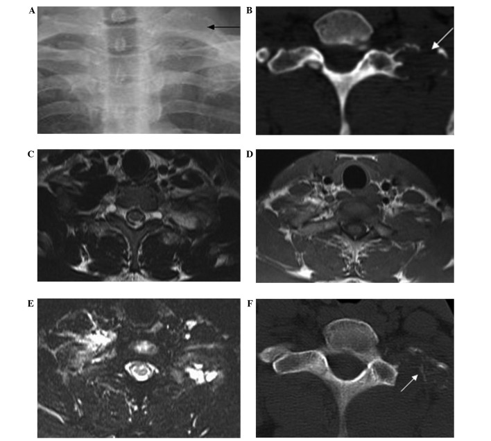
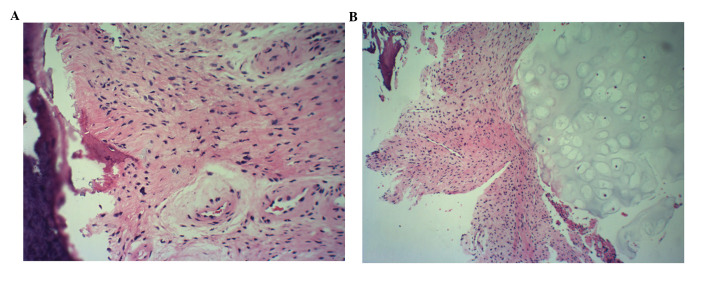
Focal fibrocartilaginous dysplasia (FFCD) is a rare, paraneoplastic disease that often presents in children and teenagers. Previous studies have reported cases of lesions in the proximal tibia and distal femur, as well as lesions in the upper extremities. The present study describes a case of FFCD on the transverse process and the rib. The imaging findings were found to correspond with the typical observations of FFCD and a biopsy from the nidus revealed pathological results similar to those of previous reports. Thus, the present study demonstrated that FFCD affects tubular bones as well as flat bones. Further studies are required to investigate the underlying mechanism and treatment of FFCD.
Keywords: focal fibrocartilaginous dysplasia; rib; thoracic vertebra.
Figures
Similar articles
-
Spontaneous resolution of angular deformity of the distal femur in focal fibrocartilaginous dysplasia: a case report.J Pediatr Orthop B. 2010 Mar;19(2):161-3. doi: 10.1097/BPB.0b013e3283361b11. J Pediatr Orthop B. 2010. PMID: 20051915
-
Spontaneous resolution of focal fibrocartilaginous dysplasia of femur on long-term follow-up: case report and review of literature.J Pediatr Orthop B. 2019 Mar;28(2):127-131. doi: 10.1097/BPB.0000000000000570. J Pediatr Orthop B. 2019. PMID: 30444750 Review.
-
Unilateral tibia vara in a toddler caused by focal fibrocartilaginous dysplasia.J Radiol Case Rep. 2009;3(9):14-7. doi: 10.3941/jrcr.v3i9.280. Epub 2009 Sep 1. J Radiol Case Rep. 2009. PMID: 22470683 Free PMC article.
-
Two unusual presentations of focal fibrocartilaginous dysplasia.J Pediatr Orthop B. 2013 Jul;22(4):367-71. doi: 10.1097/BPB.0b013e3283563732. J Pediatr Orthop B. 2013. PMID: 22751479
-
Correction of Angular Deformities Due to Focal Fibrocartilaginous Dysplasia Using Guided Growth: A Preliminary Report.J Pediatr Orthop. 2017 Apr/May;37(3):e183-e187. doi: 10.1097/BPO.0000000000000785. J Pediatr Orthop. 2017. PMID: 27261964 Review.
Cited by
-
Tibia vara caused by focal fibrocartilaginous dysplasia: A rare case report.SAGE Open Med Case Rep. 2024 Mar 4;12:2050313X241236150. doi: 10.1177/2050313X241236150. eCollection 2024. SAGE Open Med Case Rep. 2024. PMID: 38444693 Free PMC article.
-
Ulnar focal cortical indentation: a progressive, deforming variant of focal fibrocartilaginous dysplasia.Pediatr Radiol. 2019 Feb;49(2):187-195. doi: 10.1007/s00247-018-4294-6. Epub 2018 Nov 15. Pediatr Radiol. 2019. PMID: 30443667
References
-
- Bell SN, Campbell PE, Cole WG, Menelaus MB. Tibia vara caused by focal fibrocartilaginous dysplasia. Three case reports. J Bone Joint Surg Br. 1985;67:780–784. - PubMed
-
- Ohno I, Shimizu N, Nakase T, Yoshikawa H. Adult case of tibia vara associated with focal fibrocartilaginous dysplasia. J Orthop Sci. 2005;10:328–330. - PubMed
-
- Hermann G, Klein M, Abdelwahab IF, Kenan S. Fibrocartilaginous dysplasia. Skeletal Radiol. 1996;25:509–511. - PubMed
-
- Beaty JH, Barrett IR. Unilateral angular deformity of the distal end of the femur secondary to a focal fibrous tether. A report of four cases. J Bone Joint Surg Am. 1989;71:440–445. - PubMed
-
- Choi IH, Kim CJ, Cho TJ, et al. Focal fibrocartilaginous dysplasia of long bones: report of eight additional cases and literature review. J Pediatr Orthop. 2000;20:421–427. - PubMed
LinkOut - more resources
Full Text Sources
Other Literature Sources