

Synchronous unilateral basal cell adenoma of the parotid gland: A case report
- PMID: 25202417
- PMCID: PMC4156274
- DOI: 10.3892/ol.2014.2334
Synchronous unilateral basal cell adenoma of the parotid gland: A case report
Abstract
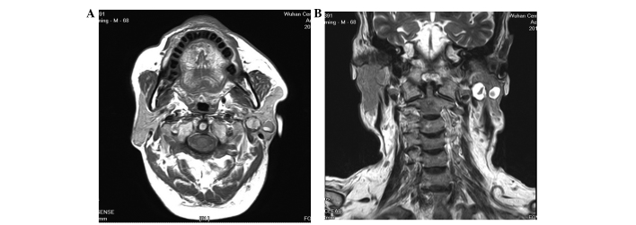
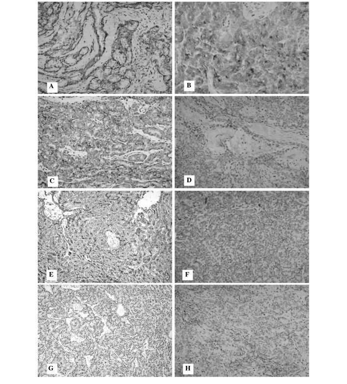
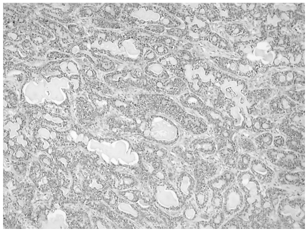
The current study reports the case of a 68-year-old male who presented with a 4-month history of a painless slow-growing mass in the left parotid region. Magnetic resonance imaging revealed two independent, round lesions in the superficial and deep lobes of the parotid gland on the left side, respectively. A total parotidectomy was performed and basal cell adenomas (BCAs) were identified by histopathological examination. At the 6-month follow-up examination, no sign of recurrence was found. This study describes the clinical features of a rare case of synchronous unilateral BCA in the parotid gland and also provides a review of the literature.
Keywords: basal cell adenoma; parotid gland; synchronous.
Figures
Similar articles
-
Synchronous bilateral multifocal basal cell adenomas of the parotid gland-a case report.BMC Oral Health. 2022 Jul 29;22(1):314. doi: 10.1186/s12903-022-02339-3. BMC Oral Health. 2022. PMID: 35906614 Free PMC article.
-
Unilateral parotid gland involvement with synchronous multiple Basal cell adenomas.J Craniofac Surg. 2007 Nov;18(6):1470-3. doi: 10.1097/SCS.0b013e3181534a9e. J Craniofac Surg. 2007. PMID: 17993904
-
Synchronous bilateral pleomorphic adenomas of the parotid gland.J Investig Clin Dent. 2012 Aug;3(3):225-7. doi: 10.1111/j.2041-1626.2012.00123.x. Epub 2012 Mar 19. J Investig Clin Dent. 2012. PMID: 22431298
-
Synchronous bilateral multifocal acinic cell carcinoma of parotid gland: case report and review of the literature.J Oral Maxillofac Surg. 2012 Oct;70(10):e574-80. doi: 10.1016/j.joms.2012.06.006. Epub 2012 Aug 3. J Oral Maxillofac Surg. 2012. PMID: 22868030 Review.
-
A retrospective study of 30 basal cell adenomas of the salivary gland in a Brazilian population and literature review.Eur Arch Otorhinolaryngol. 2021 Jul;278(7):2447-2454. doi: 10.1007/s00405-020-06331-x. Epub 2020 Sep 4. Eur Arch Otorhinolaryngol. 2021. PMID: 32886182 Review.
Cited by
-
Synchronous bilateral multifocal basal cell adenomas of the parotid gland-a case report.BMC Oral Health. 2022 Jul 29;22(1):314. doi: 10.1186/s12903-022-02339-3. BMC Oral Health. 2022. PMID: 35906614 Free PMC article.
References
-
- Kleinsasser O, Klein HJ. Basal cell adenoma of the salivary glands. Arch Klin Exp Ohr Nas Kehlkopsheilkd. 1967;189:302–316. (In German) - PubMed
-
- Seifert G, Sobin LH, editors. World Health Organization International Histological Classification of Tumours. Histological Typing of Salivary Gland Tumors. Springer-verlag; Berlin: 1991.
-
- Foote FW, Jr, Frazell EL. Tumors of the major salivary glands. Cancer. 1953;6:1065–1133. - PubMed
-
- Lam KH, Ho HC, Ho CM, Wei WI. Multifocal nature of adenolymphoma of the parotid. Br J Surg. 1994;81:1612–1614. - PubMed
-
- Maiorano E, Lo Muzio L, Favia G, Piattelli A. Warthin’s tumour: a study of 78 cases with emphasis on bilaterality, multifocality and association with other malignancies. Oral Oncol. 2002;38:35–40. - PubMed
LinkOut - more resources
Full Text Sources
Other Literature Sources