Review
. 2015 Feb;36(1):1-24.
doi: 10.1210/er.2014-1020. Epub 2014 Sep 9.

Intraovarian control of early folliculogenesis

Affiliations
Review

Intraovarian control of early folliculogenesis

Aaron J W Hsueh et al. Endocr Rev. 2015 Feb.

Abstract

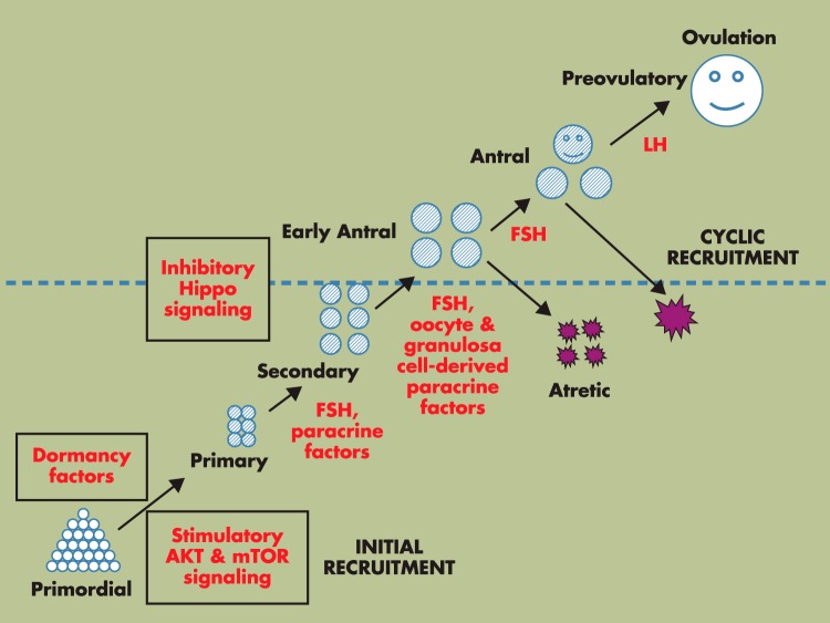
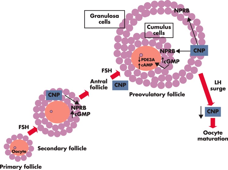
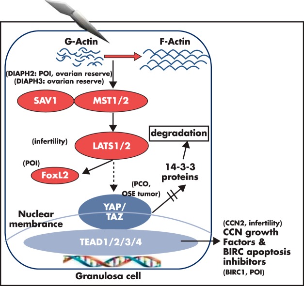
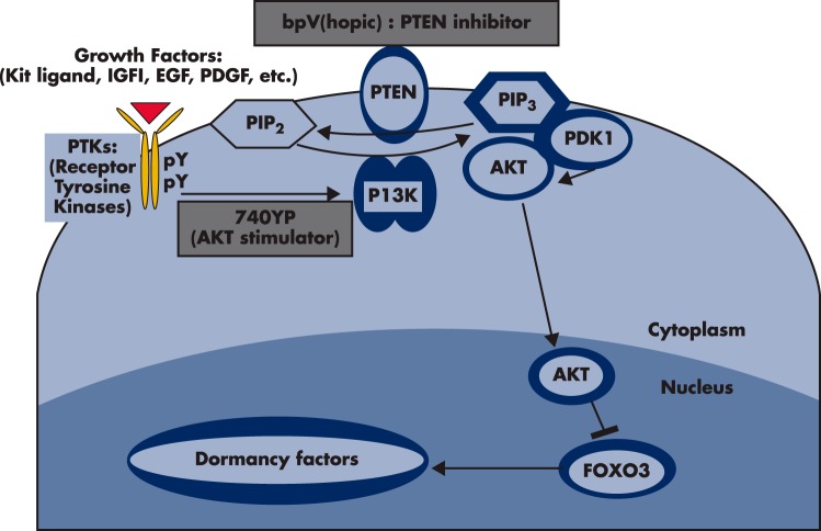
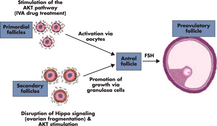
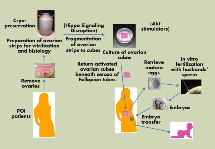
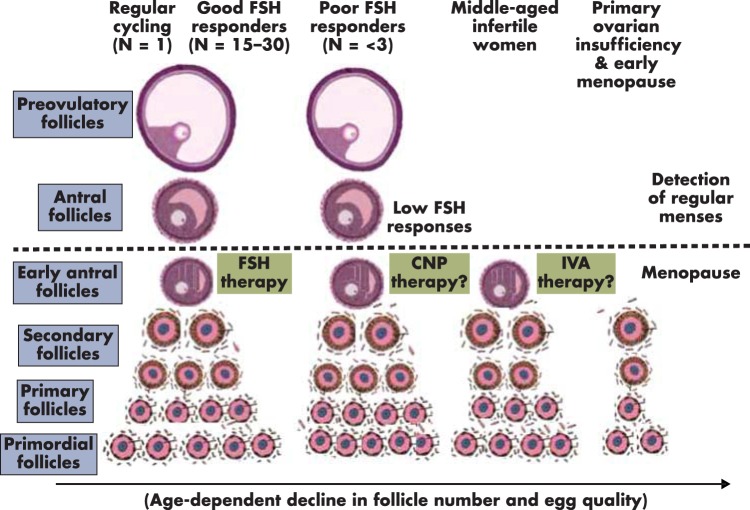
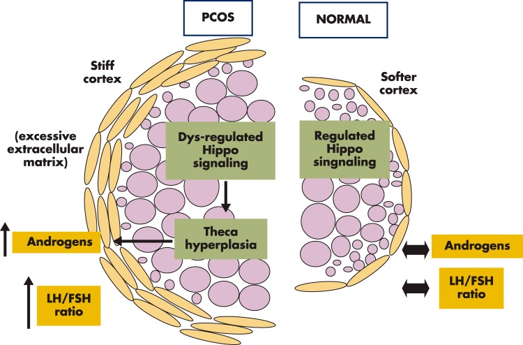
Although hormonal regulation of ovarian follicle development has been extensively investigated, most studies concentrate on the development of early antral follicles to the preovulatory stage, leading to the successful use of exogenous FSH for infertility treatment. Accumulating data indicate that preantral follicles are under stringent regulation by FSH and local intraovarian factors, thus providing the possibility to develop new therapeutic approaches. Granulosa cell-derived C-type natriuretic factor not only suppresses the final maturation of oocytes to undergo germinal vesicle breakdown before ovulation but also promotes preantral and antral follicle growth. In addition, several oocyte- and granulosa cell-derived factors stimulate preantral follicle growth by acting through wingless, receptor tyrosine kinase, receptor serine kinase, and other signaling pathways. In contrast, the ovarian Hippo signaling pathway constrains follicle growth and disruption of Hippo signaling promotes the secretion of downstream CCN growth factors capable of promoting follicle growth. Although the exact hormonal factors involved in primordial follicle activation has yet to be elucidated, the protein kinase B (AKT) and mammalian target of rapamycin signaling pathways are important for the activation of dormant primordial follicles. Hippo signaling disruption after ovarian fragmentation, combined with treating ovarian fragments with phosphatase and tensin homolog (PTEN) inhibitors and phosphoinositide-3-kinase stimulators to augment AKT signaling, promote the growth of preantral follicles in patients with primary ovarian insufficiency, leading to a new infertility intervention for such patients. Elucidation of intraovarian mechanisms underlying early folliculogenesis may allow the development of novel therapeutic strategies for patients diagnosed with primary ovarian insufficiency, polycystic ovary syndrome, and poor ovarian response to FSH stimulation, as well as for infertile women of advanced reproductive age.


Figures

Figure 1.
Figure 1.
Hormonal regulation of preantral follicle growth. Selective primordial follicles develop to the primary stage under the control of AKT and mTOR signaling (initial recruitment), whereas most primordial follicles remain arrested by dormancy factors. Once initiated to growth, primordial follicles develop through primary and secondary stages before acquiring an antral cavity. Although most early antral follicles undergo atresia, select antral follicles supported by cyclic changes in pituitary FSH and LH reach the preovulatory stage, capable of releasing mature oocytes after ovulation for fertilization (cyclic recruitment) (1). In addition to the well-studied role of FSH on antral follicle growth (above the dashed line), FSH also regulates preantral follicle growth, together with a large number of oocyte- and granulosa cell-derived paracrine factors (below the dashed line). Furthermore, development of preantral and antral follicle is restrained by the inhibitory Hippo signaling pathway.
Figure 2.
Figure 2.
CNP is an intraovarian factor important for preantral and antral follicle growth as well as for oocyte maturation inhibition. Based on murine studies, CNP is secreted by granulosa cells of secondary and antral follicles in response to FSH stimulation. CNP acts through its receptor NPRB, expressed in granulosa cells of secondary follicles, to increase cGMP production and to stimulate follicle development (38). In addition, CNP acts through its receptor, expressed in cumulus cells of antral and preovulatory follicles, to increase cGMP production. Cumulus cell-produced cGMP, after transporting to oocytes through gap junctions, inhibits the activity of the phosphodiesterase 3A (PDE3A) enzyme, leading to increased intra-oocyte cAMP levels, thus suppressing oocyte maturation (30). The preovulatory LH surge decreases CNP levels in the preovulatory follicles and allows meiotic maturation of preovulatory oocytes (32).
Figure 3.
Figure 3.
Actin polymerization disrupts ovarian Hippo signaling and promotes nuclear YAP actions to increase downstream CCN growth factors and apoptosis inhibitors involvement in POI, PCOS, follicle reserve, and infertility. The Hippo serine/threonine kinase signaling cascade is conserved from flies to mammals to restrict tissue growth. Mammalian MST1 and MST2 are orthologous to the fly Hpo (Hippo) gene. MST1/2, acting in concert with Sav1, phosphorylates downstream LATS1/2 to suppress YAP and TAZ actions by phosphorylating YAP and TAZ, leading to the degradation of phospho-YAP/TAZ after binding to cytoplasmic 14–3-3 proteins. Ovarian fragmentation induces actin polymerization (conversion of G-actin to F-actin) and disruption of the Hippo signaling pathway, leading to decreases in YAP phosphorylation and increased nuclear levels of YAP (103). Nuclear YAP interacts with cotranscriptional factors TEAD1/2/3/4 to stimulate the expression of downstream CCN growth factors and BIRC apoptosis inhibitors, leading to cell proliferation. Genomic and genetic studies underscore the important roles of the ovarian Hippo signaling pathway. DIAPH genes are important for actin polymerization. Disruption of DIAPH2 has been found in a familiar case of POI (126), whereas this gene is also important for ovarian reserve in a genome-wide association study (127). Likewise, DIAPH3 has been implicated in regulating ovarian reserve in a genome-wide association study (230). For Hippo pathway genes, LATS1 deletion in mice leads to infertility and ovarian tumorigenesis (129), whereas LATS1 regulates the activity of FOXL2 (130), a gene defective in some POI patients. Genome-wide association studies also implicate the Hippo effector gene YAP in PCO patients (131), whereas overexpression of YAP was found in ovarian surface epithelium (OSE) tumors (132). For downstream genes, conditional deletion of CCN2 in granulosa cells leads to subfertility (133), whereas gene copy changes for the BIRC1 apoptosis inhibitor gene is associated with POI in patients (134).
Figure 4.
Figure 4.
The PTEN-PI3K-AKT pathway in oocytes regulates primordial follicle activation. Mouse models were used to investigate the regulation of primordial follicle dormancy. The FOXO3 gene in primordial oocytes serves as a break to prevent the initiation of follicle growth. Activation of upstream RTKs by their cognate ligands (kit ligand, IGF-1, EGF, platelet-derived growth factor [PDGF], VEGF, etc) stimulates the autophosphorylation of intracellular regions of these receptors. Activated receptors then stimulate PI3K activity, leading to increases in PIP3 levels and AKT stimulation. Activated AKT then migrates to the cell nucleus and suppresses FOXO3 actions to promote primordial follicle growth. The PTEN gene encodes an enzyme that converts PIP3 to PIP2, thus damping the actions of PI3K. Oocyte-specific deletion of the PTEN gene leads to global activation of primordial follicles (156) whereas treatment with PTEN inhibitors, bpV(hopic) [(5-hydroxy-2-pyridinecarboxylato-kN1, kO2)oxodiperoxy-vanadate (2-), dipotassium], promotes primordial follicle activation (165). Although the exact ligand-receptor pairs responsible for the activation of primordial follicles during physiological conditions are unknown, treatment of ovaries with a cell membrane-permeable peptide (740Y-P, designed based on the phosphorylated intracellular region of the PDGF receptor) stimulates AKT signaling and promotes the activation of primordial follicles (165).
Figure 5.
Figure 5.
Ovarian fragmentation and IVA drug treatment promote follicle growth via different mechanisms. Incubation of ovaries with PTEN inhibitors and PI3K stimulators activate dormant primordial follicles by increasing oocyte AKT activity to promote nuclear exclusion of FOXO3, thus preventing its suppression of primordial follicle growth. For secondary follicles, ovarian fragmentation disrupts Hippo signaling to increase downstream CCN growth factors and BIRC apoptosis inhibitors, leading to follicle growth. In addition, concomitant treatment with PTEN inhibitor and PI3K stimulators leads to additive promotion of follicle growth by increasing AKT activity in granulosa cells of secondary follicles.
Figure 6.
Figure 6.
Ovarian fragmentation/AKT stimulation followed by autografting promotes follicle growth in POI patients to generate mature oocytes for IVF embryo transfer, pregnancy, and delivery. Under laparoscopic surgery, one or both ovaries from POI patients were removed and cut into strips before vitrification. After thawing, strips were fragmented into 1- to 2-mm2 cubes, before treatment with AKT stimulators (PTEN inhibitors and PI3K stimulator). Two days later, cubes were autografted under laparoscopic surgery beneath the serosa of Fallopian tubes. Follicle growth was monitored weekly or biweekly via transvaginal ultrasound and based on serum estrogen levels. After detection of antral follicles, patients were treated with FSH followed by human chorionic gonadotropin when preovulatory follicles were found. Mature oocytes were then retrieved and fertilized with husbands' sperm in vitro before cyropreservation of 4-cell-stage embryos. Patients then received hormonal treatments to prepare the endometrium for implantation followed by transferring of thawed embryos and pregnancy. [Modified from K. Kawamura et al: Hippo signaling disruption and Akt stimulation of ovarian follicles for infertility treatment. Proc Natl Acad Sci U S A. 2013;110:17474 (103), with permission. © National Academy of Sciences of the United States of America.]
Figure 7.
Figure 7.
Diagrammatic representation of hypothesized follicle dynamics under different clinical conditions. In women with regular menstrual cyclicity (left panel), ∼20 early antral follicles are present during the early follicular phase and produce low amounts of sex steroids. During the first half of the menstrual cycle, development of a single antral and then preovulatory follicle (n = 1) confers the secretion of sufficient estrogens and progesterone to induce uterine changes, resulting in regular menses. Treatment of these women with exogenous gonadotropins promotes development of a large number [15 to 30] of preovulatory follicles (good FSH responders). Ovarian activity above the dashed line denotes sufficient ovarian sex steroid secretion to affect uterine functions, leading to regular menstrual cycles. For poor FSH responders, early antral follicles may be present, but insufficient expression of functional FSH receptors could lead to the development of fewer (<3) preovulatory follicles after exogenous FSH stimulation. These patients could benefit from CNP therapies. For middle-aged infertile women with irregular menstrual cycles, few early antral follicles are present, but many preantral follicles still exist. These patients could benefit from IVA therapy or treatment with ovarian paracrine factors, including CNP. For POI patients and infertile women showing early menopause before 51 years of age, residual preantral follicles could still be present in the ovary. The IVA procedure promotes primordial follicle activation as well as stimulates secondary follicle growth by combining Hippo signaling disruption and AKT stimulation. The arrow at the bottom emphasizes the age-dependent decline in follicle number and egg quality under both physiological and pathophysiological conditions.
Figure 8.
Figure 8.
Hypothesized ovarian structural abnormality in PCOS. Aberrant extracellular matrix and Hippo signaling defects lead to thecal hyperplasia and the PCO phenotype. Normal ovaries have softer cortexes, and Hippo signaling restrains most follicles from overgrowth, leading to physiological levels of ovarian androgen secretion and serum LH to FSH ratios. A subgroup of PCOs could have defects in extracellular matrix regulation, leading to rigid and sclerotic cortexes. The stiff cortex of these ovaries could lead to dysregulation of Hippo signaling and excessive proliferation of stromal, theca, and granulosa cells. Thecal cell hyperplasia could increase androgen production and elevate ratios of serum LH to FSH levels, followed by the arrest of a high number of early antral follicles and enlarged ovaries characteristic of PCOS.

References

    1. McGee EA, Hsueh AJ. Initial and cyclic recruitment of ovarian follicles. Endocr Rev. 2000;21:200–214. - PubMed
    1. Hsueh AJ, Billig H, Tsafriri A. Ovarian follicle atresia: a hormonally controlled apoptotic process. Endocr Rev. 1994;15:707–724. - PubMed
    1. Zeleznik AJ. Follicle selection in primates: “many are called but few are chosen”. Biol Reprod. 2001;65:655–659. - PubMed
    1. Richards JS. Maturation of ovarian follicles: actions and interactions of pituitary and ovarian hormones on follicular cell differentiation. Physiol Rev. 1980;60:51–89. - PubMed
    1. Goodman A, Hodgen G. The ovarian triad of the primate menstrual cycle. Recent Prog Horm Res. 1983;39:1. - PubMed

Publication types

MeSH terms

Substances