

doi: 10.1016/j.cub.2014.06.044.
Stentor coeruleus
Affiliations
- PMID: 25202864
- PMCID: PMC5036449
- DOI: 10.1016/j.cub.2014.06.044
Item in Clipboard
Stentor coeruleus
Curr Biol.
.
No abstract available
Figures
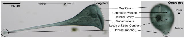
Brightfield images of both an extended cell (left) and a contracted cell (right) highlighting a few key features. The oral cilia are at the anterior and constantly beat to create a vortex to capture food particles in the buccal cavity. The contractile vacuole can be seen beneath the cell membrane and is also associated with scars in the membrane called contractile vacuole pores. The cell’s macronucleus is stretched along its length and packaged into distinct nodes, giving it the appearance of ‘beads on a string’. The cortical stripes are also visible; the clear stripes represent microtubule bundles known as Km fibers and the blue/green stripes contain dense fields of pigment granules. Asymmetric spacing of the rows results in a point at which the wide stripes meet the narrow stripes and is called the locus of stripe contrast. The posterior of the cell is defined by an anchoring structure known as the holdfast.
References
-
- Lillie FR. On the smallest parts of Stentor capable of regeneration. J Morphol. 1896;12:239–249.
-
- Morgan TH. Regeneration of proportionate structures in Stentor. The Biological Bulletin 1901
-
- Tartar V. International Series of Monographs on Pure and Applied Biology. Vol. 5. Pergamon Press; 1961. The Biology of Stentor; pp. 1–434.
-
- De Terra N. Cytoskeletal discontinuities in the cell body cortex initiate basal body assembly and oral development in the ciliate Stentor. J Embryol Exp Morph. 1985;87:249–257. - PubMed
MeSH terms
Grants and funding
LinkOut - more resources
Full Text Sources
Other Literature Sources
