Review
. 2014 Sep 8;24(17):R815-25.
doi: 10.1016/j.cub.2014.06.068.

Epithelial homeostasis

Affiliations
Review

Epithelial homeostasis

Ian G Macara et al. Curr Biol. .

Abstract

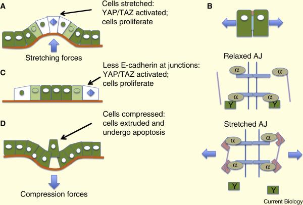
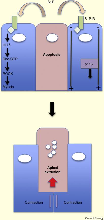
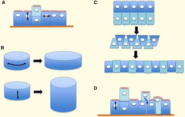
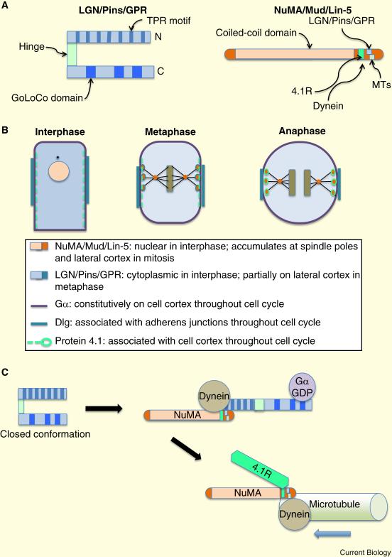
Epithelia form intelligent, dynamic barriers between the external environment and an organism's interior. Intercellular cadherin-based adhesions adapt and respond to mechanical forces and cell density, while tight junctions flexibly control diffusion both within the plasma membrane and between adjacent cells. Epithelial integrity and homeostasis are of central importance to survival, and mechanisms have evolved to ensure these processes are maintained during growth and in response to damage. For instance, cell competition surveys the fitness of cells within epithelia and removes the less fit; extrusion or delamination can remove apoptotic or defective cells from the epithelial sheet and can restore homeostasis when an epithelial layer becomes too crowded; spindle orientation ensures two-dimensional growth in simple epithelia and controls stratification in complex epithelia; and transition to a mesenchymal phenotype enables active escape from an epithelial layer. This review will discuss these various mechanisms and consider how they are subverted in disease.


Figures

Figure 1
Figure 1
Effects of intercellular tension on epithelial homeostasis. A. Cell stretching reduces intercellular tension and activates proliferation through YAP/TAZ signaling (YAP marked green, showing nuclear localization in stretched cells). B. A speculative model for YAP/TAZ activation in which stretched adherens junctions bind vinculin (pink rectangles) and actin (purple lines) and release YAP. C. At the edges of epithelial sheets there is less compression, resulting in nuclear YAP and cell proliferation. D. Compressive forces trigger cell extrusion.
Figure 2
Figure 2
Signaling involved in the extrusion of apoptotic cells. Cells undergoing apoptosis release sphingosine-1-phosphate, which activates Rho/ROCK signaling in neighboring cells, resulting in basal actomyosin contraction and apical extrusion.
Figure 3
Figure 3
Consequences of different mitotic spindle orientations in epithelial tissues. A. Divisions perpendicular to the epithelial sheet generate multilayering; those parallel to the sheet extend the surface area of the sheet. B. In ducts, divisions in the X plane will increase the diameter of the duct, while those in the Y plane will increase its length. C. Convergent extension through cell intercalation alters the shape of epithelial sheets in the absence of oriented divisions. D. Relocation of cells by migration and insertion can restore single layering of epithelial sheets.
Figure 4
Figure 4
The molecular basis for spindle orientation. A. The LGN protein and its homologues consists of 2 domains connected by a hinge. The N-terminal TPR motifs bind to NuMA and to Inscuteable; the C-terminal GoLoCo domains bind to Gα-GDP subunits. NuMA is a very large protein with multiple C-terminal motifs that bind multiple proteins involved in spindle orientation. B. Schematic showing the location of spindle orientation proteins at different phases of the cell cycle. C. In metaphase, LGN undergoes a conformational switch. In the open state it binds both to Gα at the cell cortex, and to NuMA and dynein. In anaphase LGN is replaced by the cytoskeletal protein 4.1R.
Figure 5
Figure 5
Mutually inhibitory circuits are inherently unstable but in response to small perturbations to the concentration of one of the two factors (A and B) can switch to one of 2 stable states. This type of circuit might control the decision by cells to become either mesenchymal or epithelial.
Figure 6
Figure 6
Cell competition. A. The schematic shows the consequences when a clone of less fit cells (pink), for instance that is mutant for a polarity protein, is surrounded by wild type cells (pale green). The less fit cells at the boundary undergo apoptosis (red nuclei), and are replaced by the more fit cells through compensatory proliferation. B. The cells sense relative fitness, such that a clone of wild type cells will be eliminated by “supercompetitor” neighbors (dark green). C. Expression of an oncogene such as Ras (yellow nuclei), which would by itself cause hyperproliferation but not invasive outgrowth, can drive invasion in the context of a polarity mutation. The polarity mutant cells (pink) would normally be induced to undergo apoptosis by their more fit neighbors (as in A) but the oncogene subverts the apoptotic signaling into a proliferative state that can drive invasive tumorigenesis.

Similar articles

Cited by

References

    1. Sato T, Clevers H. Growing self-organizing mini-guts from a single intestinal stem cell: mechanism and applications. Science. 2013;340:1190–1194. - PubMed
    1. Alexandre C, Baena-Lopez A, Vincent JP. Patterning and growth control by membrane-tethered Wingless. Nature. 2014;505:180–185. - PMC - PubMed
    1. Matis M, Axelrod JD. Regulation of PCP by the Fat signaling pathway. Genes Dev. 2013;27:2207–2220. - PMC - PubMed
    1. Lawrence PA, Casal J. The mechanisms of planar cell polarity, growth and the Hippo pathway: some known unknowns. Dev Biol. 2013;377:1–8. - PMC - PubMed
    1. Simoes Sde M, Blankenship JT, Weitz O, Farrell DL, Tamada M, Fernandez-Gonzalez R, Zallen JA. Rho-kinase directs Bazooka/Par-3 planar polarity during Drosophila axis elongation. Dev Cell. 2010;19:377–388. - PMC - PubMed

LinkOut - more resources