

Transient vascularization of transplanted human adult-derived progenitors promotes self-organizing cartilage
- PMID: 25202983
- PMCID: PMC4191036
- DOI: 10.1172/JCI76443
Transient vascularization of transplanted human adult-derived progenitors promotes self-organizing cartilage
Abstract
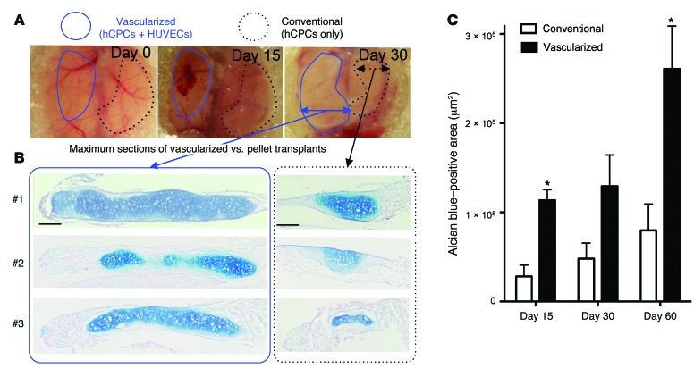
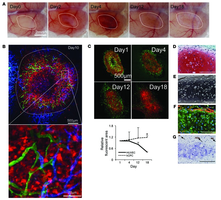
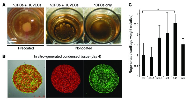
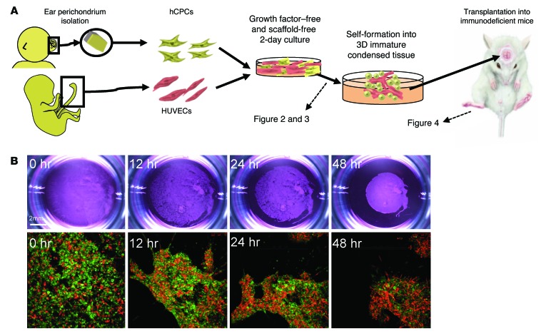
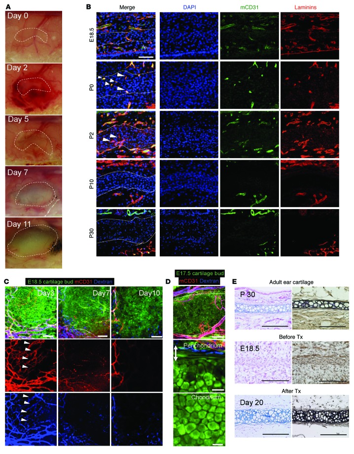
Millions of patients worldwide are affected by craniofacial deformations caused by congenital defects or trauma. Current surgical interventions have limited therapeutic outcomes; therefore, methods that would allow cartilage restoration are of great interest. A number of studies on embryonic limb development have shown that chondrogenesis is initiated by cellular condensation, during which mesenchymal progenitors aggregate and form 3D structures. Here, we demonstrated efficient regeneration of avascular elastic cartilage from in vitro-grown mesenchymal condensation, which recapitulated the early stages of chondrogenesis, including transient vascularization. After transplantation of vascularized condensed progenitors into immunodeficient mice, we used an intravital imaging approach to follow cartilage maturation. We determined that endothelial cells are present inside rudimentary cartilage (mesenchymal condensation) prior to cartilage maturation. Recreation of endothelial interactions in culture enabled a recently identified population of adult elastic cartilage progenitors to generate mesenchymal condensation in a self-driven manner, without requiring the support of exogenous inductive factors or scaffold materials. Moreover, the culture-grown 3D condensed adult-derived progenitors were amenable to storage via simple freezing methods and efficiently reconstructed 3D elastic cartilage upon transplantation. Together, our results indicate that transplantation of endothelialized and condensed progenitors represents a promising approach to realizing a regenerative medicine treatment for craniofacial deformations.
Figures
Similar articles
-
Angiogenic Potential of Human Bone Marrow-Derived Mesenchymal Stem Cells in Chondrocyte Brick-Enriched Constructs Promoted Stable Regeneration of Craniofacial Cartilage.Stem Cells Transl Med. 2017 Feb;6(2):601-612. doi: 10.5966/sctm.2016-0050. Epub 2016 Sep 14. Stem Cells Transl Med. 2017. PMID: 28191761 Free PMC article.
-
Mesenchymal stem cell-derived extracellular matrix enhances chondrogenic phenotype of and cartilage formation by encapsulated chondrocytes in vitro and in vivo.Acta Biomater. 2018 Mar 15;69:71-82. doi: 10.1016/j.actbio.2017.12.043. Epub 2018 Jan 6. Acta Biomater. 2018. PMID: 29317369 Free PMC article.
-
Progress of co-culture systems in cartilage regeneration.Expert Opin Biol Ther. 2018 Nov;18(11):1151-1158. doi: 10.1080/14712598.2018.1533116. Epub 2018 Oct 10. Expert Opin Biol Ther. 2018. PMID: 30295075 Review.
-
Fibroblast growth factor receptors in in vitro and in vivo chondrogenesis: relating tissue engineering using adult mesenchymal stem cells to embryonic development.Tissue Eng Part A. 2010 Feb;16(2):545-56. doi: 10.1089/ten.TEA.2008.0551. Tissue Eng Part A. 2010. PMID: 19728793
-
Mesenchymal stem cells for cartilage engineering.Biomed Mater Eng. 2012;22(1-3):69-80. doi: 10.3233/BME-2012-0691. Biomed Mater Eng. 2012. PMID: 22766704 Review.
Cited by
-
Rejuvenated Stem/Progenitor Cells for Cartilage Repair Using the Pluripotent Stem Cell Technology.Bioengineering (Basel). 2021 Apr 10;8(4):46. doi: 10.3390/bioengineering8040046. Bioengineering (Basel). 2021. PMID: 33920285 Free PMC article. Review.
-
Cell membrane fluidity and ROS resistance define DMSO tolerance of cryopreserved synovial MSCs and HUVECs.Stem Cell Res Ther. 2022 May 3;13(1):177. doi: 10.1186/s13287-022-02850-y. Stem Cell Res Ther. 2022. PMID: 35505370 Free PMC article.
-
Requirement of direct contact between chondrocytes and macrophages for the maturation of regenerative cartilage.Sci Rep. 2021 Nov 18;11(1):22476. doi: 10.1038/s41598-021-01437-6. Sci Rep. 2021. PMID: 34795319 Free PMC article.
-
Electron microscopic observation of human auricular chondrocytes transplanted into peritoneal cavity of nude mice for cartilage regeneration.Regen Ther. 2017 Dec 15;8:1-8. doi: 10.1016/j.reth.2017.11.002. eCollection 2018 Jun. Regen Ther. 2017. PMID: 30271859 Free PMC article.
-
Biological aspects of tissue-engineered cartilage.Histochem Cell Biol. 2018 Apr;149(4):375-381. doi: 10.1007/s00418-018-1652-2. Epub 2018 Mar 6. Histochem Cell Biol. 2018. PMID: 29511835 Review.
References
Publication types
MeSH terms
Substances
LinkOut - more resources
Full Text Sources
Other Literature Sources
Medical
