. 2014 Sep 9;9(9):e102034.
doi: 10.1371/journal.pone.0102034. eCollection 2014.

Vessel labeling in combined confocal scanning laser ophthalmoscopy and optical coherence tomography images: criteria for blood vessel discrimination

Affiliations

Vessel labeling in combined confocal scanning laser ophthalmoscopy and optical coherence tomography images: criteria for blood vessel discrimination

Jeremias Motte et al. PLoS One. .

Abstract

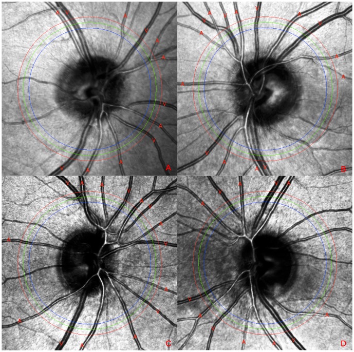
Introduction: The diagnostic potential of optical coherence tomography (OCT) in neurological diseases is intensively discussed. Besides the sectional view of the retina, modern OCT scanners produce a simultaneous top-view confocal scanning laser ophthalmoscopy (cSLO) image including the option to evaluate retinal vessels. A correct discrimination between arteries and veins (labeling) is vital for detecting vascular differences between healthy subjects and patients. Up to now, criteria for labeling (cSLO) images generated by OCT scanners do not exist.

Objective: This study reviewed labeling criteria originally developed for color fundus photography (CFP) images.

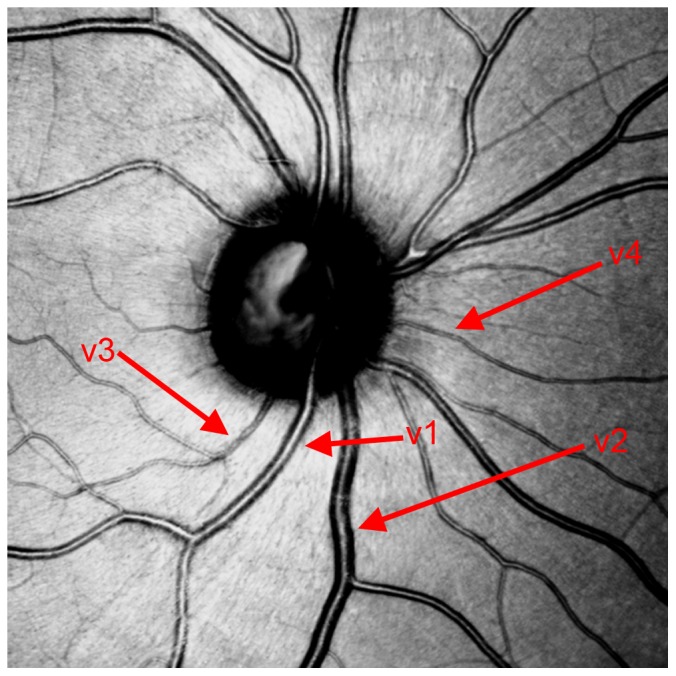
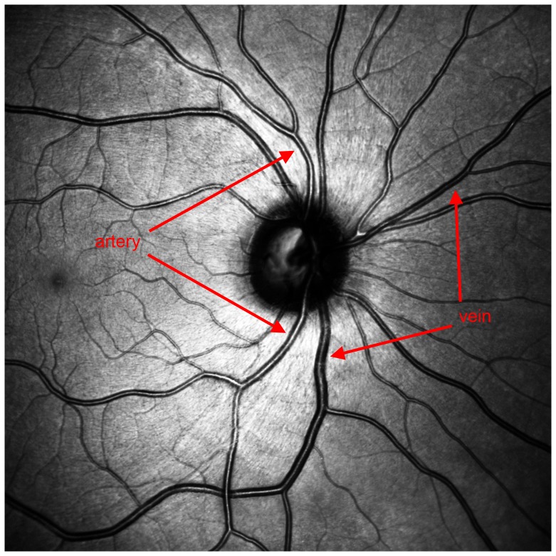
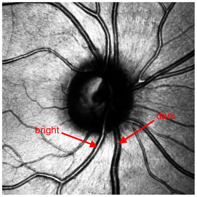
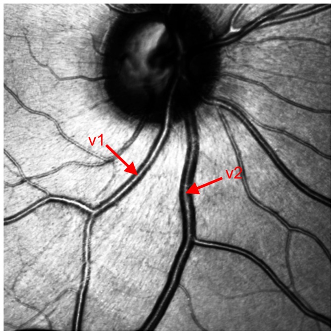
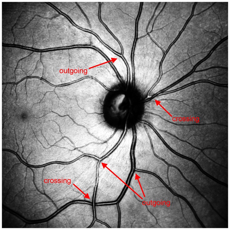
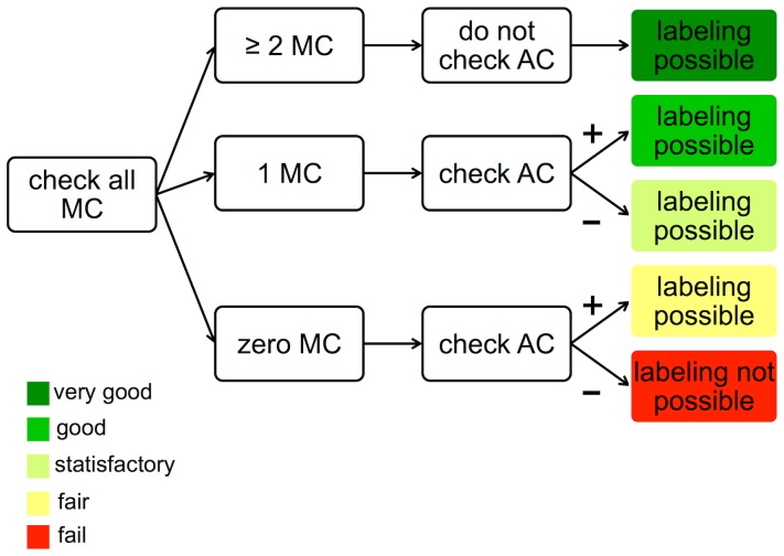
Methods: The criteria were modified to reflect the cSLO technique, followed by development of a protocol for labeling blood vessels. These criteria were based on main aspects such as central light reflex, brightness, and vessel thickness, as well as on some additional criteria such as vascular crossing patterns and the context of the vessel tree.

Results and conclusion: They demonstrated excellent inter-rater agreement and validity, which seems to indicate that labeling of images might no longer require more than one rater. This algorithm extends the diagnostic possibilities offered by OCT investigations.


Conflict of interest statement

Competing Interests: The author disclosure information is as follows: Motte J, none; Alten F, Heidelberg Engineering, Novartis, Bayer, Allergan; Ewering C, none; Osada N, none; Kadas EM, none; Brand A, none; Oberwahrenbrock T, none; Clemens CR, Heidelberg Engineering, Novartis, Bayer, Allergan; Eter N, Heidelberg Engineering, Bayer, Novartis, Allergan, Pfizer, Bausch and Lomb; Paul F, BiogenIdec, Teva, SanofiGenzyme, Merck, Novartis, Heidelberg Engineering; supported by German Research Foundation (DFG Exc 257), German Ministry for Education and Research (BMBF Competence Network Multiple Sclerosis); and Marziniak M, BiogenIdec, Teva, SanofiGenzyme, Merck, Novartis. The authors confirm that they received funding from Novartis Germany but this does not alter their adherence to all PLOS ONE policies on sharing data and materials.

Figures

Figure 1
Figure 1. Vessels 1 (v1) and 2 (v2) are larger than vessel 3 (v3) and 4 (v4).
V1 is an artery, v2 is a vein, v3 and v4 cannot be allocated clearly.
Figure 2
Figure 2. The artery shows a reflection-zone extending from the optic disc to the periphery of the retina.
The reflection of the vein cannot be traced to the periphery. Compared to the venous cross section, the reflection is smaller. The venous vessel wall appears thicker.
Figure 3
Figure 3. The darker vessel is a vein, the brighter an artery.
Figure 4
Figure 4. An example of the difference in size can be observed between vessel 1 and vessel 2.
V1 is an artery, v2 is a vein.
Figure 5
Figure 5. Three rings around the optic disc ensure vessel grading at consistent eccentricity for each vessel.
Figure 6
Figure 6. Alternate vessels with low variance of thickness and brightness.
Figure 7
Figure 7. Both the crossing and the outgoing vessels show 90° and 30°-45° angles.
Figure 8
Figure 8. Workflow for correct vessel labelling in cSLO images.
The different levels of test security are visualized by five colours. MC  =  main criteria: The central light reflex is wider in arteries and smaller in veins. Arteries are brighter than veins. Arteries are thinner than veins. AC  =  additional criteria: Arteries never cross arteries and veins never cross veins. Vessels should be seen in the context of the vessel tree.
Figure 9
Figure 9. As an example for vascular diseases cSLO images of four eyes of two patients are shown.
Picture A and B belong to a patient suffering from cerebral vasculitis. Picture C and D arise from a patient who is affected by giant-cell arteriitis. A change of vessel characteristics is not noticeable. Vessel labeling was performed using the new workflow.

References

    1. Huang D, Swanson EA, Lin CP, Schuman JS, Stinson WG, et al. (1991) Optical coherence tomography. Science 254: 1178–1181. - PMC - PubMed
    1. Frohman EM, Fujimoto JG, Frohman TC, Calabresi PA, Cutter G, et al. (2008) Optical coherence tomography: a window into the mechanisms of multiple sclerosis. Nat Clin Pract Neurol 4: 664–675. - PMC - PubMed
    1. Petzold A, de Boer JF, Schippling S, Vermersch P, Kardon R, et al. (2010) Optical coherence tomography in multiple sclerosis: a systematic review and meta-analysis. Lancet Neurol 9: 921–932. - PubMed
    1. Oberwahrenbrock T, Schippling S, Ringelstein M, Kaufhold F, Zimmermann H, et al. (2012) Retinal damage in multiple sclerosis disease subtypes measured by high-resolution optical coherence tomography. Mult Scler Int 2012: 530305. - PMC - PubMed
    1. Oberwahrenbrock T, Ringelstein M, Jentschke S, Deuschle K, Klumbies K, et al. (2013) Retinal ganglion cell and inner plexiform layer thinning in clinically isolated syndrome. Mult Scler 19: 1887–1895. - PubMed

Publication types

LinkOut - more resources