

Novel RNA hybridization method for the in situ detection of ETV1, ETV4, and ETV5 gene fusions in prostate cancer
- PMID: 25203299
- PMCID: PMC4159774
- DOI: 10.1097/PAI.0000000000000095
Novel RNA hybridization method for the in situ detection of ETV1, ETV4, and ETV5 gene fusions in prostate cancer
Abstract
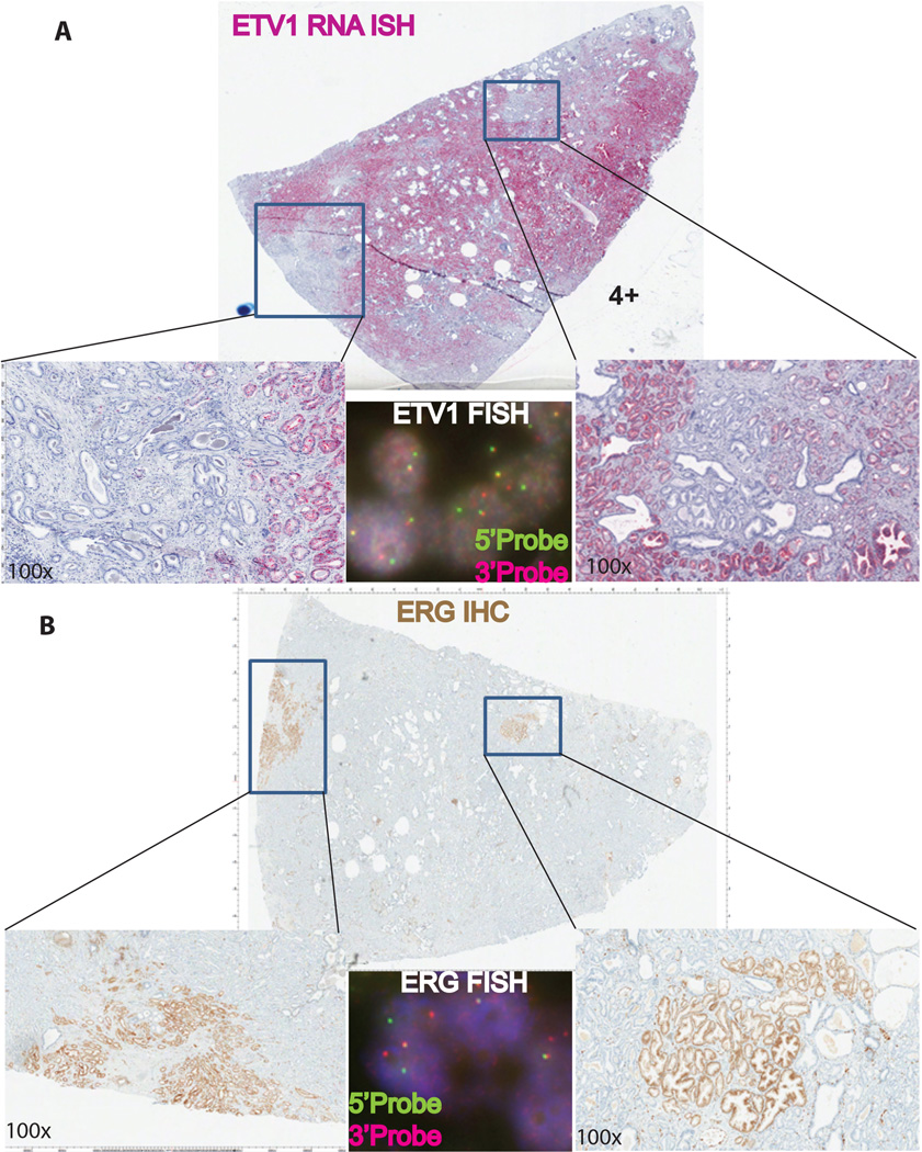
The genetic basis of 50% to 60% of prostate cancer (PCa) is attributable to rearrangements in E26 transformation-specific (ETS) (ERG, ETV1, ETV4, and ETV5), BRAF, and RAF1 genes and overexpression of SPINK1. The development and validation of reliable detection methods are warranted to classify various molecular subtypes of PCa for diagnostic and prognostic purposes. ETS gene rearrangements are typically detected by fluorescence in situ hybridization and reverse-transcription polymerase chain reaction methods. Recently, monoclonal antibodies against ERG have been developed that detect the truncated ERG protein in immunohistochemical assays where staining levels are strongly correlated with ERG rearrangement status by fluorescence in situ hybridization. However, specific antibodies for ETV1, ETV4, and ETV5 are unavailable, challenging their clinical use. We developed a novel RNA in situ hybridization-based assay for the in situ detection of ETV1, ETV4, and ETV5 in formalin-fixed paraffin-embedded tissues from prostate needle biopsies, prostatectomy, and metastatic PCa specimens using RNA probes. Further, with combined RNA in situ hybridization and immunohistochemistry we identified a rare subset of PCa with dual ETS gene rearrangements in collisions of independent tumor foci. The high specificity and sensitivity of RNA in situ hybridization provides an alternate method enabling bright-field in situ detection of ETS gene aberrations in routine clinically available PCa specimens.
Conflict of interest statement
The remaining authors declare no conflicts of interest.
Figures


References
-
- Tomlins SA, Rhodes DR, Perner S, et al. Recurrent fusion of TMPRSS2 and ETS transcription factor genes in prostate cancer. Science. 2005;310(5748):644–648. - PubMed
-
- Helgeson BE, Tomlins SA, Shah N, et al. Characterization of TMPRSS2:ETV5 and SLC45A3:ETV5 gene fusions in prostate cancer. Cancer Res. 2008;68(1):73–80. - PubMed
-
- Tomlins SA, Mehra R, Rhodes DR, et al. TMPRSS2:ETV4 gene fusions define a third molecular subtype of prostate cancer. Cancer Res. 2006;66(7):3396–400. - PubMed
Publication types
MeSH terms
Substances
Grants and funding
LinkOut - more resources
Full Text Sources
Other Literature Sources
Medical
Research Materials
Miscellaneous
