. 2014 Sep 8;26(3):390-401.
doi: 10.1016/j.ccr.2014.07.023.

Notch activation as a driver of osteogenic sarcoma

Affiliations

Notch activation as a driver of osteogenic sarcoma

Jianning Tao et al. Cancer Cell. .

Abstract

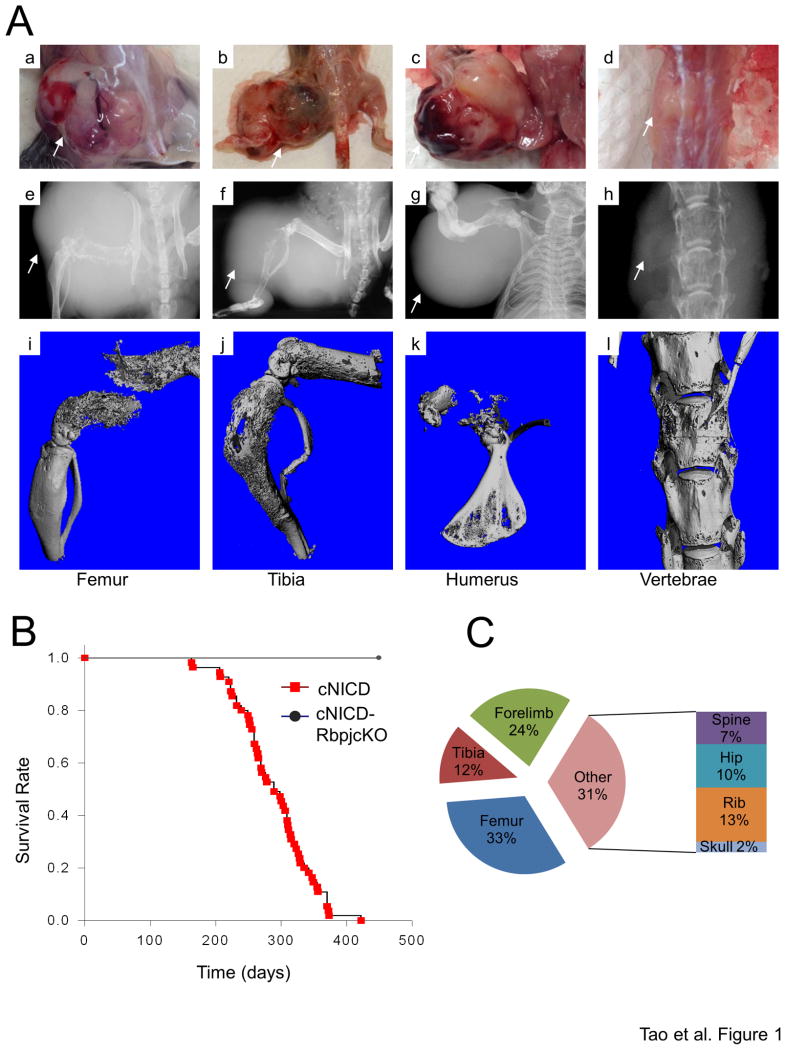
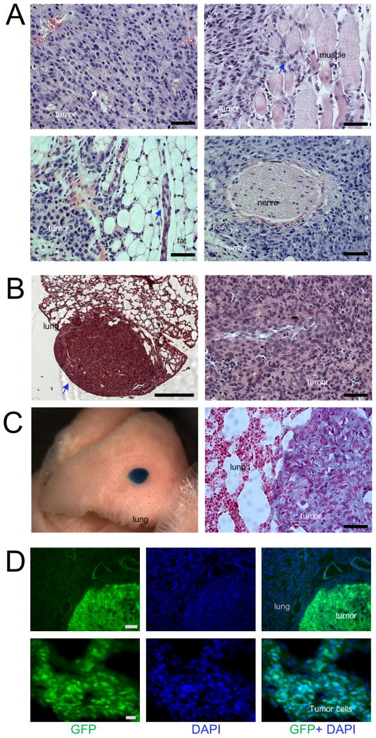
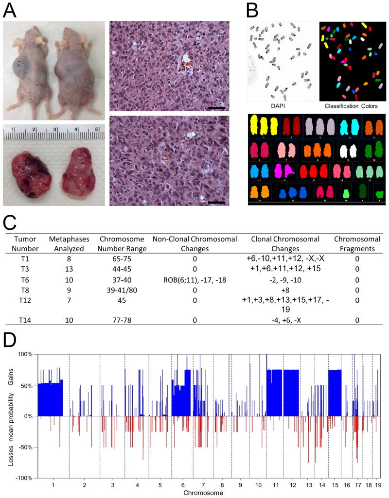
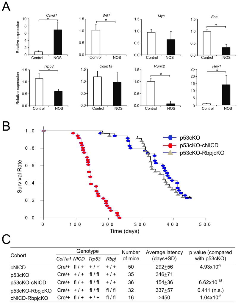
Osteogenic sarcoma (OS) is a deadly skeletal malignancy whose cause is unknown. We report here a mouse model of OS based on conditional expression of the intracellular domain of Notch1 (NICD). Expression of the NICD in immature osteoblasts was sufficient to drive the formation of bone tumors, including OS, with complete penetrance. These tumors display features of human OS; namely, histopathology, cytogenetic complexity, and metastatic potential. We show that Notch activation combined with loss of p53 synergistically accelerates OS development in mice, although p53-driven OS is not Rbpj dependent, which demonstrates a dual dominance of the Notch oncogene and p53 mutation in the development of OS. Using this model, we also reveal the osteoblasts as the potential sources of OS.


Figures

Figure 1
Figure 1. Osteosarcoma in the cNICD mice
(A) Necropsy photos (a-d), X-ray images (e-h), and micro-CT scans (i-l) of representative NOS in the femur (a, e, i), tibia (b, f, j), humerus (c, g, k), and vertebrae (d, h, l) with white arrows pointing to the tumor. (B) Kaplan-Meier tumor-free survival plots in cNICD (n=50) and cNICD-RbpjcKO (n=16) mice. (C) Location and frequency of NOS in cNICD mice. See also Figure S1.
Figure 2
Figure 2. Histopathological features of primary and metastatic NOS
(A) Histology of representative NOS tumors stained with hematoxylin and eosin (H&E) showing poorly differentiated OS with high cellularity and scattered mitoses (white arrow, top left), and invasive infiltration of tumor cells into surrounding muscle tissue (blue arrow, top right) and fat (blue arrow, bottom left), but not nerves (bottom right). Scale bars, 50 μm. (B) H&E-stained sections of representative lung metastatic OS mass (blue arrow) showing high cellularity at high magnification (right). Scale bars, 500 μm (left) and 50 μm (right). (C) Photo of a mouse lung lobe (left), which was harvested from a tri-transgenic mouse (Rosa26NICD; Col1a1 2.3kb-Cre; Rosa26LacZ) with whole mount X-gal staining shows a metastatic lesion (blue) that could not be detected macroscopically. Lung section counter-stained with eosin (right) showed that LacZ was only expressed in tumor cells (blue) but not in lung cells. Scale bars, 50 μm. (D) Immunofluorescent images stained with GFP (green) and DAPI (Blue) in lung metastatic tumor at low (top panels, scale bars, 200 μm) and high (bottom panels, 20 μm) magnification. See also Figure S2
Figure 3
Figure 3. Transplanted tumors and genomic instability in NOS
(A) Photos of representative nude mice (top left) and subcutaneous tumors (bottom left) obtained from the mice 14 days after a single s.c. injection with the NOS-T12 cell line into their flanks and H&E-stained section of primary OS T12 tumor (top right) and transplanted tumors (bottom right) derived from the injected mice. Scale bars, 50 μm. (B) Representative spectral karyotyping (SKY) evaluation of harvested cells from NOS-T12 cell line. (C) Summary of SKY results for the indicated tumors. (D) Summary of percentages of gains (positive axis) and losses (negative axis) for chromosomes 1–19, as determined by analysis of copy number variation (CNV), compared tumor to non-tumor controls using whole genome sequencing (CNV-seq). See also Figure S3.
Figure 4
Figure 4. Genetic interaction of Notch activation and p53 loss of function in OS development
(A) Quantitative RT-PCR analysis of genes associated with human OS comparing Notch-induced OS with normal bone (mean ± SD of three mice per group). *p<0.05 (Student's t test). (B) Kaplan-Meier survival plots of p53cKO, p53cKO-cNICD and p53cKO-RbpjcKO mice. (C) Summary of genetic studies from each cohort of mice. Mice without bone tumors were sacrificed by 450 days of age in cohorts of p53cKO (8 of 35), p53cKO-RbpjcKO (8 of 34), and cNICD-RbpjcKO (16 of 16). The p values were calculated using SigmaPlot through all Pairwise Multiple Comparison Procedures (Holm-Sidak method). fl, Flox; n.s, not significant. See also Figure S4
Figure 5
Figure 5. Expression signature of NOS compared to p53-loss-induced OS
(A) Unsupervised hierarchical clustering of mouse samples and heat map of RNA-sequencing data between Notch OS or p53-loss-induced tumors (p53 OS) (B) Ingenuity Pathways Analysis (IPA) of cancer canonical signaling pathways associated with significantly regulated genes (p<0.05) in Notch OS versus control. Significantly changed pathways only observed in NOS are colored in light blue, and those changed in both NOS and p53-induced OS are shown in black. (C) GSEA results for each of the indicated human sarcomas for NOS primary malignant tumors. Asterisks indicate statistical significance. NOM p val, nominal p value; FDR, false discovery rate; FWER, family-wise error rate. See also Figure S5, Tables S1, S2, S3, and S4.
Figure 6
Figure 6. Candidate cells of origin in murine OS models
(A) Kaplan-Meier survival plots of p53cKO, Osxp53cKO and Prxp53cKO mice. (B) Summary of genetic studies from each cohort of mice. The p values were calculated using SigmaPlot (Holm-Sidak method). *There is no significant difference (p=0.153) between Osxp53cKO and Prxp53cKO. (C) Summary of results from various genetic experiments. Loss of function of p53 can induce OS at the early and late stage of MSC differentiation, but Notch gain of function can only induce OS at a late stage.

References

    1. Allen TD, Rodriguez EM, Jones KD, Bishop JM. Activated Notch1 induces lung adenomas in mice and cooperates with Myc in the generation of lung adenocarcinoma. Cancer research. 2011;71:6010–6018. - PMC - PubMed
    1. Baird K, Davis S, Antonescu CR, Harper UL, Walker RL, Chen Y, Glatfelter AA, Duray PH, Meltzer PS. Gene expression profiling of human sarcomas: insights into sarcoma biology. Cancer research. 2005;65:9226–9235. - PubMed
    1. Berman SD, Calo E, Landman AS, Danielian PS, Miller ES, West JC, Fonhoue BD, Caron A, Bronson R, Bouxsein ML, et al. Metastatic osteosarcoma induced by inactivation of Rb and p53 in the osteoblast lineage. Proceedings of the National Academy of Sciences of the United States of America. 2008;105:11851–11856. - PMC - PubMed
    1. Beverly LJ, Capobianco AJ. Perturbation of Ikaros isoform selection by MLV integration is a cooperative event in Notch(IC)-induced T cell leukemogenesis. Cancer cell. 2003;3:551–564. - PubMed
    1. Cai Y, Mohseny AB, Karperien M, Hogendoorn PC, Zhou G, Cleton-Jansen AM. Inactive Wnt/beta-catenin pathway in conventional high-grade osteosarcoma. The Journal of pathology. 2009;220:24–33. - PubMed

Publication types

MeSH terms