

Chronic thromboembolic pulmonary hypertension: do not miss the chance for an early diagnosis
- PMID: 25203436
- PMCID: PMC4159243
- DOI: 10.12659/AJCR.891014
Chronic thromboembolic pulmonary hypertension: do not miss the chance for an early diagnosis
Abstract
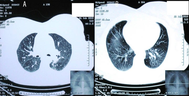
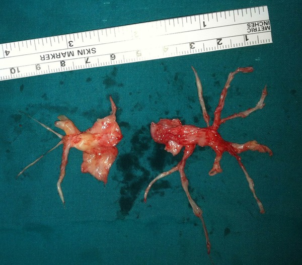
Background: Chronic thromboembolic pulmonary hypertension most often results from obstruction of the pulmonary vascular bed by nonresolving thromboemboli. Misdiagnosis of the disease is common because patients often present with subtle or nonspecific symptoms. Furthermore, some features in chest imaging may mimic parenchymal lung disease. The most clinically important mimic in high-resolution chest tomography is air trapping, which can be seen in a variety of small airway diseases.
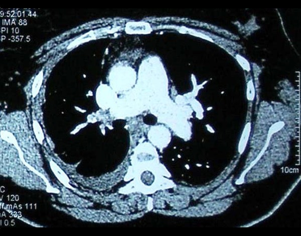
Case report: We present the case of a 45-year-old woman with a long history of dyspnea and exercise intolerance, misdiagnosed with allergic alveolitis. The diagnosis of CTEPH was finally established with computed tomography (CT) angiography and hemodynamics.
Conclusions: Chronic thromboembolism is under-diagnosed and also frequently misdiagnosed in clinical practice. The present report aims to increase the awareness of clinicians towards an accurate diagnosis of the disease, which is necessary for the early referral of CTEPH patients for operability.
Figures
Similar articles
-
Imaging techniques in chronic thromboembolic pulmonary hypertension.Curr Opin Pulm Med. 2013 Sep;19(5):562-74. doi: 10.1097/MCP.0b013e3283645a00. Curr Opin Pulm Med. 2013. PMID: 23880705 Review.
-
Diagnosis of chronic thromboembolic pulmonary hypertension.Eur Respir Rev. 2017 Mar 15;26(143):160108. doi: 10.1183/16000617.0108-2016. Print 2017 Jan. Eur Respir Rev. 2017. PMID: 28298387 Free PMC article. Review.
-
Chronic Thromboembolic Pulmonary Hypertension: Pearls and Pitfalls of Diagnosis.Methodist Debakey Cardiovasc J. 2016 Oct-Dec;12(4):199-204. doi: 10.14797/mdcj-12-4-199. Methodist Debakey Cardiovasc J. 2016. PMID: 28289494 Free PMC article. Review.
-
[Successful operative case of chronic thromboembolic pulmonary hypertension clinically diagnosed as bronchial asthma].Nihon Kokyuki Gakkai Zasshi. 2010 Nov;48(11):836-41. Nihon Kokyuki Gakkai Zasshi. 2010. PMID: 21141063 Japanese.
-
Recent progress in the diagnosis and management of chronic thromboembolic pulmonary hypertension.Respir Investig. 2013 Sep;51(3):134-46. doi: 10.1016/j.resinv.2013.02.005. Epub 2013 Apr 30. Respir Investig. 2013. PMID: 23978639 Review.
Cited by
-
A Review of Clinical Trial Endpoints of Patients with Pulmonary Arterial Hypertension and Chronic Thromboembolic Pulmonary Hypertension and How They Relate to Patient Outcomes in the United States.J Manag Care Spec Pharm. 2017 Jan;23(1):92-104. doi: 10.18553/jmcp.2017.23.1.92. J Manag Care Spec Pharm. 2017. PMID: 28025931 Free PMC article. Review.
References
-
- Hoeper MM, Mayer E, Simonneau G, Rubin LJ. Chronic thromboembolic pulmonary hypertension. Circulation. 2006;113:2011–20. - PubMed
-
- Pengo V, Lensing AW, Prins MH, et al. Thromboembolic Pulmonary Hypertension Study Group. Incidence of chronic thromboembolic pulmonary hypertension after pulmonary embolism. N Engl J Med. 2004;350:2257–64. - PubMed
-
- Pepke-Zaba J, Delcroix M, Lang I, et al. Chronic thromboembolic pulmonary hypertension (CTEPH): results from an international prospective registry. Circulation. 2011;124:1973–81. - PubMed
-
- Lang IM, Klepetko W. Chronic thromboembolic pulmonary hypertension: an updated review. Curr Opin Cardiol. 2008;23:555–59. - PubMed
-
- Coulden R. State-of-the-art imaging techniques in chronic thromboembolic pulmonary hypertension. Proc Am Thorac Soc. 2006;3:577–83. - PubMed
Publication types
MeSH terms
LinkOut - more resources
Full Text Sources
Other Literature Sources
Medical
