. 2014 Sep 9;9(9):e107398.
doi: 10.1371/journal.pone.0107398. eCollection 2014.

A diffusion-tensor-based white matter atlas for rhesus macaques

Affiliations

A diffusion-tensor-based white matter atlas for rhesus macaques

Elizabeth Zakszewski et al. PLoS One. .

Abstract

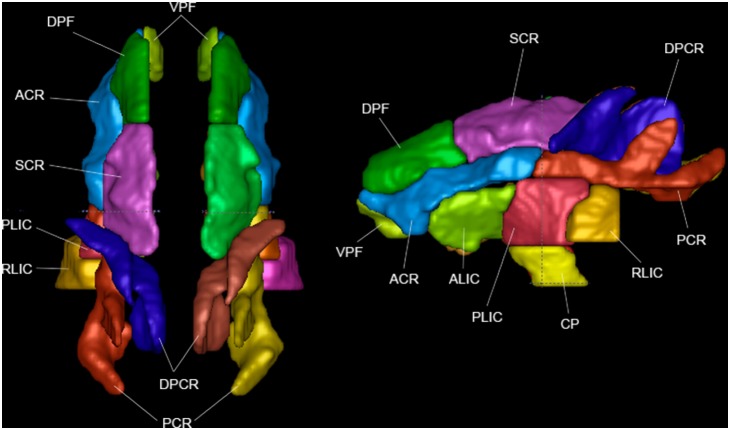
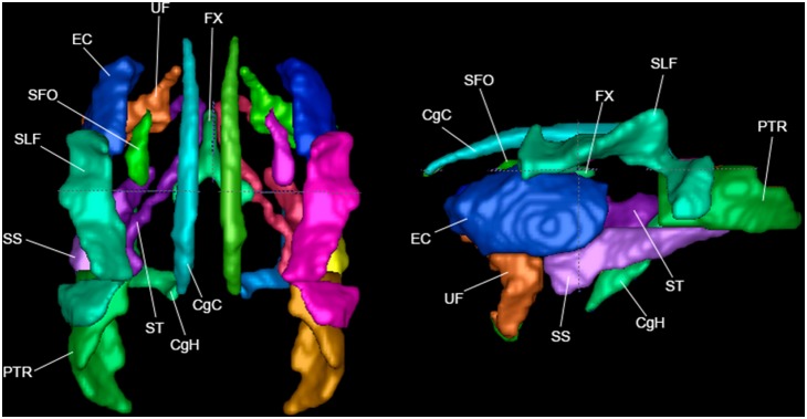
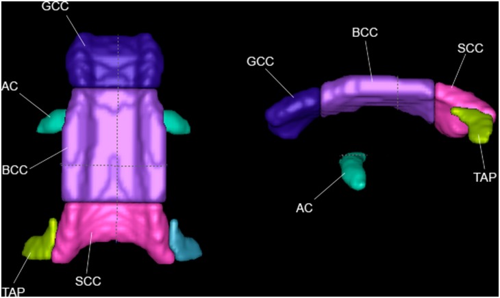
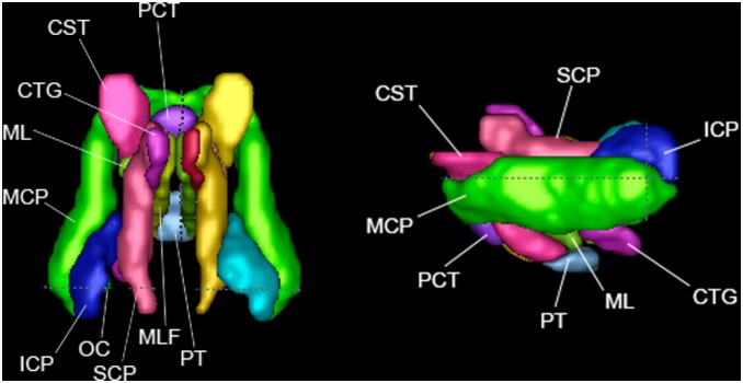
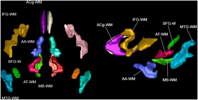
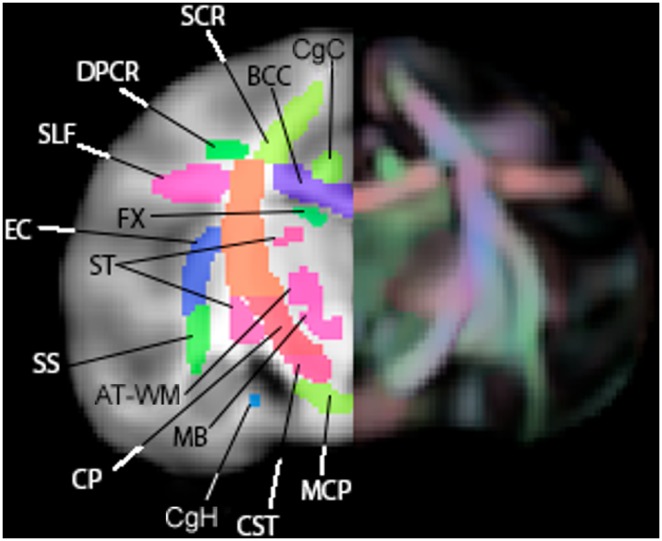
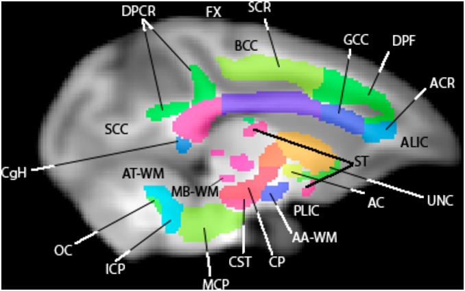
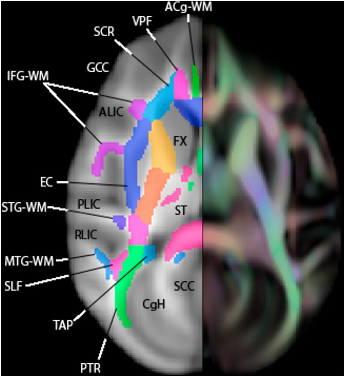
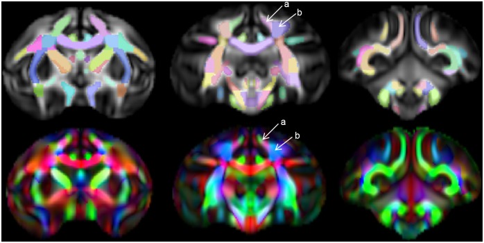
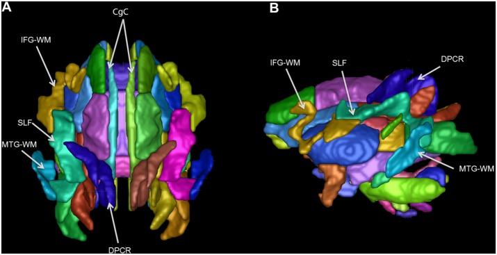
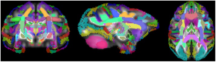
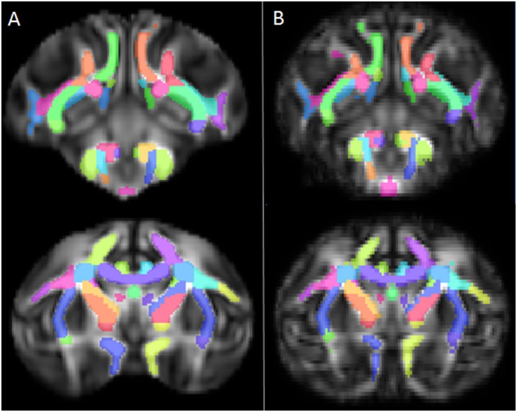
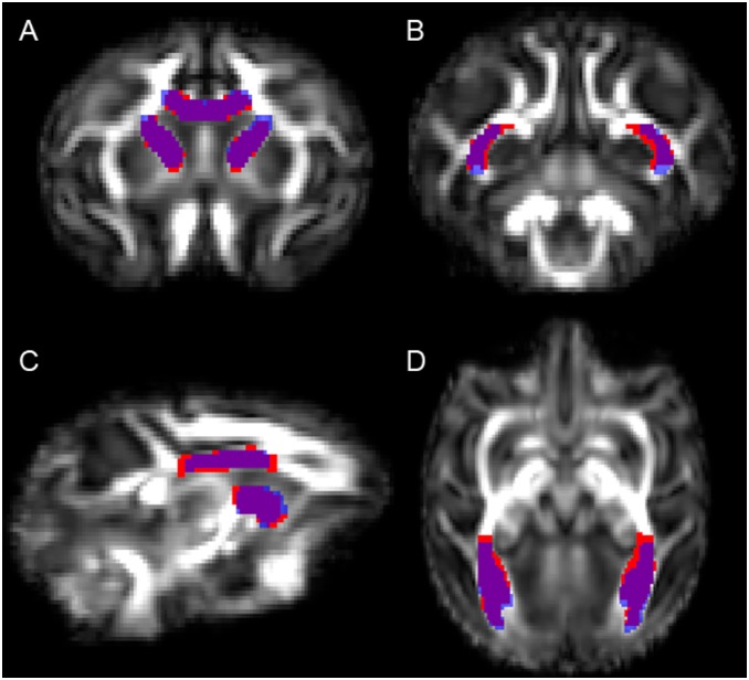
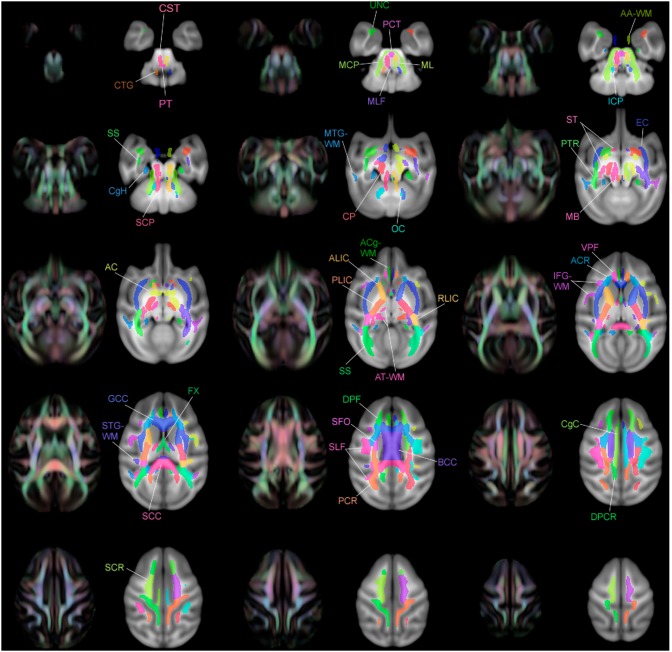
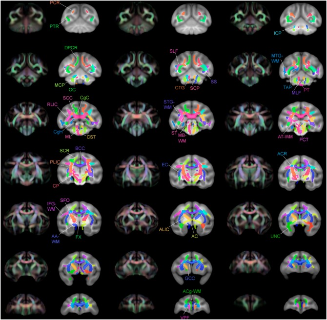
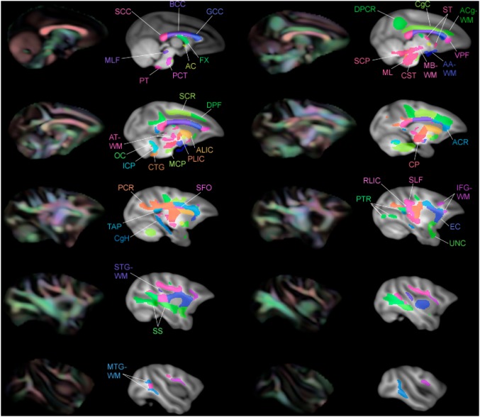
Atlases of key white matter (WM) structures in humans are widely available, and are very useful for region of interest (ROI)-based analyses of WM properties. There are histology-based atlases of cortical areas in the rhesus macaque, but none currently of specific WM structures. Since ROI-based analysis of WM pathways is also useful in studies using rhesus diffusion tensor imaging (DTI) data, we have here created an atlas based on a publicly available DTI-based template of young rhesus macaques. The atlas was constructed to mimic the structure of an existing human atlas that is widely used, making results translatable between species. Parcellations were carefully hand-drawn on a principle-direction color-coded fractional anisotropy image of the population template. The resulting atlas can be used as a reference to which registration of individual rhesus data can be performed for the purpose of white-matter parcellation. Alternatively, specific ROIs from the atlas may be warped into individual space to be used in ROI-based group analyses. This atlas will be made publicly available so that it may be used as a resource for DTI studies of rhesus macaques.


Conflict of interest statement

Competing Interests: The authors have declared that no competing interests exist.

Figures

Figure 1
Figure 1. Projection fibers, in top view and left view.
Figure 2
Figure 2. Association fibers in top view and left view.
Figure 3
Figure 3. Commissural fibers in top view and left view.
Figure 4
Figure 4. Brainstem fibers in top view and left view.
Figure 5
Figure 5. Short range WM regions in top and left view.
Figure 6
Figure 6. A coronal slice of the WM atlas.
Overlaid on a T1-W template (left) and an FA color map (right). AT-WM = adjacent thalamus, BCC = body of corpus callosum, CgC = superior cingulum bundle, CgH = perihippocampal cingulum tract, CP = cerebral peduncle, CST = corticospinal tract, DPCR = dorsal posterior corona radiata, EC = external capsule, FX = fornix, MCP = middle cerebellar peduncle, SCR = superior corona radiata, SLF = superior longitudinal fasciculus, SS = sagittal striatum, ST = stria terminalus.
Figure 7
Figure 7. A sagittal slice of the WM atlas.
Overlaid on a T1-W template. AC = anterior commissure, AT-WM = adjacent thalamus, AA-WM = adjacent amygdala, ALIC = anterior limb of the internal capsule, BCC = body of corpus callosum, CgH = perihippocampal cingulum tract, CP = cerebral peduncle, CST = corticospinal tract, DPCR = dorsal posterior corona radiata, DPF = dorsal prefrontal, FX = fornix, GCC = genu of corpus callosum, ICP = inferior cerebellar peduncle, MB-WM = midbrain, MCP = middle cerebellar peduncle, OC = olivocerebellar tract, PLIC = posterior limb of the internal capsule, SCC = splenium of corpus callosum, ST = stria terminalis, UNC = uncinate fasciculus.
Figure 8
Figure 8. An axial slice of the WM atlas.
Overlaid on a T1-W template (left) and an FA color map (right). ACg-WM = anterior cingulum, ALIC = anterior limb of the internal capsule, CgC = superior cingulum bundle, CgH = perihippocampal cingulum tract, EC = external capsule, FX = fornix, GCC = genu of corpus callosum, IFG-WM = inferior frontal gyrus WM, MTG-WM = middle temporal gyrus WM, PLIC = posterior limb of the internal capsule, PTR = posterior thalamic radiation, RLIC = retrolenticular limb of the internal capsule, SCC = splenium of corpus callosum, SCR = superior corona radiata, SLF = superior longitudinal fasciculus, ST = stria terminalus, STG-WM = superior temporal gyrus WM, TAP = tapetum.
Figure 9
Figure 9. Delineation between tracts.
Top: FA maps of select slices of template (anterior on left, posterior on right) with atlas ROIs overlaid. Bottom: Color FA map of matching slices, showing delineation between tracts. Arrows indicate the division between the SCR (a) and the DPCR (b), and the directional (color) changes that guided the decision of where to put this boundary.
Figure 10
Figure 10. 3D renderings of atlas.
(A) Superior 3D surface rendered view of atlas. (B) Left 3D surface rendered view of atlas. The superior cingulum bundle (CgC) and many peripheral WM tracts are pointed out. IFG-WM = inferior frontal gyrus WM, SLF = superior longitudinal fasciculus, MTG-WM = middle temporal gyrus WM, DPCR = dorsal posterior corona radiata.
Figure 11
Figure 11. WM atlas and digital Paxinos atlas of GM regions overlaid on UWRMAC-DTI271 template FA map.
Figure 12
Figure 12. Registering to a different template.
Two representative coronal slices of the atlas in its default space (A) and after registering with nearest-neighbor interpolation to a template made from a smaller number of young rhesus macaques (B), which were not a part of the original DTI template used to draw the atlas. White matter coverage is equally good in the registered atlas, qualitatively.
Figure 13
Figure 13. Image showing overlap between manual (blue) and automatic (red) methods of defining tracts.
Manual definitions of the corpus callosum and ALIC (anterior limb of the internal capsule (A, C) and the PTR (posterior thalamic radiation) (B, D) were drawn on the different template and compared to automatically defined regions. Overlap agreement appears in purple. Slices were selected that illustrated areas where the two segmentation methods disagreed.
Figure 14
Figure 14. Multiple slices of atlas overlaid on T1-W template beside directional FA color image, axial view.
Figure 15
Figure 15. Multiple slices of atlas overlaid on T1-W template beside directional FA color image, coronal view.
Figure 16
Figure 16. Multiple slices of atlas overlaid on T1-W template beside directional FA color image, sagittal view.

References

    1. Baur V, Hänggi J, Rufer M, Delsignore A, Jäncke L, et al. (2011) White matter alterations in social anxiety disorder. J Psychiatr Res 45: 1366–1372 10.1016/j.jpsychires.2011.05.007 - DOI - PubMed
    1. Korgaonkar MS, Grieve SM, Koslow SH, Gabrieli JD, Gordon E, et al. (2011) Loss of white matter integrity in major depressive disorder: Evidence using tract-based spatial statistical analysis of diffusion tensor imaging. Hum Brain Mapp 32: 2161–2171. - PMC - PubMed
    1. Motzkin JC, Newman JP, Kiehl KA, Koenigs M (2011) Reduced prefrontal connectivity in psychopathy. J Neurosci 31: 17348–17357. - PMC - PubMed
    1. Mak-Fan KM, Morris D, Vidal J, Anagnostou E, Roberts W, et al. (2012) White matter and development in children with an autism spectrum disorder. Autism 17(5): 541–557. - PubMed
    1. Stricker N, Schweinsburg B, Delano-Wood L, Wierenga C, Bangen K, et al. (2009) Decreased white matter integrity in late-myelinating fiber pathways in Alzheimer’s disease supports retrogenesis. Neuroimage 45: 10–16. - PMC - PubMed

Publication types

MeSH terms

LinkOut - more resources