

Protein microarray for complex apoptosis monitoring of dysplastic oral keratinocytes in experimental photodynamic therapy
- PMID: 25204017
- PMCID: PMC4125699
- DOI: 10.1186/0717-6287-47-33
Protein microarray for complex apoptosis monitoring of dysplastic oral keratinocytes in experimental photodynamic therapy
Abstract
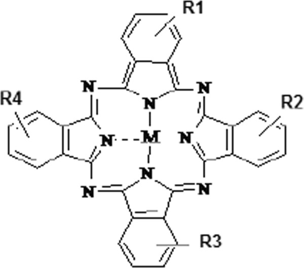
Background: Photodynamic therapy is an alternative treatment of muco-cutaneous tumors that uses a light source able to photoactivate a chemical compound that acts as a photosensitizer. The phthalocyanines append to a wide chemical class that encompasses a large range of compounds; out of them aluminium-substituted disulphonated phthalocyanine possesses a good photosensitizing potential.
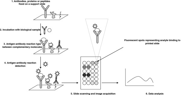
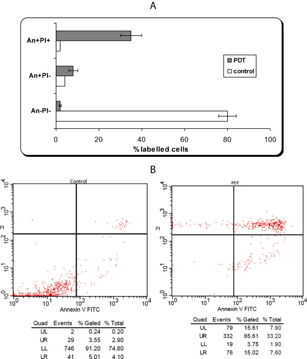
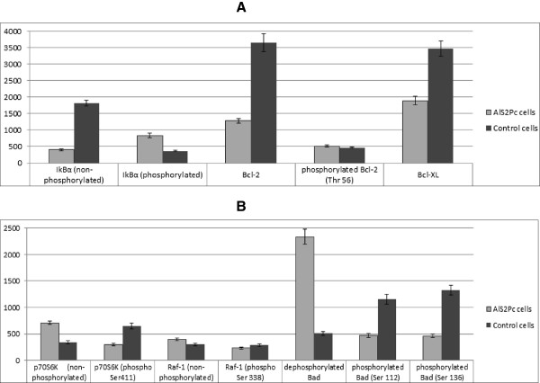
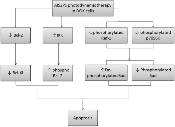
Results: The destructive effects of PDT with aluminium-substituted disulphonated phthalocyanine are achieved by induction of apoptosis in tumoral cells as assessed by flow cytometry analysis. Using protein microarray we evaluate the possible molecular pathways by which photodynamic therapy activates apoptosis in dysplastic oral keratinocytes cells, leading to the tumoral cells destruction. Among assessed analytes, Bcl-2, P70S6K kinase, Raf-1 and Bad proteins represent the apoptosis related biomolecules that showed expression variations with the greatest amplitude.
Conclusions: Up to date, the intimate molecular apoptotic mechanisms activated by photodynamic therapy with this type of phthalocyanine in dysplastic human oral keratinocytes are not completely elucidated. With protein microarray as high-throughput proteomic approach a better understanding of the manner in which photodynamic therapy leads to tumoral cell destruction can be obtained, by depicting apoptotic molecules that can be potentially triggered in future anti-tumoral therapies.
Figures
References
-
- Silva JN, Filipe P, Morlière P, Mazière JC, Freitas JP, Gomes MM, Santus R. Photodynamic therapy: dermatology and ophthalmology as main fields of current applications in clinic. Biomed Mater Eng. 2008;18(4–5):319–327. - PubMed
-
- Ion R-M. Photosensitizers in Medicine, Environment, and Security. Netherlands: Springer; 2012. The use of Phthalocyanines and Related Complexes in Photodynamic Therapy; pp. 315–349.
-
- Matei C, Tampa M, Ion RM, Neagu M, Constantin C. Photodynamic properties of aluminium sulphonated phthalocyanines in human dysplazic oral keratinocytes experimental model. Digest Journal of Nanomaterials and Biostructures. 2012;7(4):1535–1547.
Publication types
MeSH terms
Substances
LinkOut - more resources
Full Text Sources
Medical
Molecular Biology Databases
Research Materials
Miscellaneous