

Isoform-specific SCF(Fbw7) ubiquitination mediates differential regulation of PGC-1α
- PMID: 25204433
- PMCID: PMC4596538
- DOI: 10.1002/jcp.24812
Isoform-specific SCF(Fbw7) ubiquitination mediates differential regulation of PGC-1α
Abstract
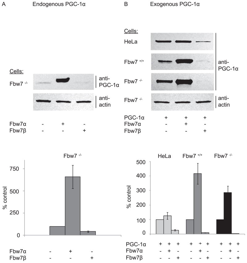
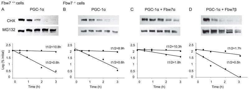
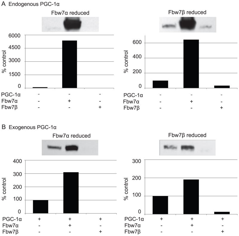
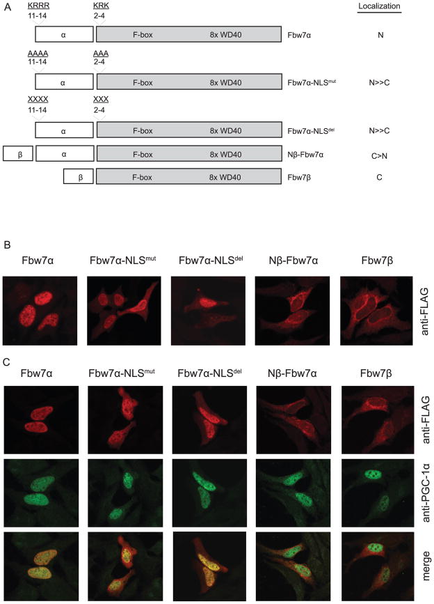
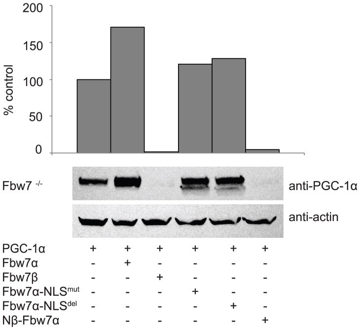
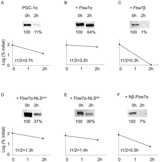
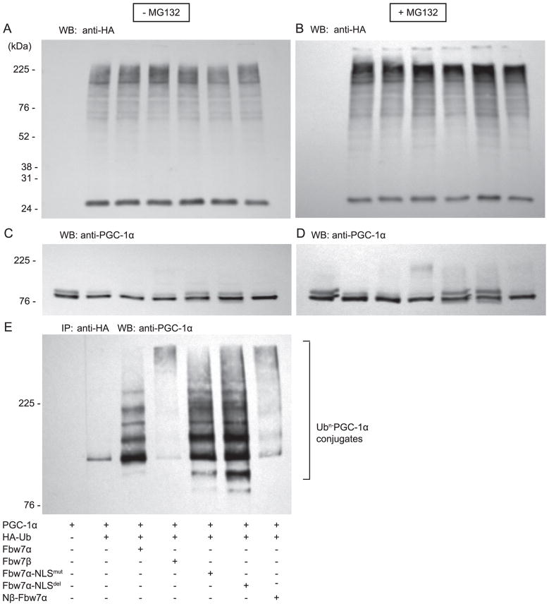
The E3 ubiquitin ligase and tumor suppressor SCF(Fbw7) exists as three isoforms that govern the degradation of a host of critical cell regulators, including c-Myc, cyclin E, and PGC-1α. Peroxisome proliferator activated receptor-gamma coactivator 1α (PGC-1α) is a transcriptional coactivator with broad effects on cellular energy metabolism. Cellular PGC-1α levels are tightly controlled in a dynamic state by the balance of synthesis and rapid degradation via the ubiquitin-proteasome system. Isoform-specific functions of SCF(Fbw7) are yet to be determined. Here, we show that the E3 ubiquitin ligase, SCF(Fbw7), regulates cellular PGC-1α levels via two independent, isoform-specific, mechanisms. The cytoplasmic isoform (SCF(Fbw7β)) reduces cellular PGC-1α levels via accelerated ubiquitin-proteasome degradation. In contrast, the nuclear isoform (SCF(Fbw7α)) increases cellular PGC-1α levels and protein stability via inhibition of ubiquitin-proteasomal degradation. When nuclear Fbw7α proteins are redirected to the cytoplasm, cellular PGC-1α protein levels are reduced through accelerated ubiquitin-proteasomal degradation. We find that SCF(Fbw7β) catalyzes high molecular weight PGC-1α-ubiquitin conjugation, whereas SCF(Fbw7α) produces low molecular weight PGC-1α-ubiquitin conjugates that are not effective degradation signals. Thus, selective ubiquitination by specific Fbw7 isoforms represents a novel mechanism that tightly regulates cellular PGC-1α levels. Fbw7 isoforms mediate degradation of a host of regulatory proteins. The E3 ubiquitin ligase, Fbw7, mediates PGC-1α levels via selective isoform-specific ubiquitination. Fbw7β reduces cellular PGC-1α via ubiquitin-mediated degradation, whereas Fbw7α increases cellular PGC-1α via ubiquitin-mediated stabilization.
© 2014 Wiley Periodicals, Inc.
Conflict of interest statement
Conflict of Interest Disclosure: No conflicts to disclose (JST-A, MA, AO, ALS).
Figures
Similar articles
-
Stringent Control of NFE2L3 (Nuclear Factor, Erythroid 2-Like 3; NRF3) Protein Degradation by FBW7 (F-box/WD Repeat-containing Protein 7) and Glycogen Synthase Kinase 3 (GSK3).J Biol Chem. 2015 Oct 23;290(43):26292-302. doi: 10.1074/jbc.M115.666446. Epub 2015 Aug 25. J Biol Chem. 2015. PMID: 26306035 Free PMC article.
-
Fbw7α and Fbw7γ collaborate to shuttle cyclin E1 into the nucleolus for multiubiquitylation.Mol Cell Biol. 2013 Jan;33(1):85-97. doi: 10.1128/MCB.00288-12. Epub 2012 Oct 29. Mol Cell Biol. 2013. PMID: 23109421 Free PMC article.
-
Regulation of GATA-binding protein 2 levels via ubiquitin-dependent degradation by Fbw7: involvement of cyclin B-cyclin-dependent kinase 1-mediated phosphorylation of THR176 in GATA-binding protein 2.J Biol Chem. 2015 Apr 17;290(16):10368-81. doi: 10.1074/jbc.M114.613018. Epub 2015 Feb 10. J Biol Chem. 2015. PMID: 25670854 Free PMC article.
-
Physiological Functions of FBW7 in Metabolism.Horm Metab Res. 2022 May;54(5):280-287. doi: 10.1055/a-1816-8903. Epub 2022 May 9. Horm Metab Res. 2022. PMID: 35533672 Review.
-
Role of the ubiquitin ligase Fbw7 in cancer progression.Cancer Metastasis Rev. 2012 Jun;31(1-2):75-87. doi: 10.1007/s10555-011-9330-z. Cancer Metastasis Rev. 2012. PMID: 22124735 Review.
Cited by
-
The hitchhiker's guide to PGC-1α isoform structure and biological functions.Diabetologia. 2015 Sep;58(9):1969-77. doi: 10.1007/s00125-015-3671-z. Epub 2015 Jun 25. Diabetologia. 2015. PMID: 26109214 Review.
-
FBXW7 alleviates hyperglycemia-induced endothelial oxidative stress injury via ROS and PARP inhibition.Redox Biol. 2022 Dec;58:102530. doi: 10.1016/j.redox.2022.102530. Epub 2022 Nov 15. Redox Biol. 2022. PMID: 36427396 Free PMC article.
-
Peroxisome Proliferator-activated Receptor γ Coactivator-1 α Isoforms Selectively Regulate Multiple Splicing Events on Target Genes.J Biol Chem. 2016 Jul 15;291(29):15169-84. doi: 10.1074/jbc.M115.705822. Epub 2016 May 26. J Biol Chem. 2016. PMID: 27231350 Free PMC article.
-
ERK kinase phosphorylates and destabilizes the tumor suppressor FBW7 in pancreatic cancer.Cell Res. 2015 May;25(5):561-73. doi: 10.1038/cr.2015.30. Epub 2015 Mar 10. Cell Res. 2015. PMID: 25753158 Free PMC article.
-
PGC1α: Friend or Foe in Cancer?Genes (Basel). 2018 Jan 22;9(1):48. doi: 10.3390/genes9010048. Genes (Basel). 2018. PMID: 29361779 Free PMC article. Review.
References
-
- Arany Z, Foo SY, Ma Y, Ruas JL, Bommi-Reddy A, Girnun G, Cooper M, Laznik D, Chinsomboon J, Rangwala SM, Baek KH, Rosenzweig A, Spiegelman BM. HIF-independent regulation of VEGF and angiogenesis by the transcriptional coactivator PGC-1 alpha. Nature. 2008;451:1008–1012. - PubMed
Publication types
MeSH terms
Substances
Grants and funding
LinkOut - more resources
Full Text Sources
Other Literature Sources
